Visual Fields, Retinal Duplex, and Photoreceptors - Neurophysiology and Perception Spring 2026
 Call Kai
 Learn
Learn Practice Test
 Spaced Repetition
 Match
Match Flashcards
 Knowt Play
1/126

There's no tags or description
Looks like no tags are added yet.
Name | Mastery | Learn | Test | Matching | Spaced | Call with Kai |
|---|
No analytics yet
Send a link to your students to track their progress
127 Terms
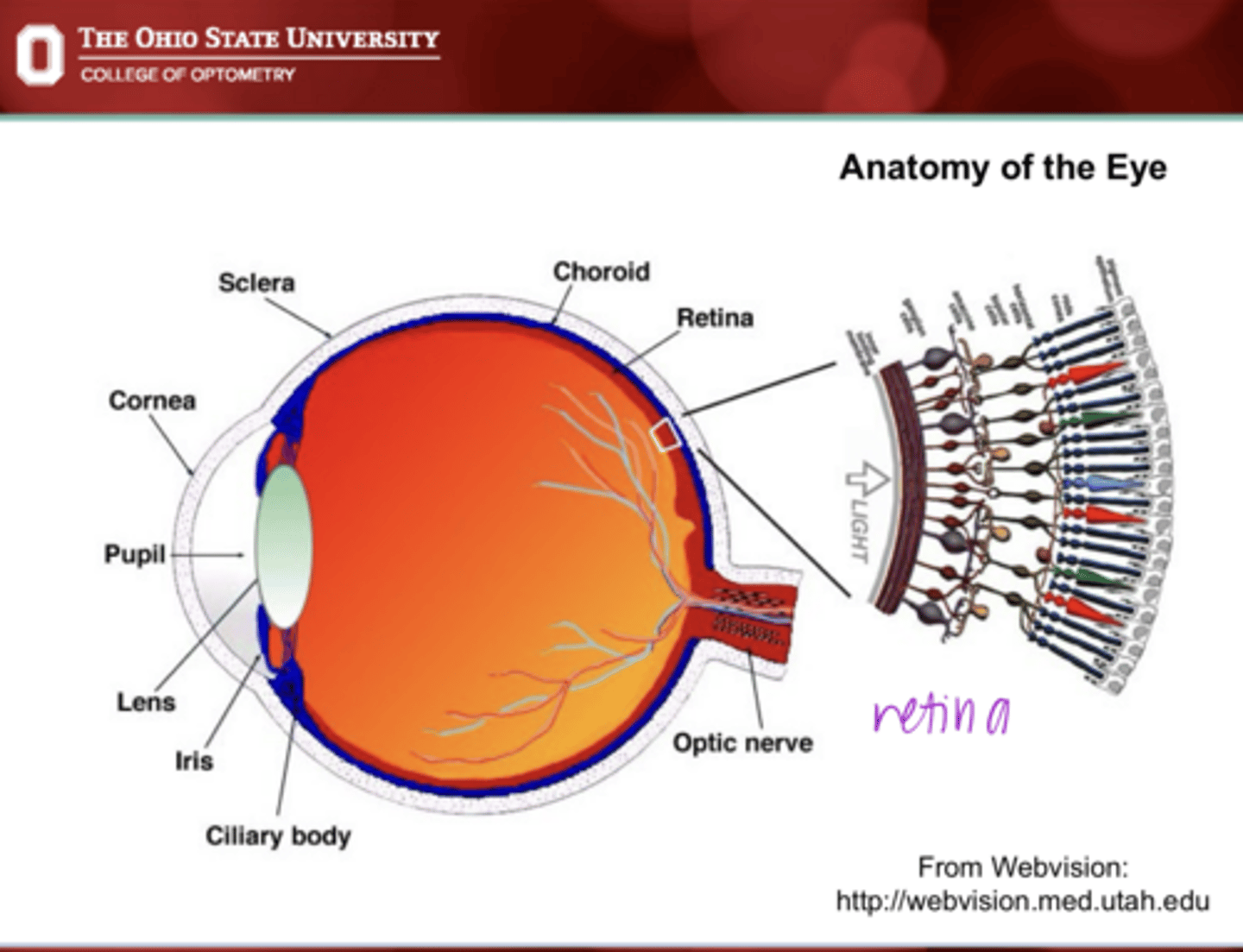
Anatomy of the Eye Pic
Anatomy of the Eye Pic
nervous system containing the brain and the spinal cord
What does the CNS include?
cranial nerves, spinal nerves, and autonomic nervous system
What does the PNS include?
No, the retina is a part of the CNS and an extension of the brain. Defined area of the brain that we can see
Because the optic nerve is a cranial nerve, should it be considered part of the PNS?
central nervous system (CNS)
The optic nerve (CN II) is more like a tract of the _____ system than a peripheral nerve
neural tube -- diencephalon
(same embryological origin as the thalamus of the brain)
The retina and the optic nerve develop from what?
oligodendrocytes
Myelin (insulation material) surrounding the optic nerve is produced by what?
blood-retina barrier
There is a specialized _____ that separates the vascular and the nervous system. It is hard to get drugs to target the retina and brain d/t this.
narrow
Retinal synapses, where NT is released, are (narrow/thick)
glial cells
What are the retinal synapses ensheathed by?
will NOT
The optic nerve (will/will not) naturally regenerate when damaged
yes
Can peripheral nerves (PNS) regenerate?
1) transduce visual images into electrochemical signals
2) perform initial processing of this signal
3) convert the processed signal into signal that can be sent and interpreted by the brain
What are the 3 principal functions of the retina?
yes
Does the retina have a organized design with multiple layers?
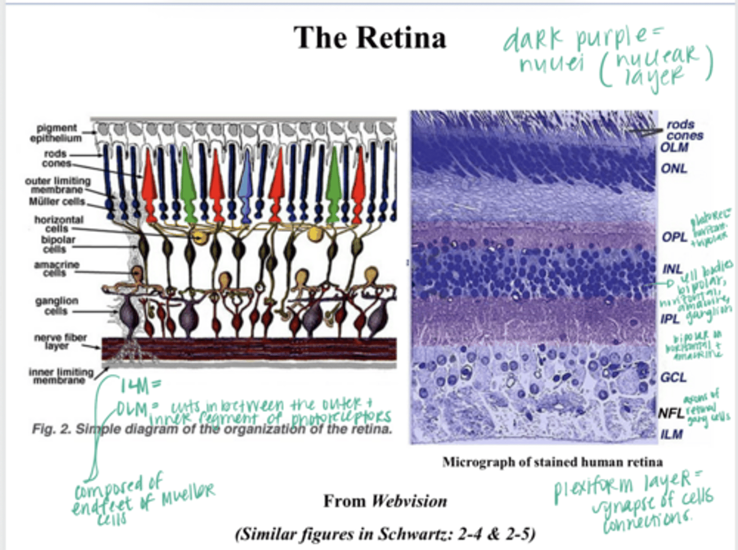
RPE
What structure of the retina is furthest from the lens and cornea?
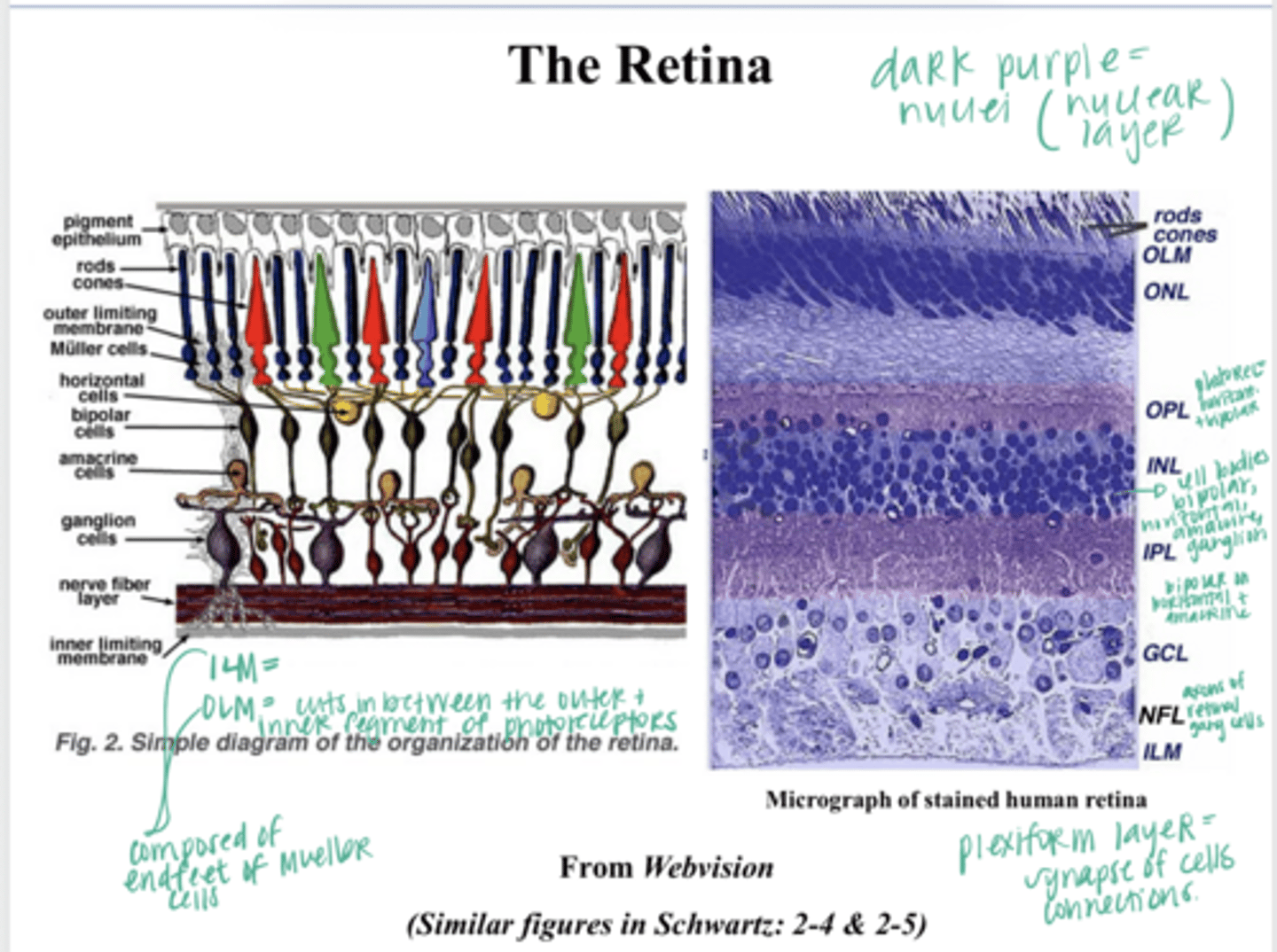
to bipolar cells
Where do photoreceptors directly send their impulses?
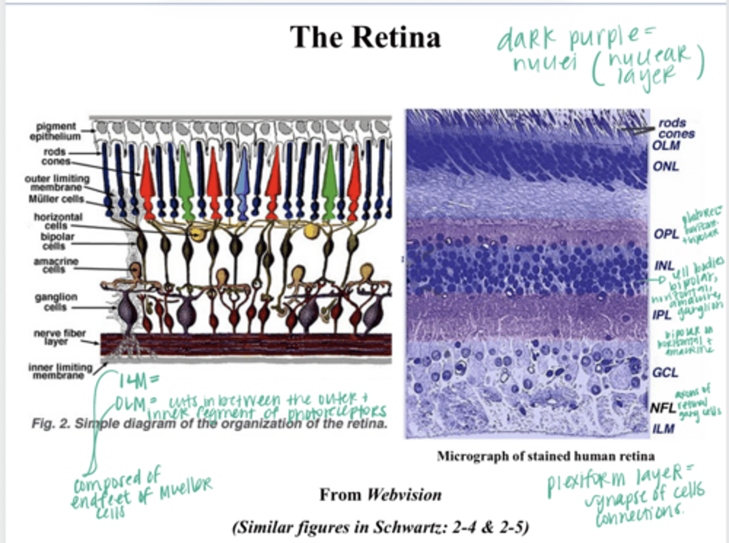
to ganglion cells (inner retina)
Where do bipolar cells send their impulses?
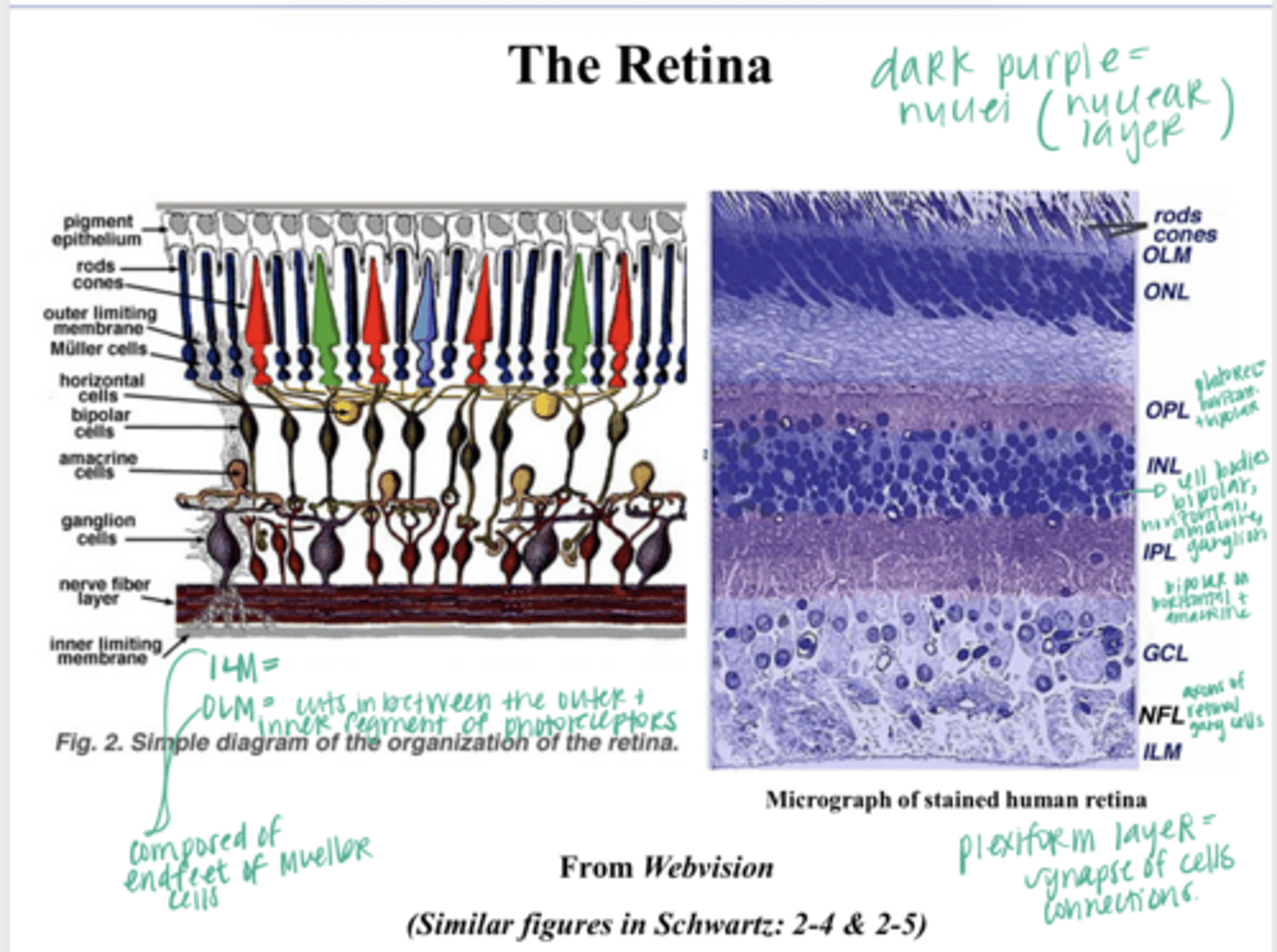
horizontal and amacrine cells
What are the lateral cells of the retina that modify the signals from photoreceptors-->bipolar--> ganglion cells?
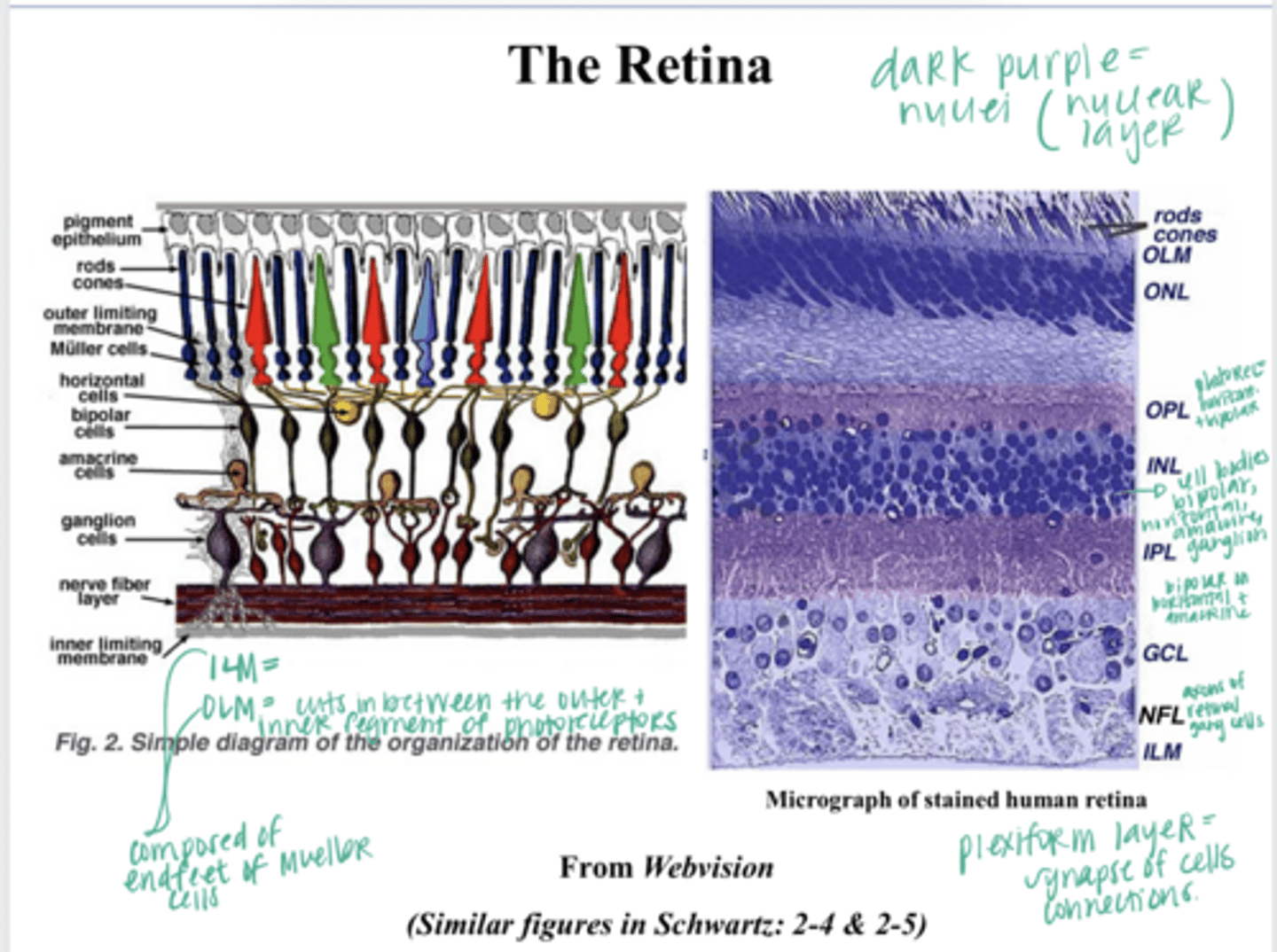
-cell body/nuclei of the neurons
-Nuclear layer --> nuclei present
When you stain the retina, where do the dark purple structures represent? Where will these be found?
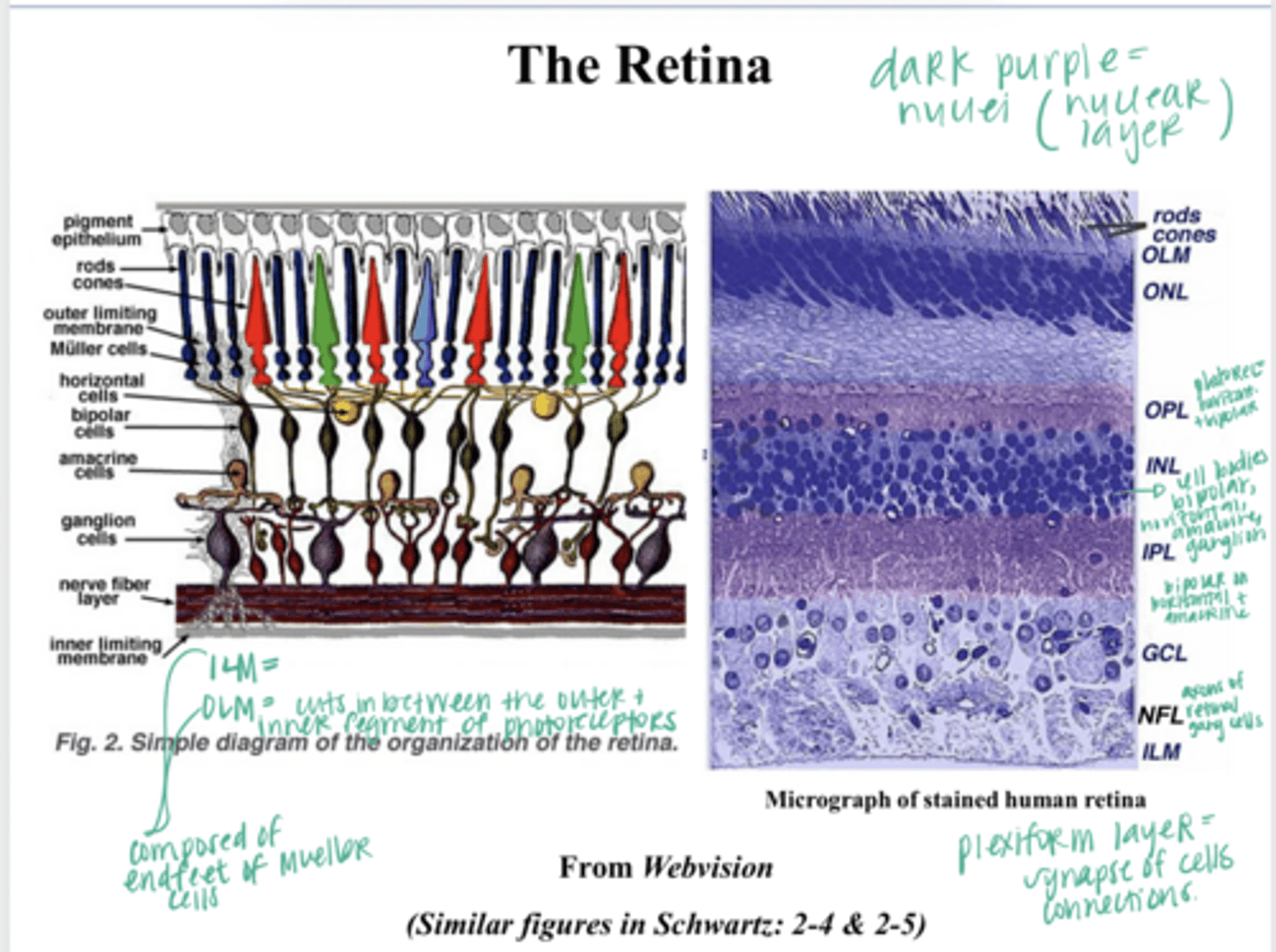
cell bodies of the photoreceptors
What cell bodies will be present in the outer nuclear layer?
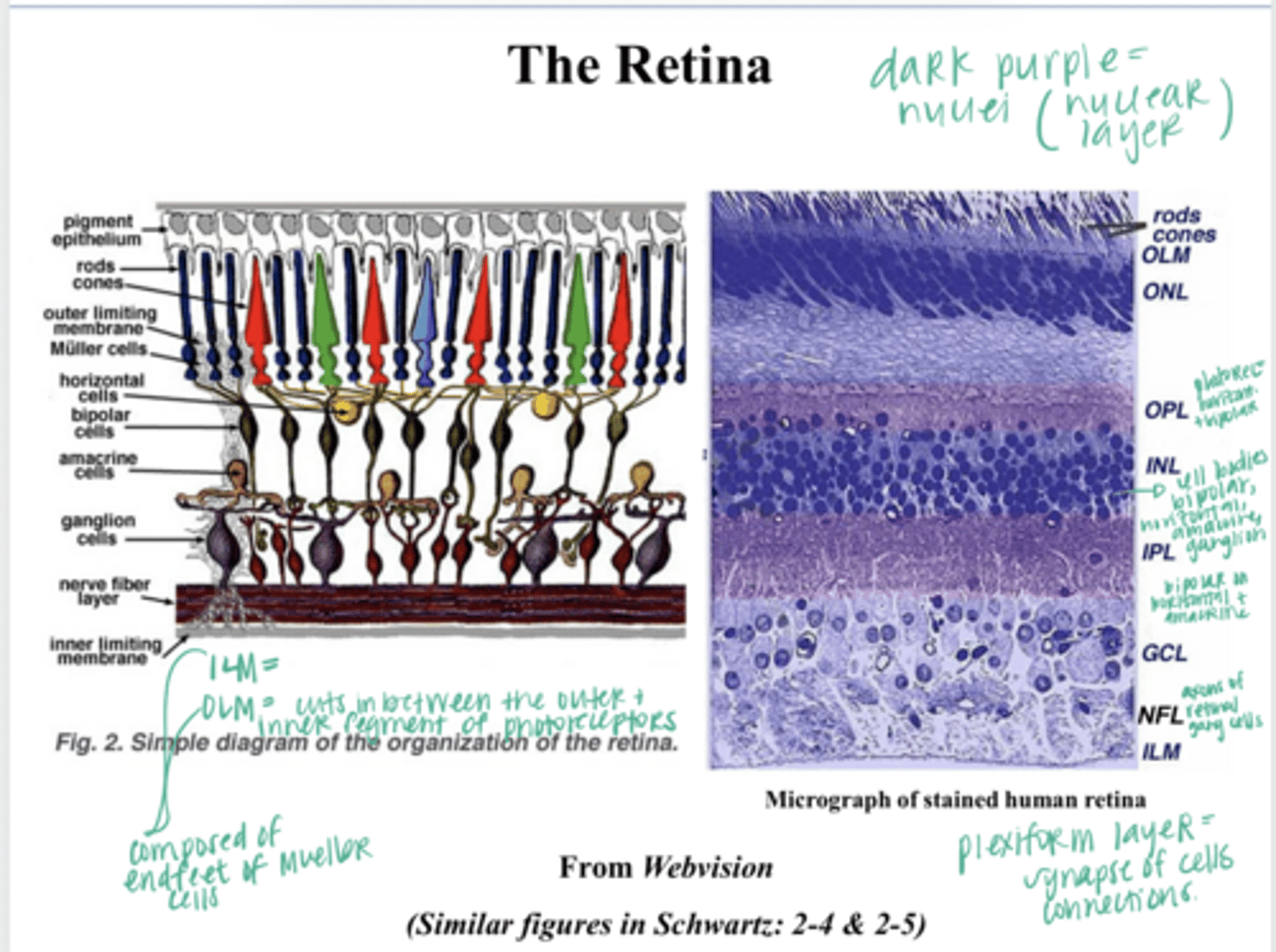
cell bodies of the bipolar cells/horizontal cells/amacrine cells/ganglion cells
What cell bodies will be present in the inner nuclear layer?
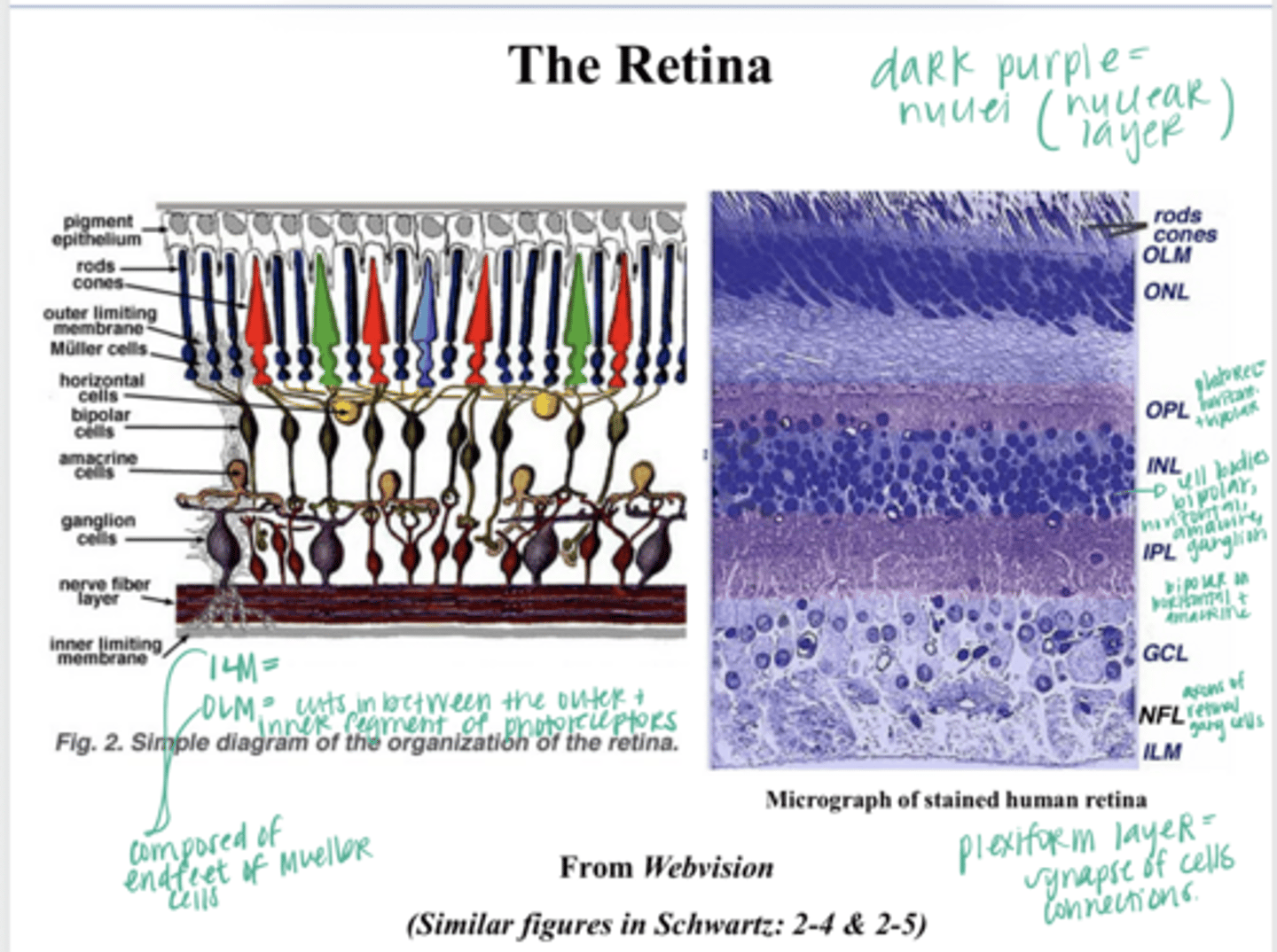
where synapses will occur
What is present in a plexiform layer of the retina?
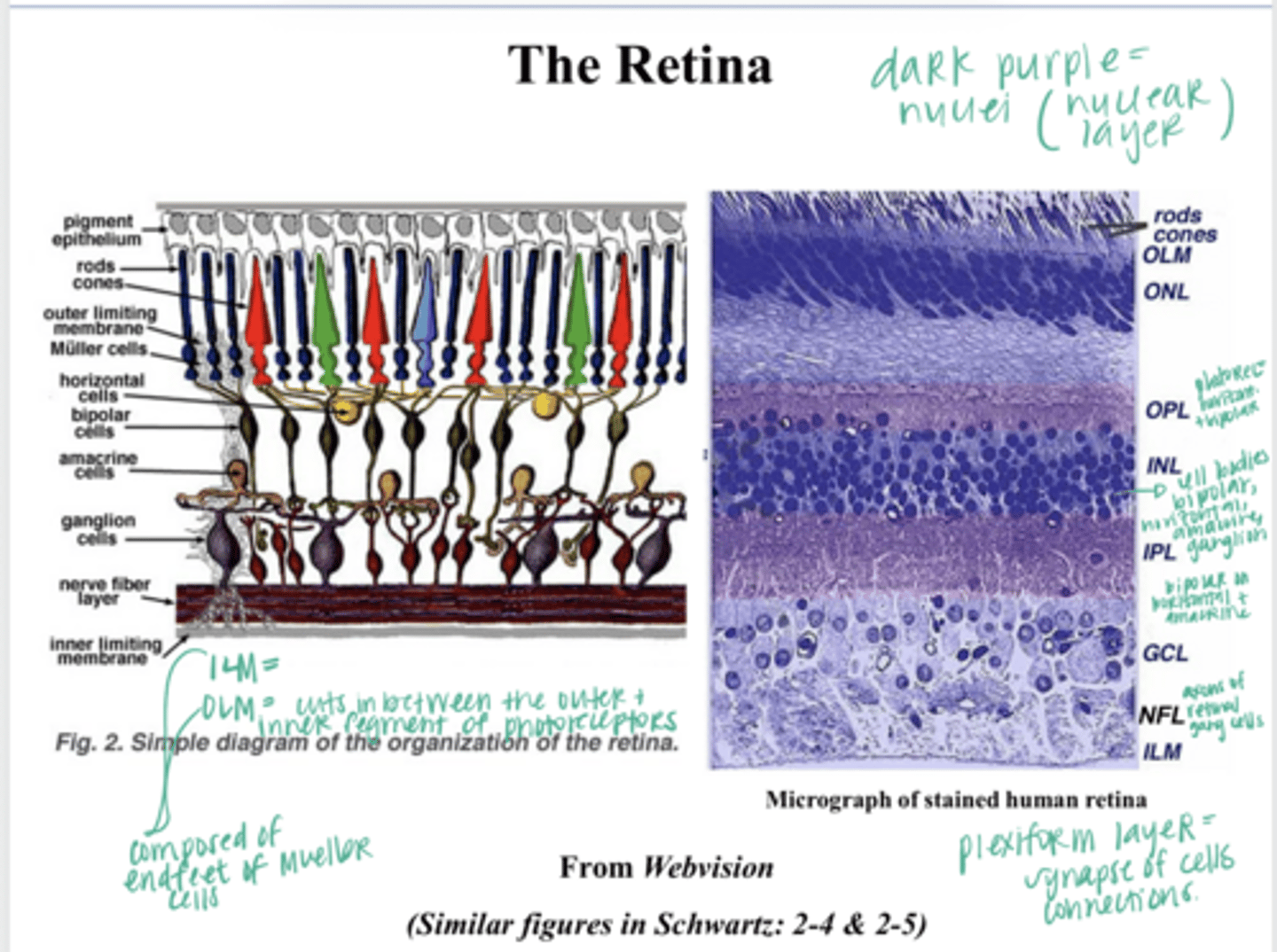
photoreceptors synapse on horizontal and bipolar cells
What is present in the outer plexiform layer?
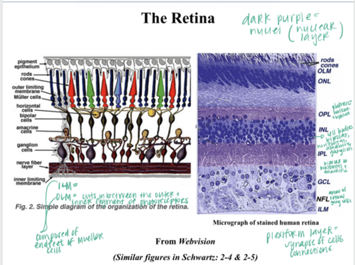
bipolar cells synapse onto ganglion and amacrine cells
What is present in the inner plexiform layer?
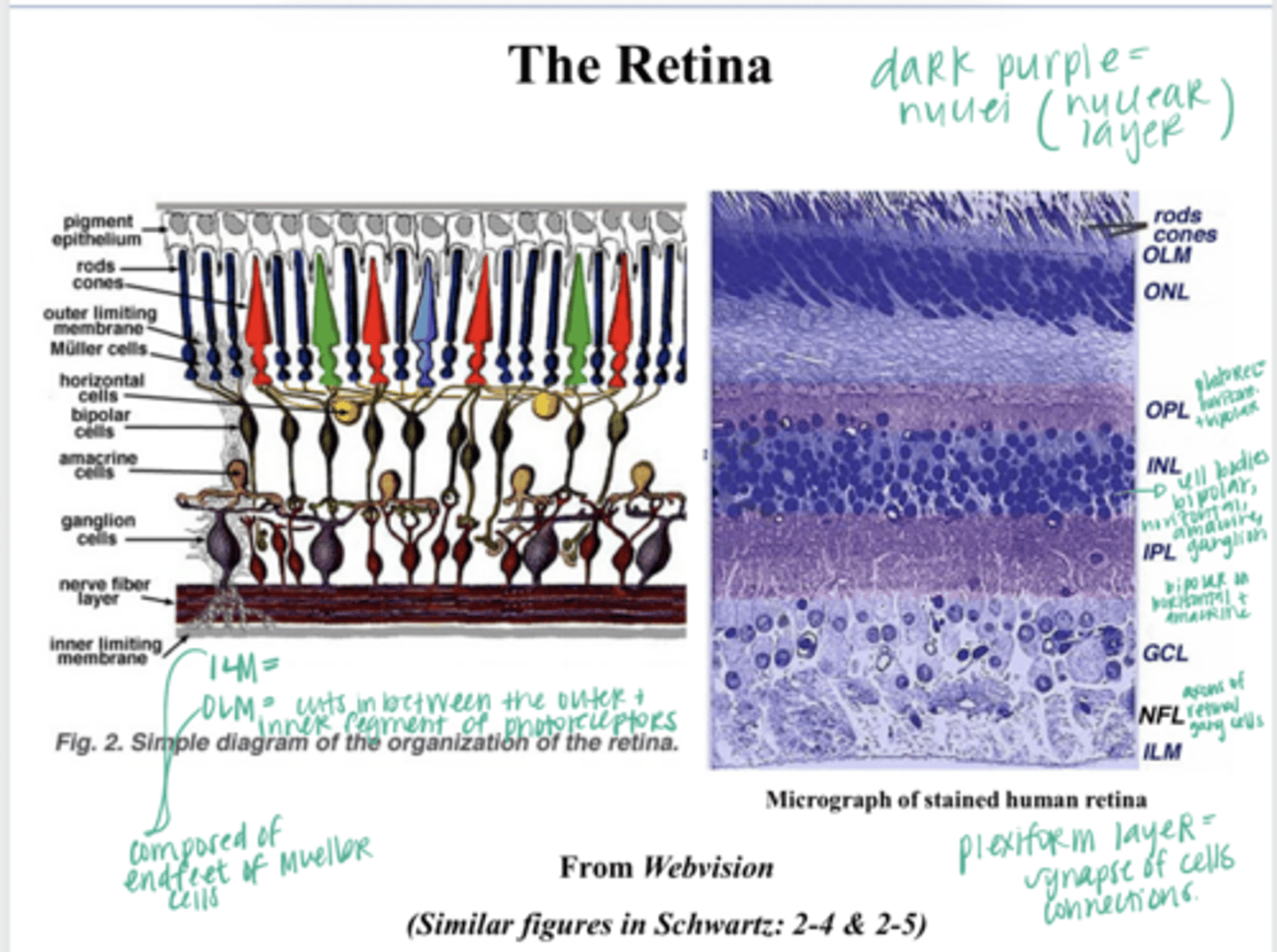
ganglion cell bodies, amacrine cells
What is present in the ganglion cell layer?
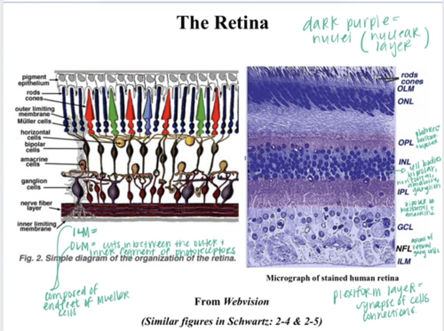
axons of the retinal ganglion cells --> axons leave the eye and head to the brain
What is present in the Nerve fiver layer?
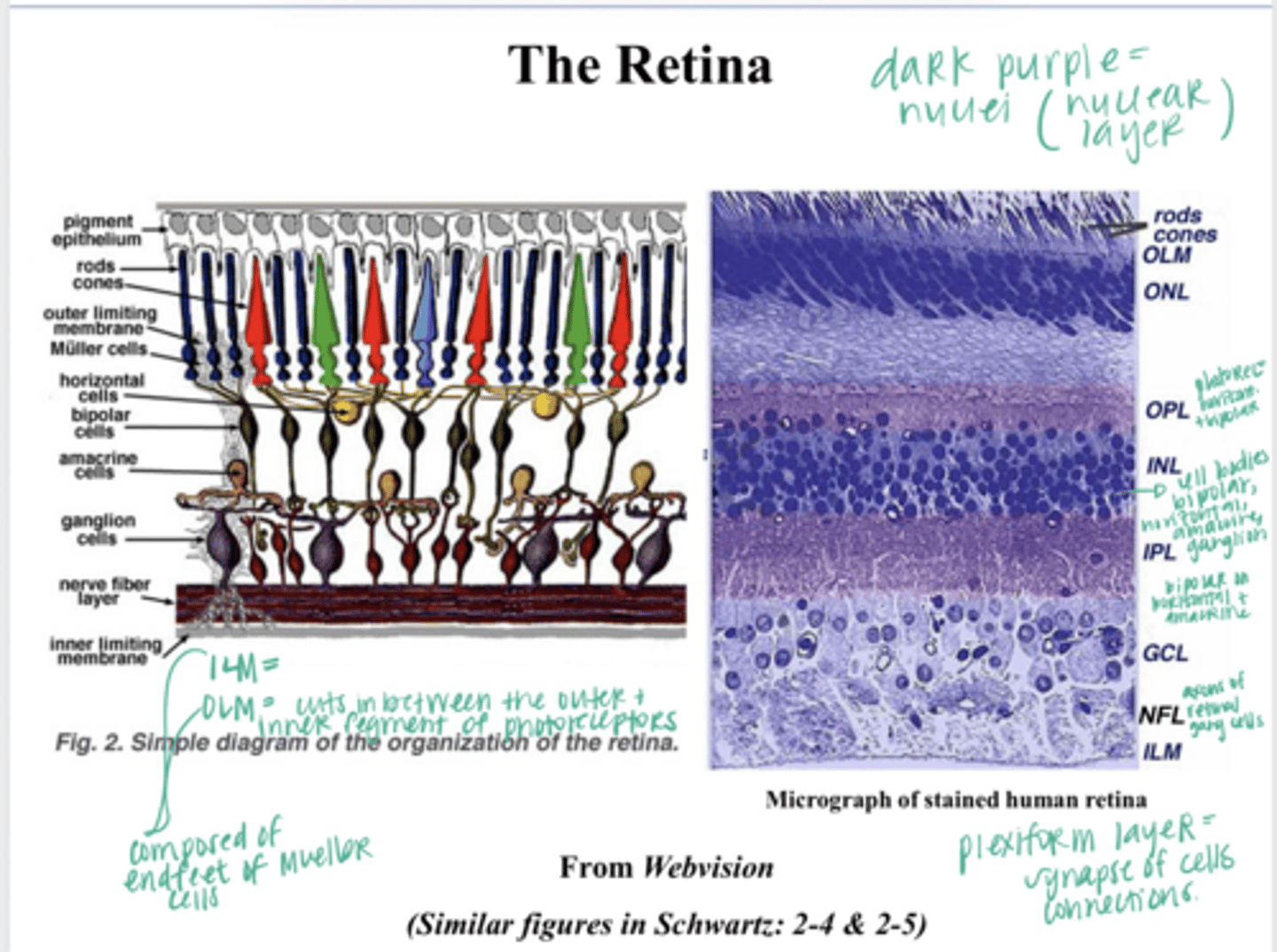
on the inner layer of the nerve fiber layer
Where does the ILM lie?
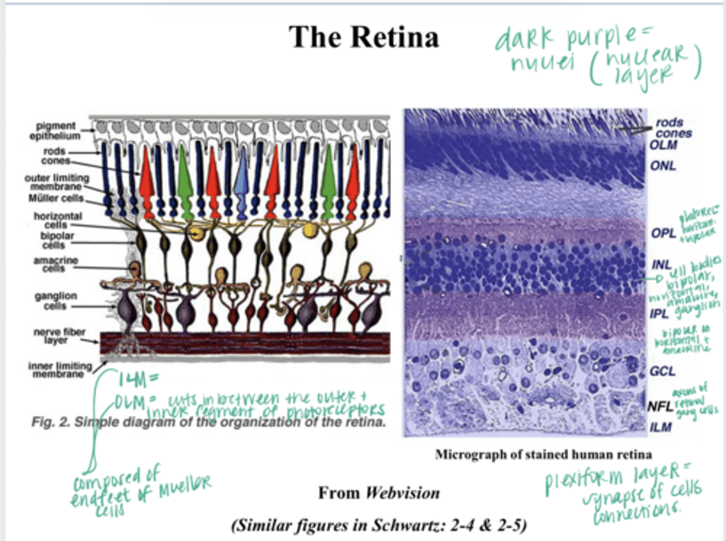
in between the outer and inner segments of the photoreceptors
Where does the OLM lie?
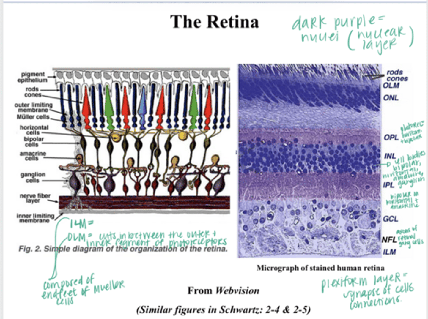
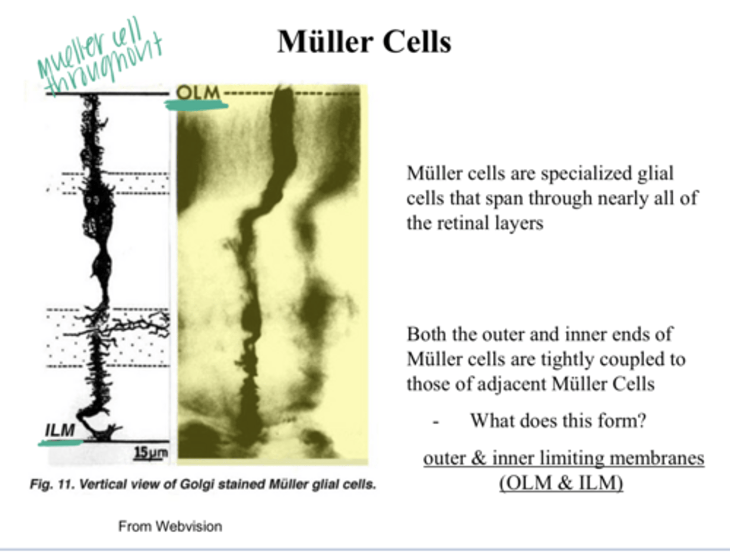
specialized glial cells that span through nearly all of the retinal layers
What are Muller Cells?
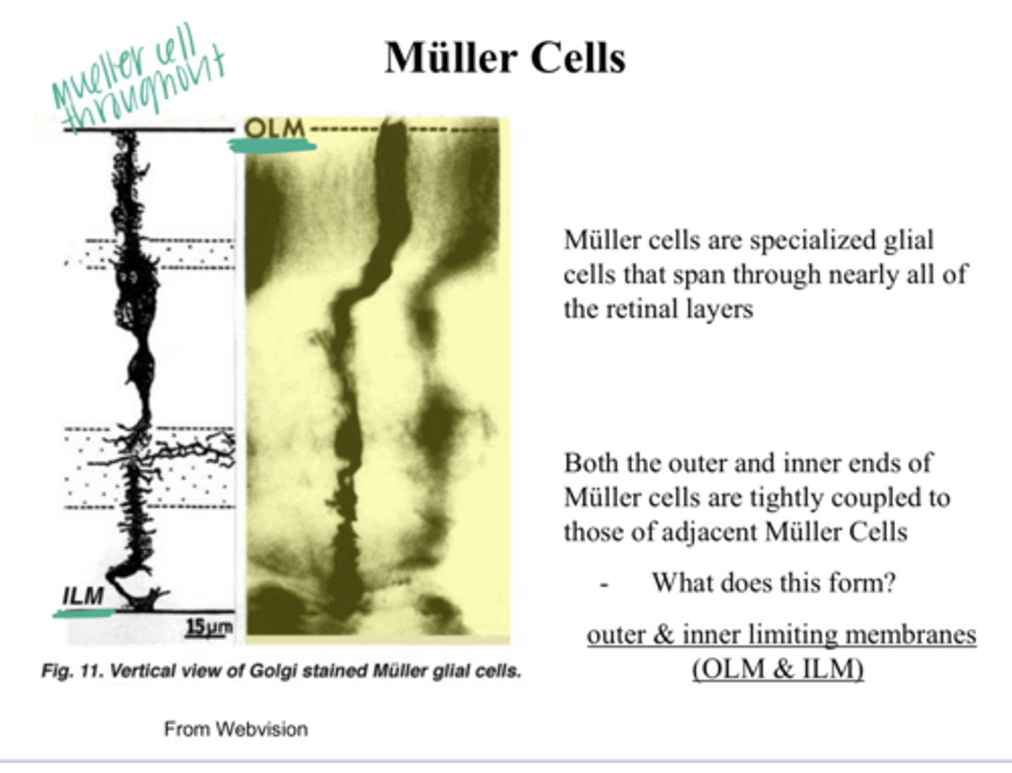
ILM and OLM
Both the outer and inner ends of Muller Cells are tightly coupled to those of adjacent Muller cells. What does this form?
photoreceptors --> bipolar cells --> ganglion cells
Basic vertical visual pathway within the retina?
photoreceptors (rods and cones)
these convert light energy into electrical signals
receive visual info synaptically from the photoreceptors and transmit this signal on to the retinal ganglion cells
What is the function of the bipolar cells of the retina?
-output neurons of the retina (project to various brain centers)
-the axons form the optic nerve
What is the function of the ganglion cells of the retina?
retinal ganglion cells
The axons of the ____ form the optic nerve
-retinal neurons that modulate the horizontal transmission between the photoreceptors, bipolar cells and ganglion cells
What are the function of horizontal and amacrine cells?
at the optic disk
the axons of the retinal ganglion cells converge where to exit the eye?
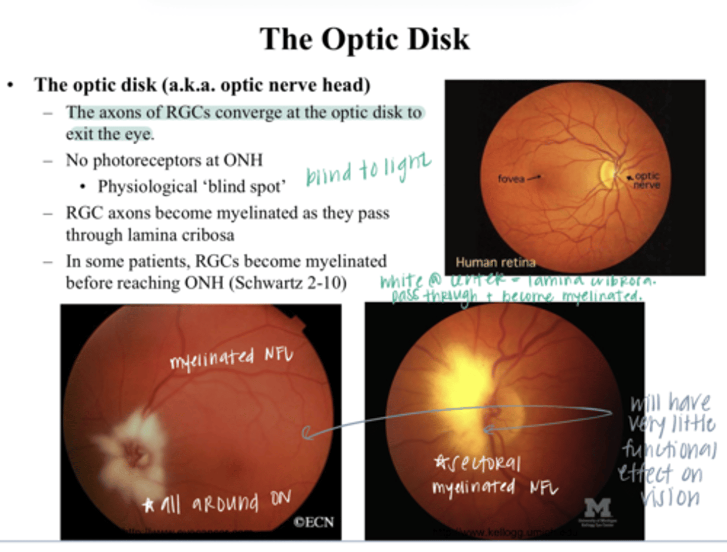
no -- this is a physiological blind spot
Are there any photoreceptors at the ONH?
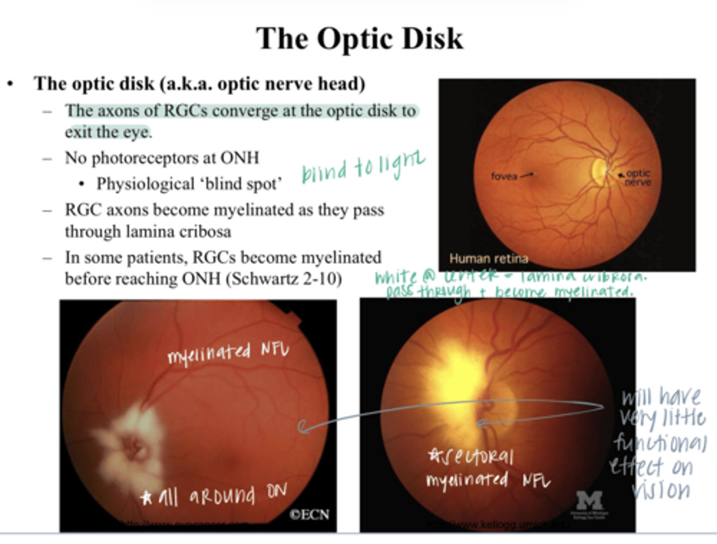
myelinated
Retinal ganglion cells become _____ as they pass through the lamina cribosa
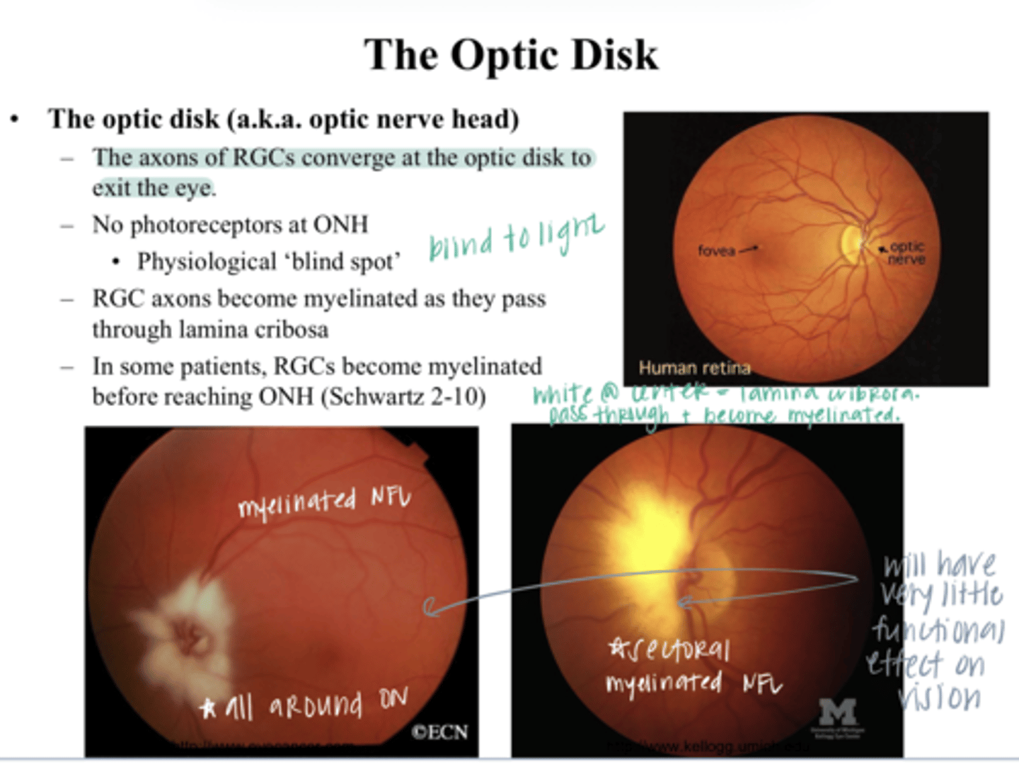
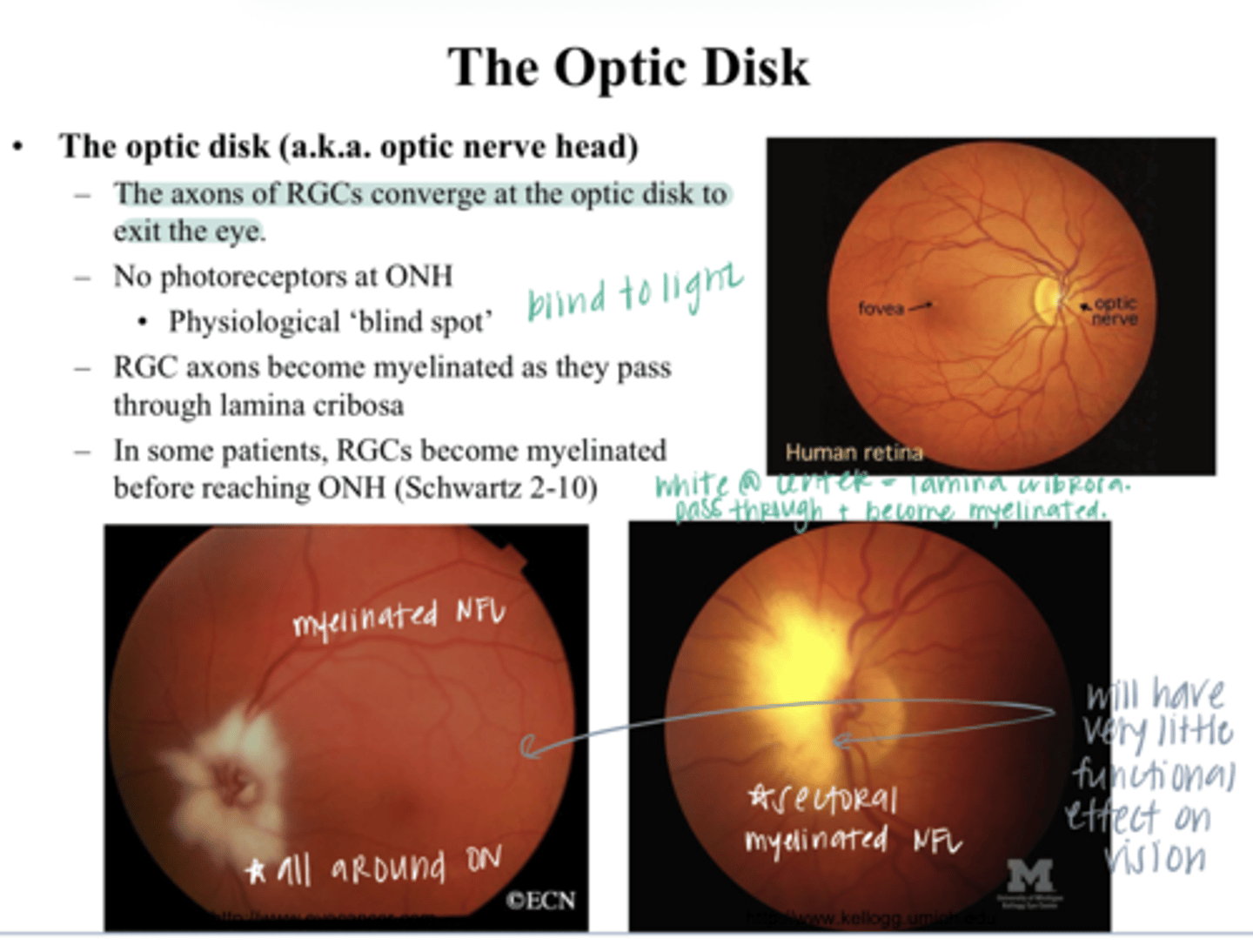
yes -- very little functional effect on vision
Can retinal ganglion cells become myelinated before reaching the ONH? Is this dangerous?
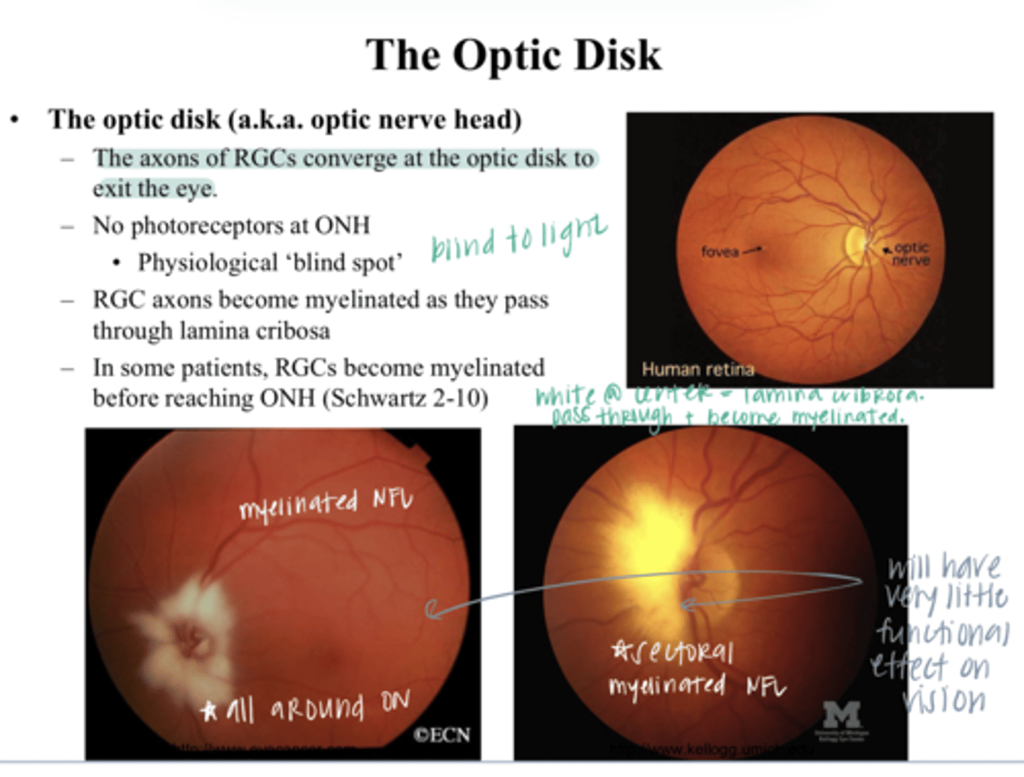
light would have to pass through the myelin in order to get to the photoreceptors underneath. Only want myelin to be present AFTER the lamina cribosa
Why do you not want the entire RGC to be myelinated?
on the retina in an INVERTED image
Due to the optical properties of the eye, the image of the visual field is focused where?
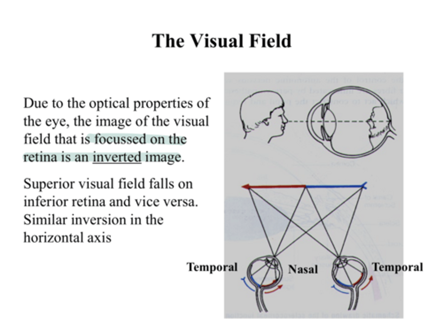
inferior retina
Superior visual field falls where on the retina?
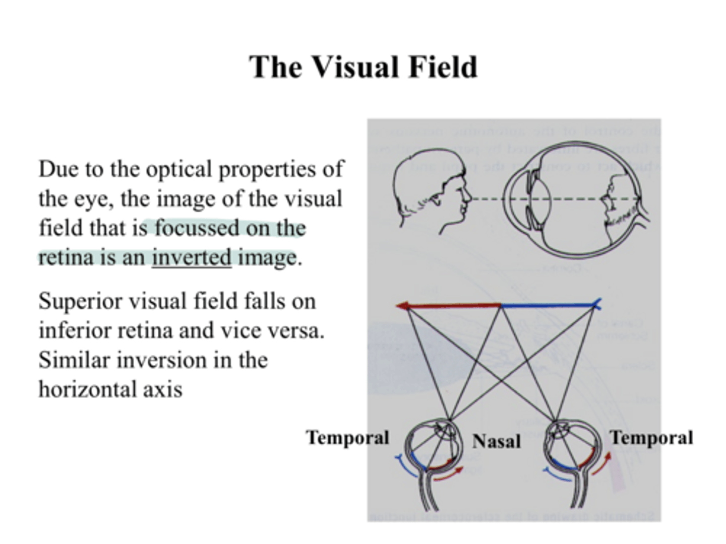
superior retina
Inferior visual field falls where on the retina?
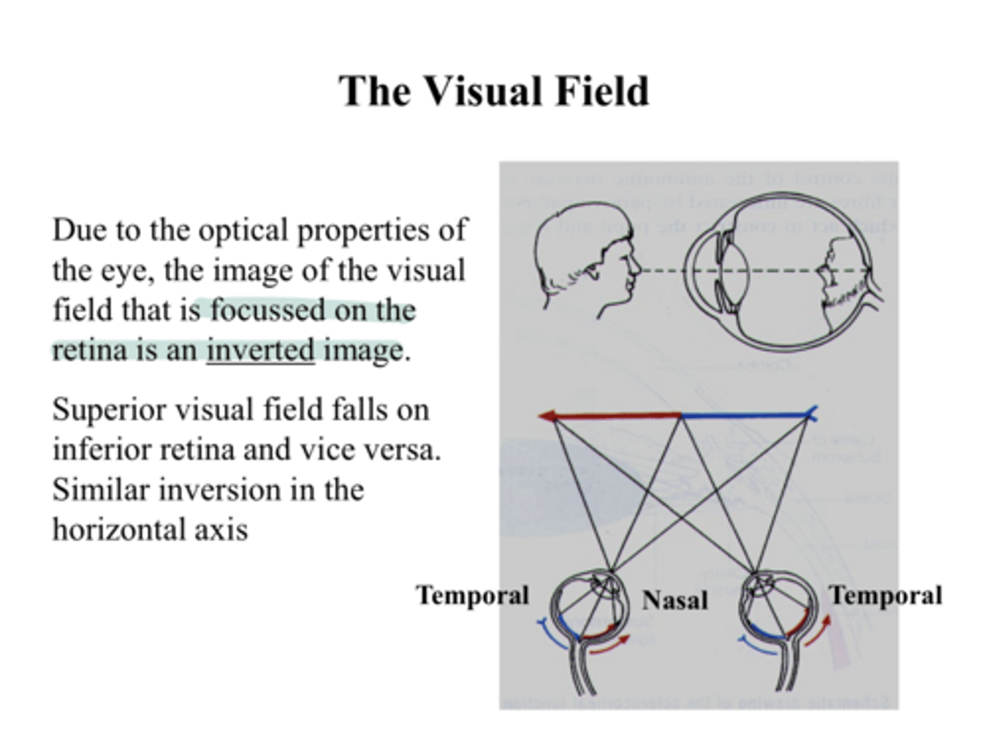
temporal retina
Nasal visual field falls where on the retina?
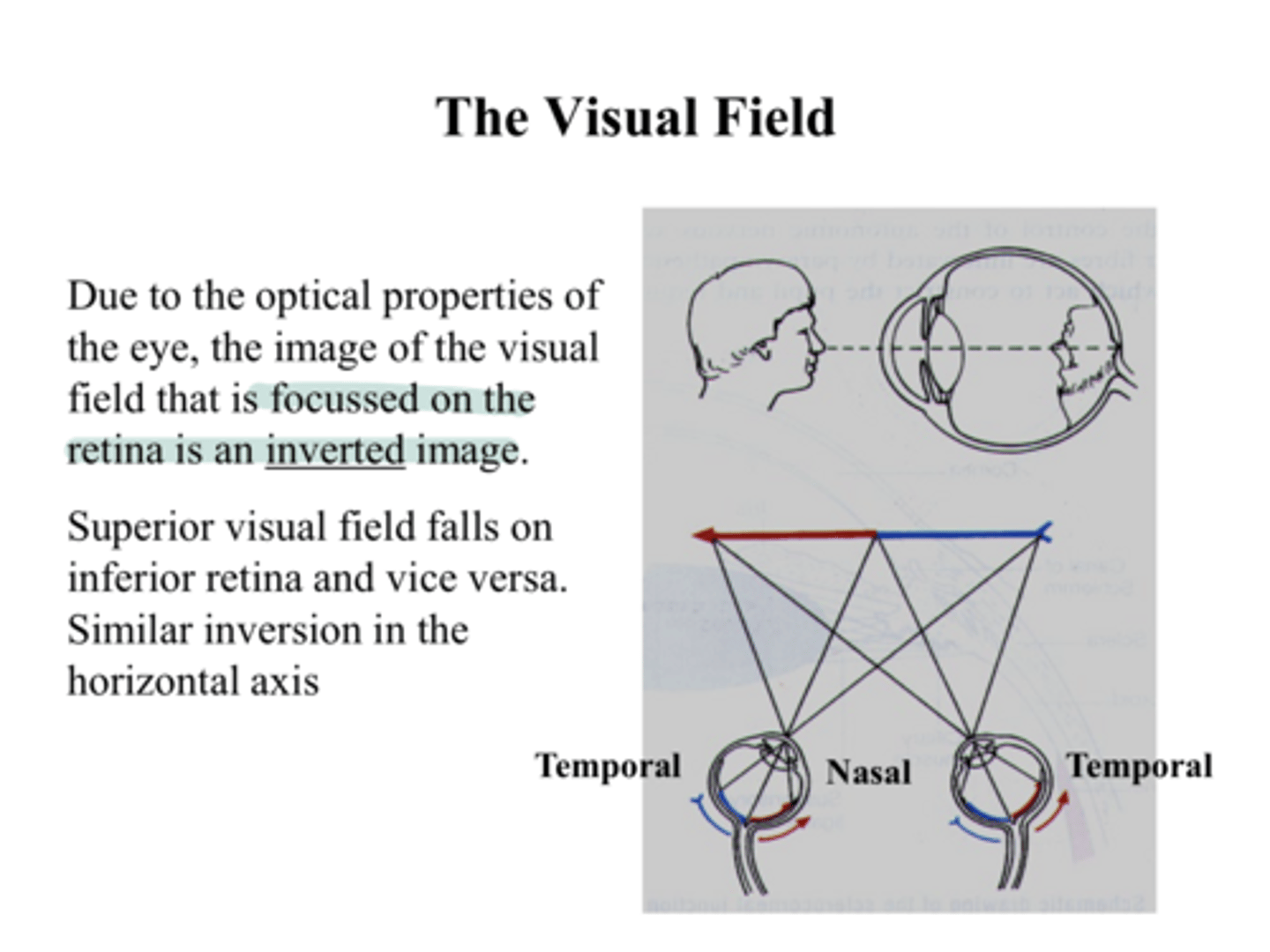
nasal retina
Temporal visual field falls where on the retina?
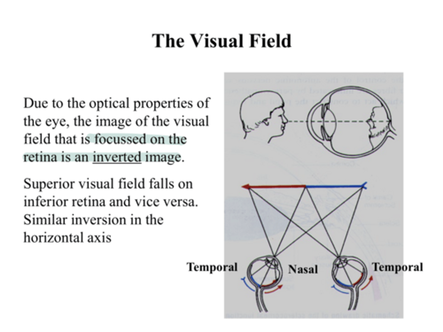
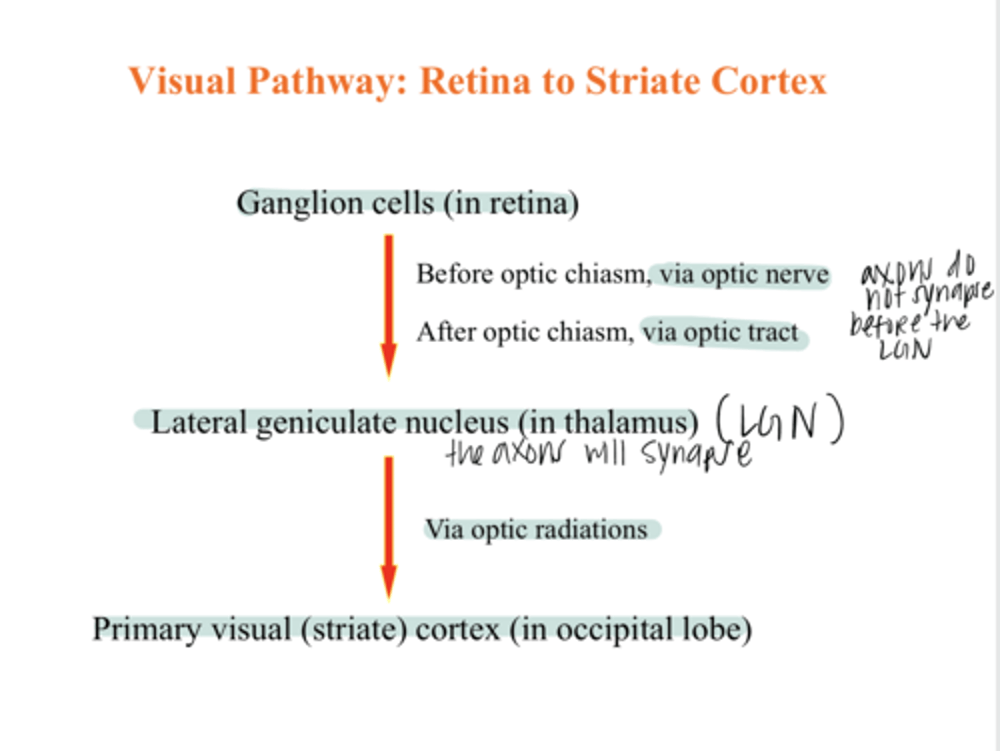
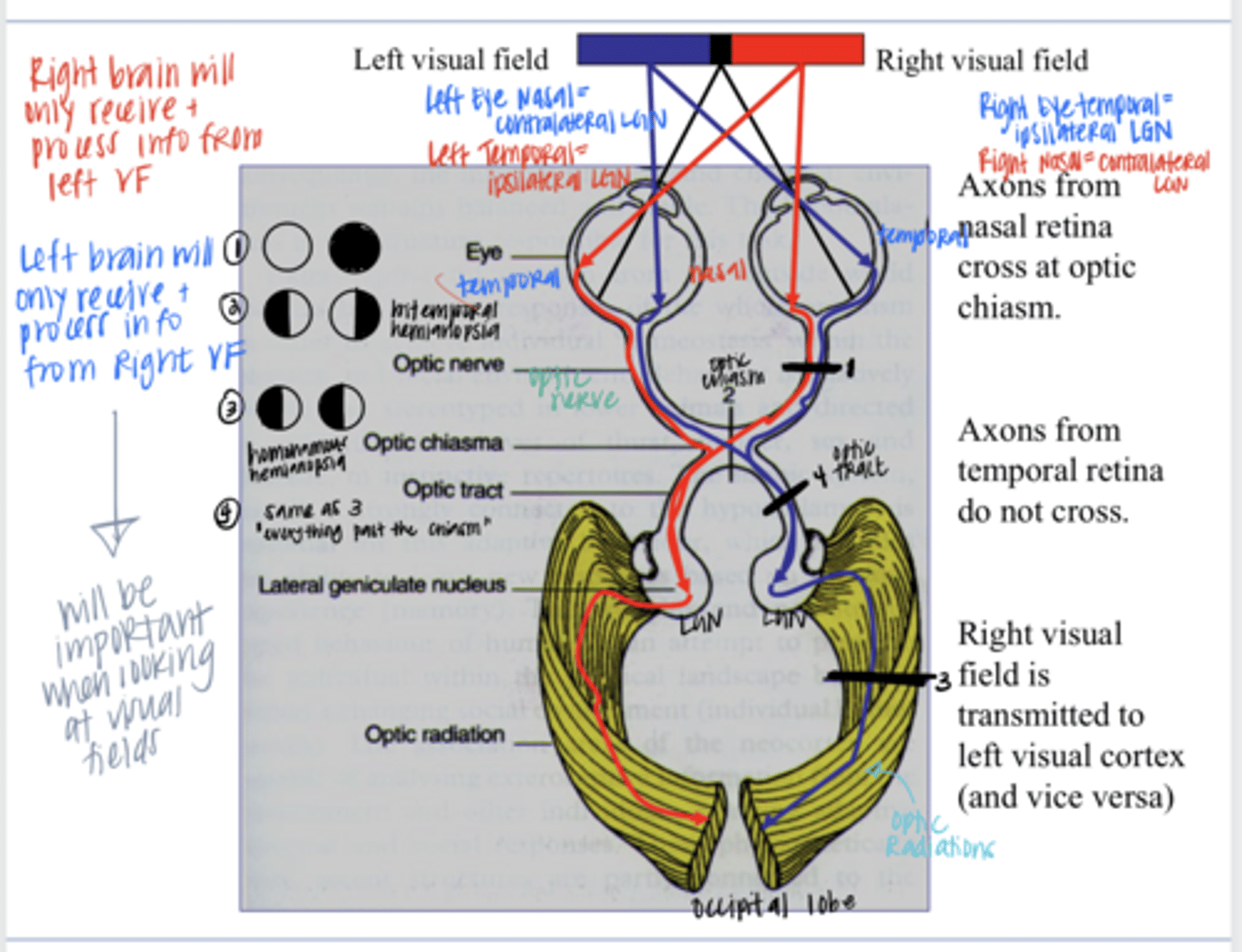
ganglion cells (in the retina) --> LGN (in the thalamus) --> primary visual cortex in the occipital lobe
What is the basic visual pathway from the retina to the striate cortex?
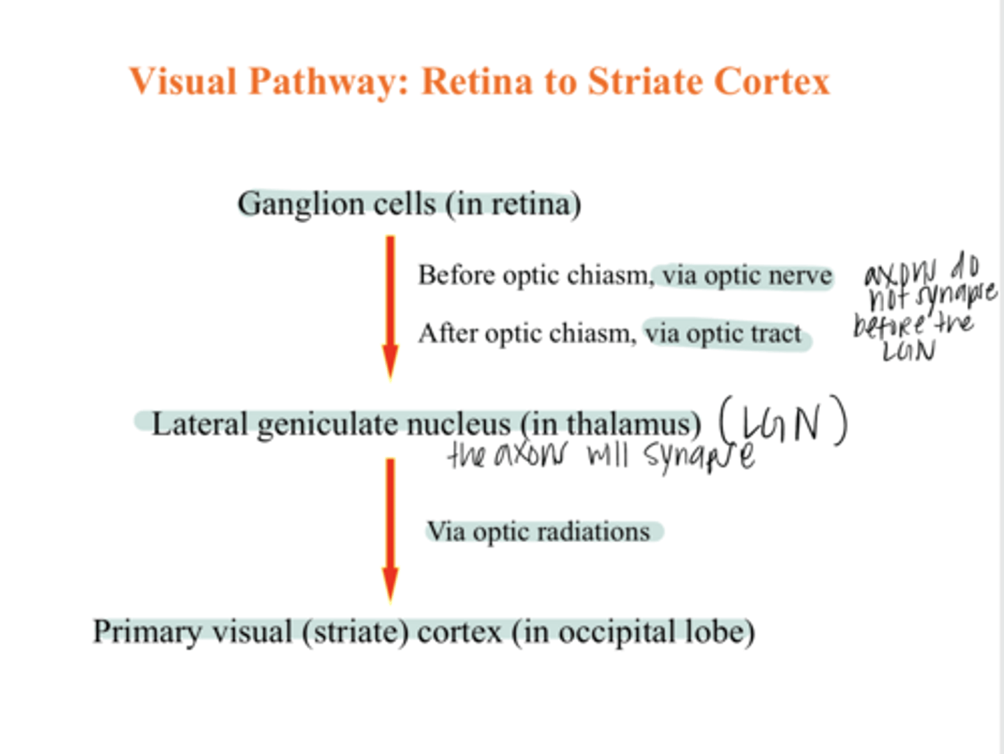
optic nerve
Before the optic chiasm, what are the ganglion cell axons called?
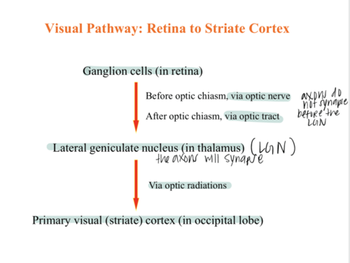
optic tract
After the optic chiasm, what are the ganglion cell axons called?
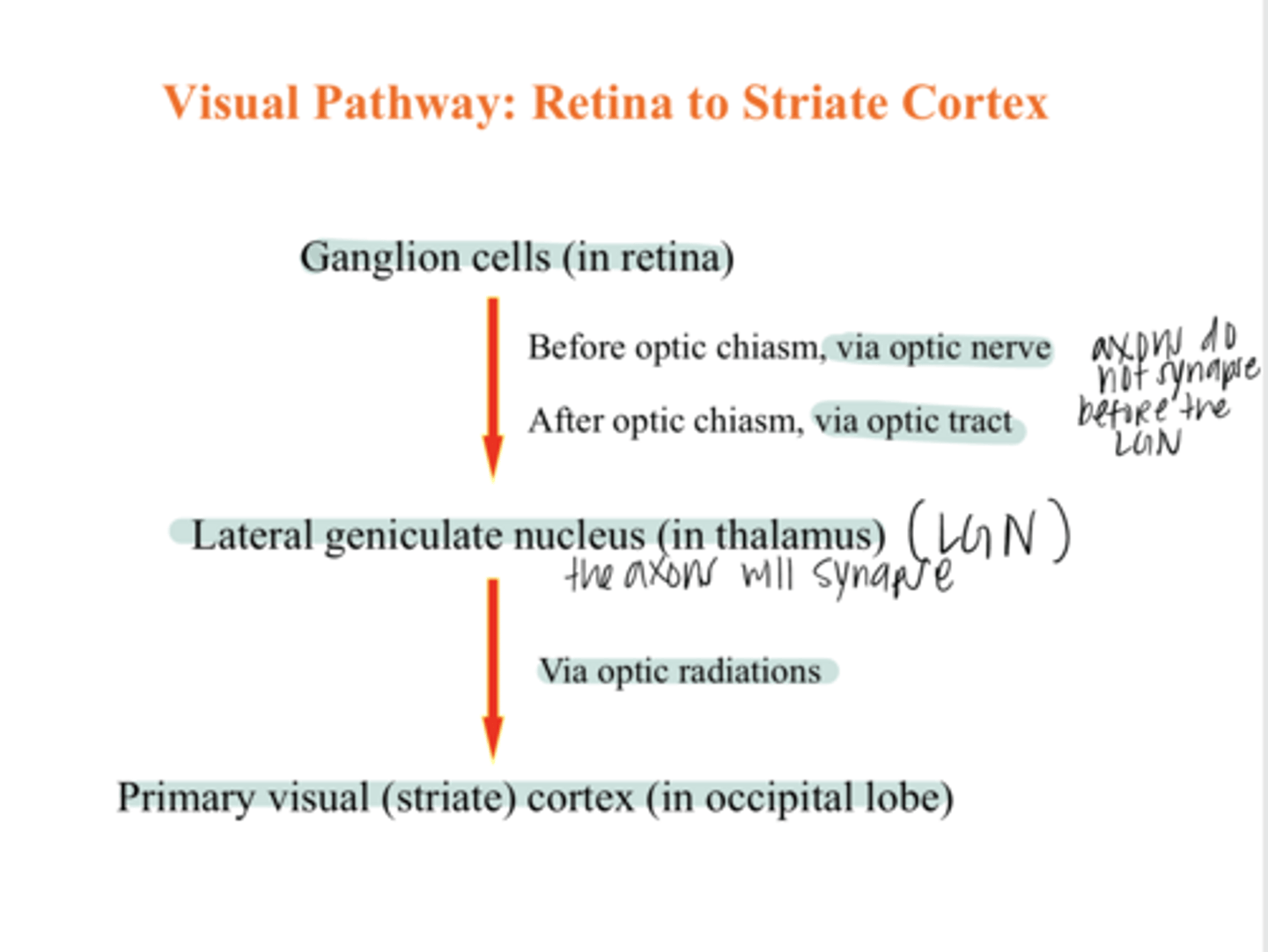
no
Does the bundle of ganglion cell axons synapse before the LGN of the brain?
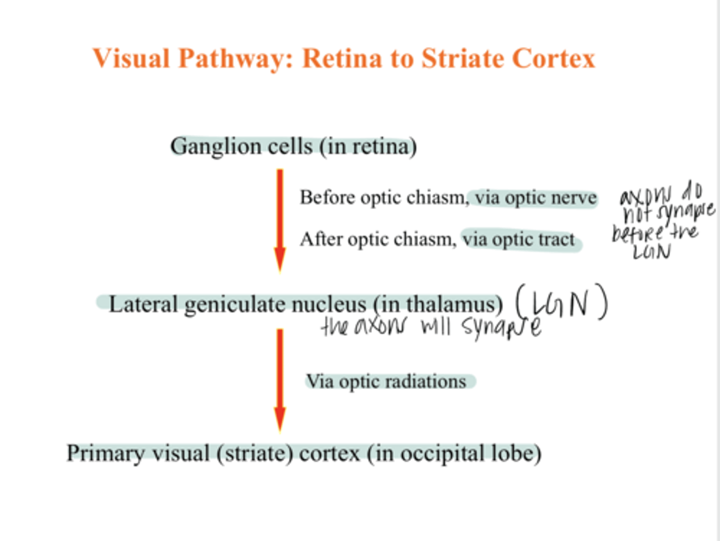
optic radiations
After the LGN where the ganglion cell axons synapse, what are the ganglion cell axons called?
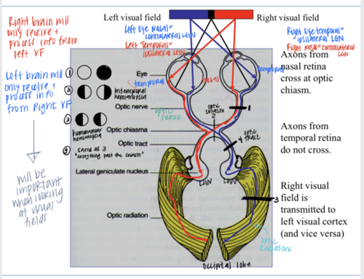
they cross
Axons from the nasal retina (temporal VF) do what at the optic chiasm?
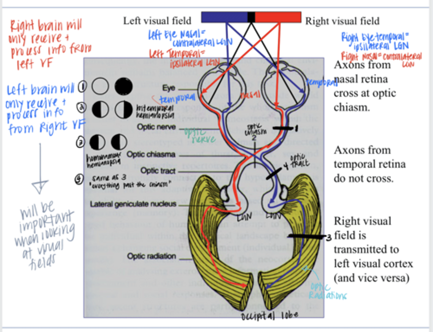
they do not cross
Axons from the temporal retina (nasal VF) do what at the optic chiasm?
left
The Right VF is transmitted to the (right/left) LGN & visual cortex
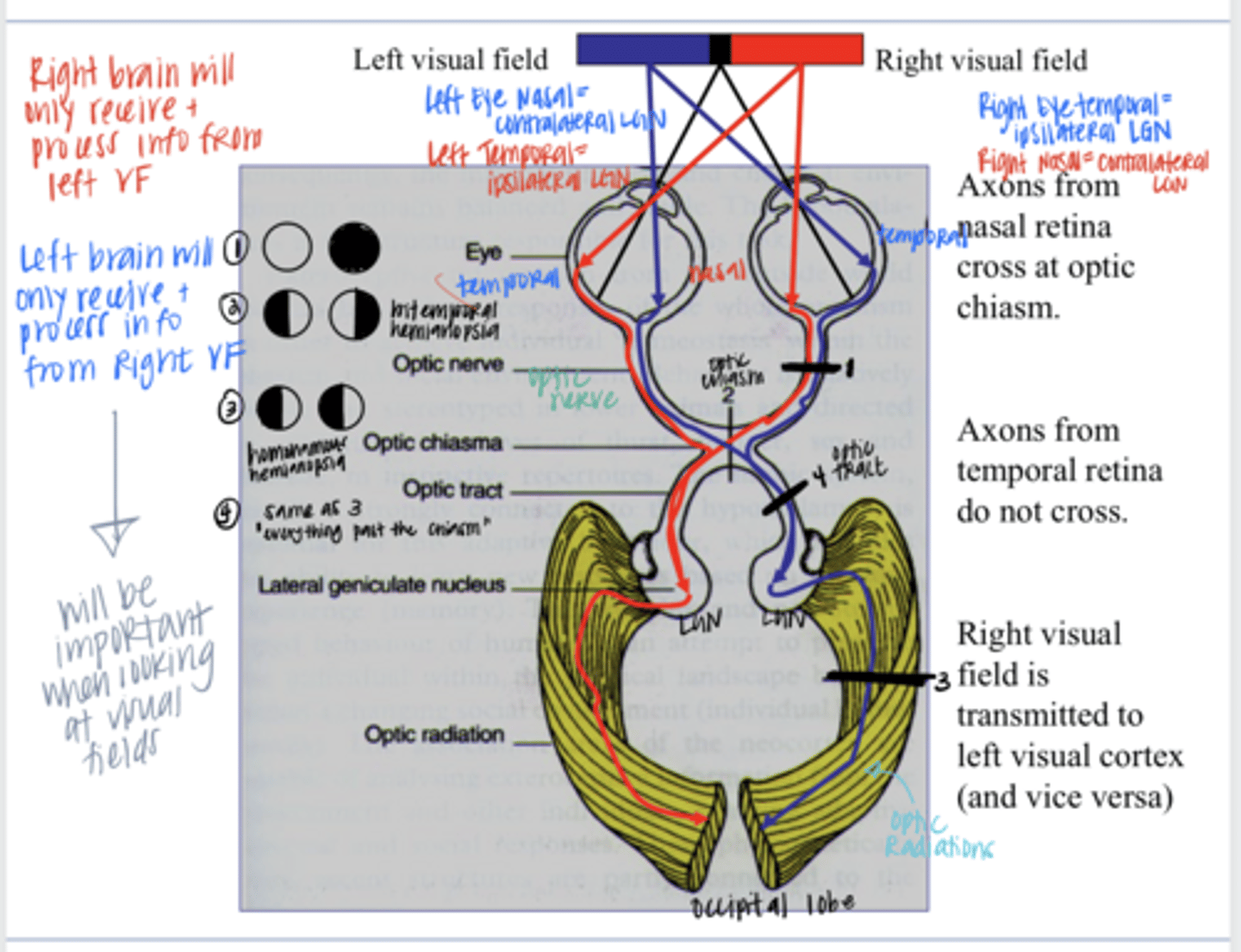
right
The Left VF is transmitted to the (right/left) LGN & visual cortex
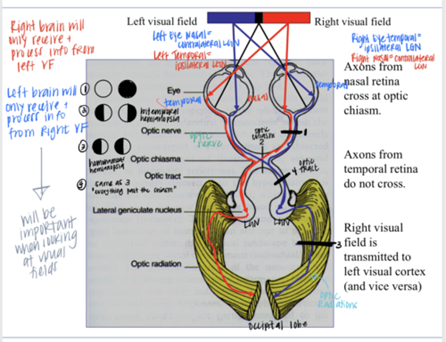
optic chiasm
Once you get past the ____, the processing of vision is segregated down the midline.
true
True or False:
If you know neuroanatomy, you can look at a VF and know where a lesion is based on a defect
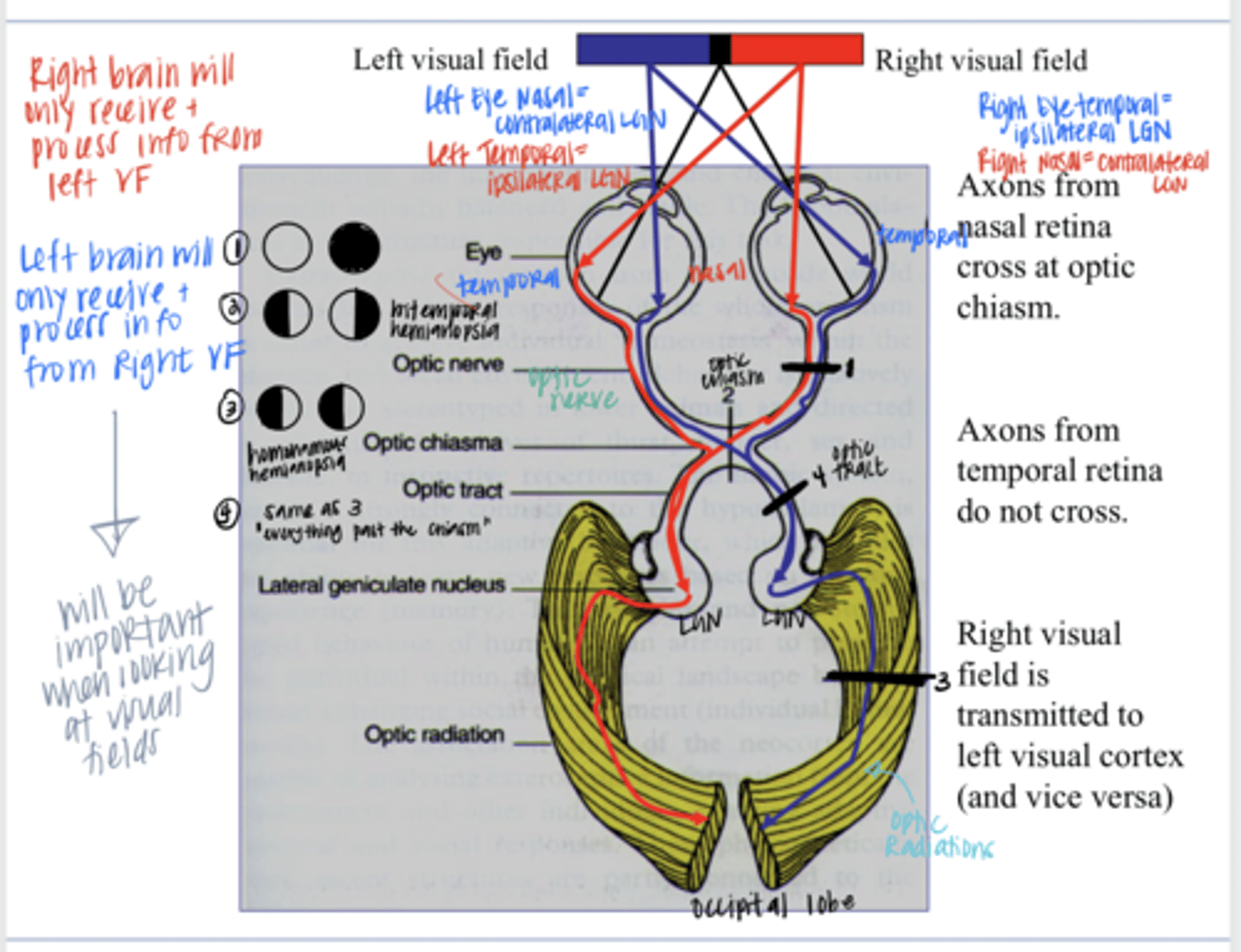
OD VF completely gone
If there was an injury that completely severed the R optic nerve, what would the corresponding VF look like?
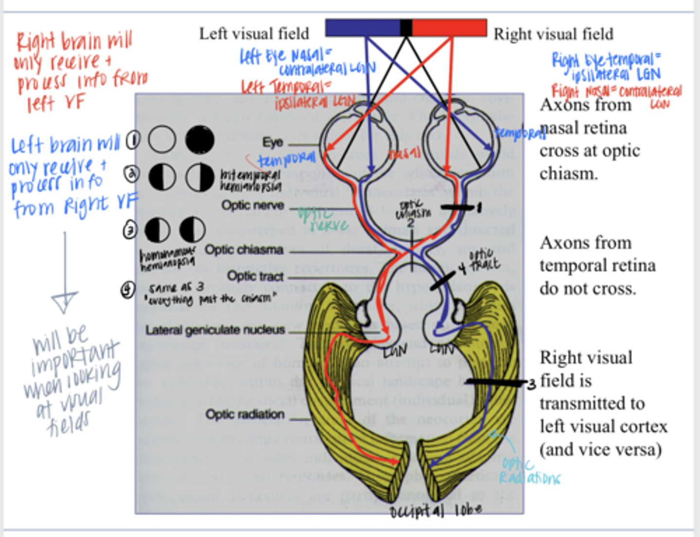
Bitemporal hemianopsia
If there was a trans section through the middle of the optic chiasm, what would the corresponding VF look like?
pituitary adenoma
What disease would present as a bitemporal hemianopsia?
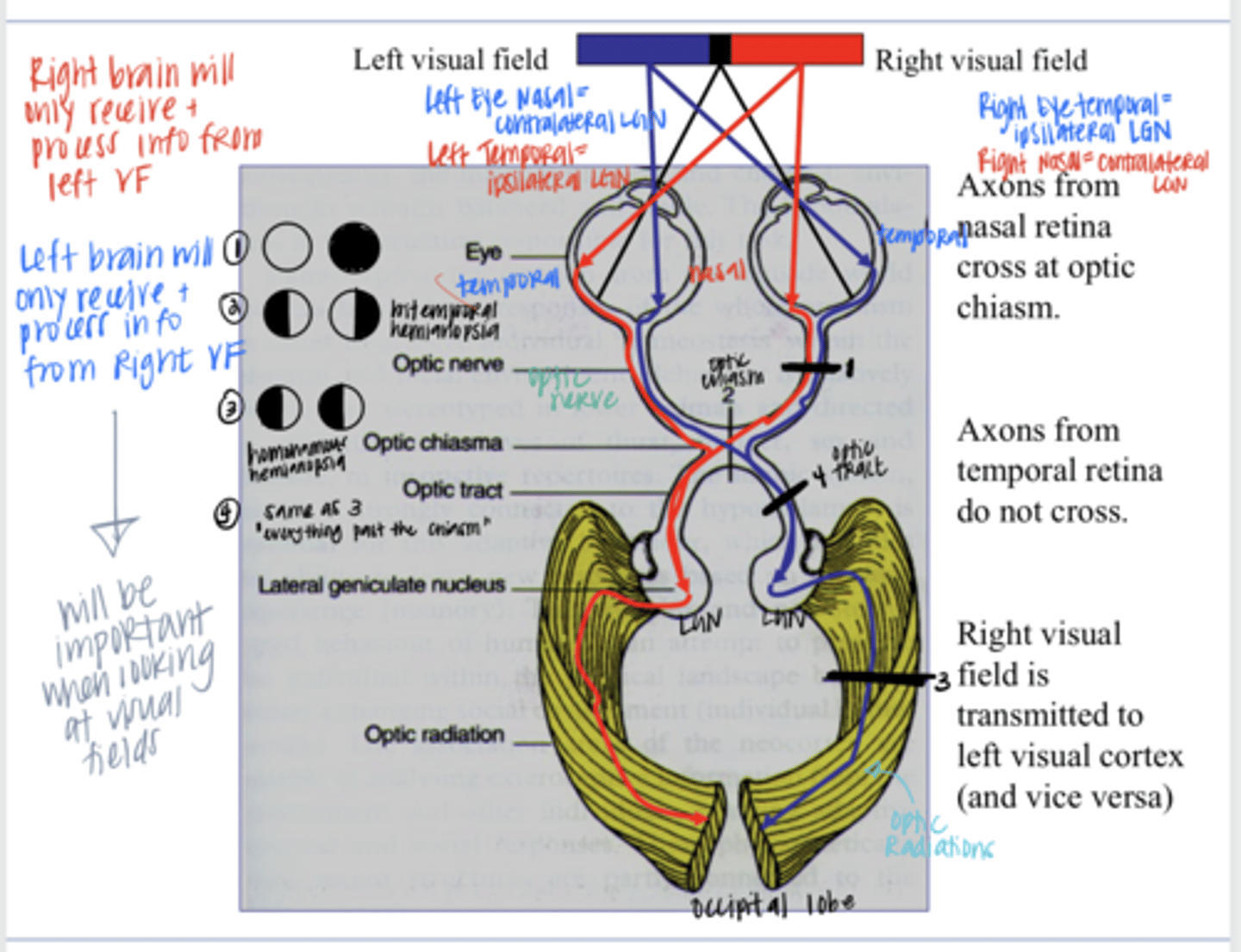
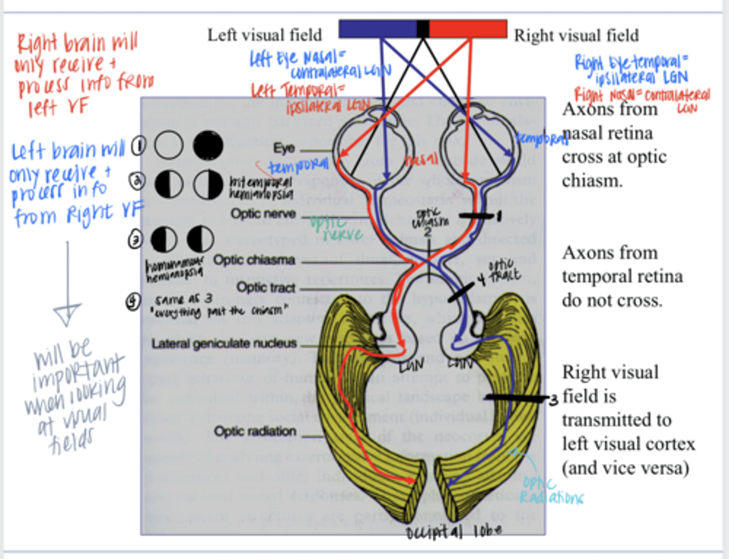
L homonymous hemianopia (Left VF OU)
If there was a trans section through the optic radiations of the R visual cortex, what would the corresponding VF look like?
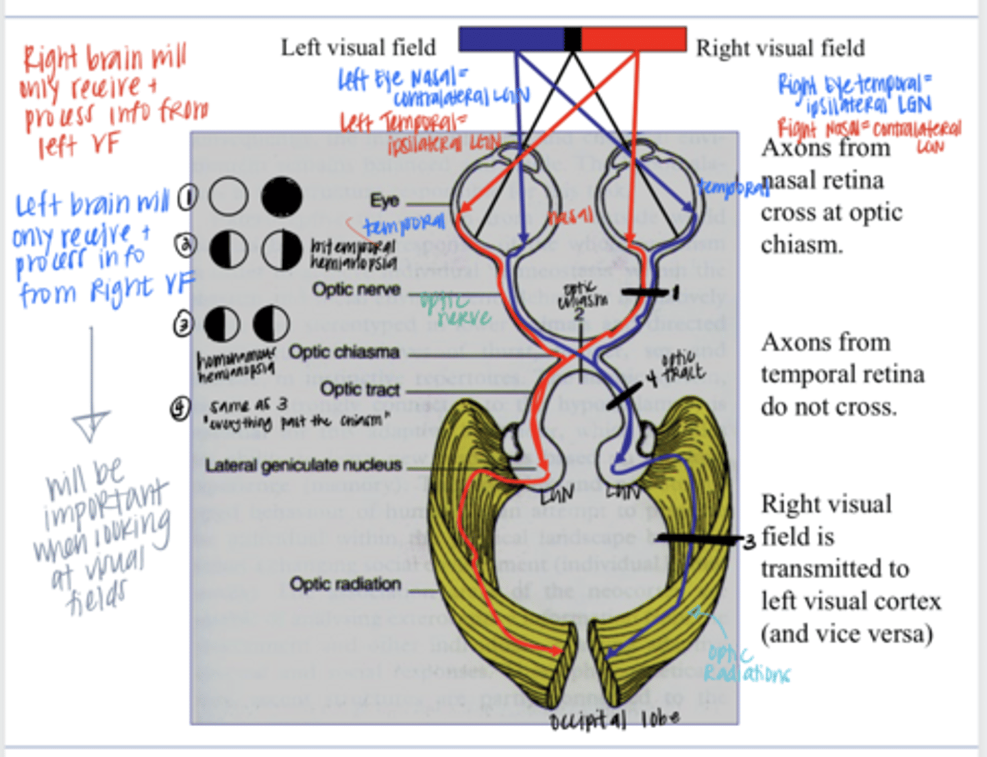
L homonymous hemianopia (Left VF OU)
If there was a trans section through the optic tract (chiasm to LGN) of the Right side, what would the corresponding VF look like?
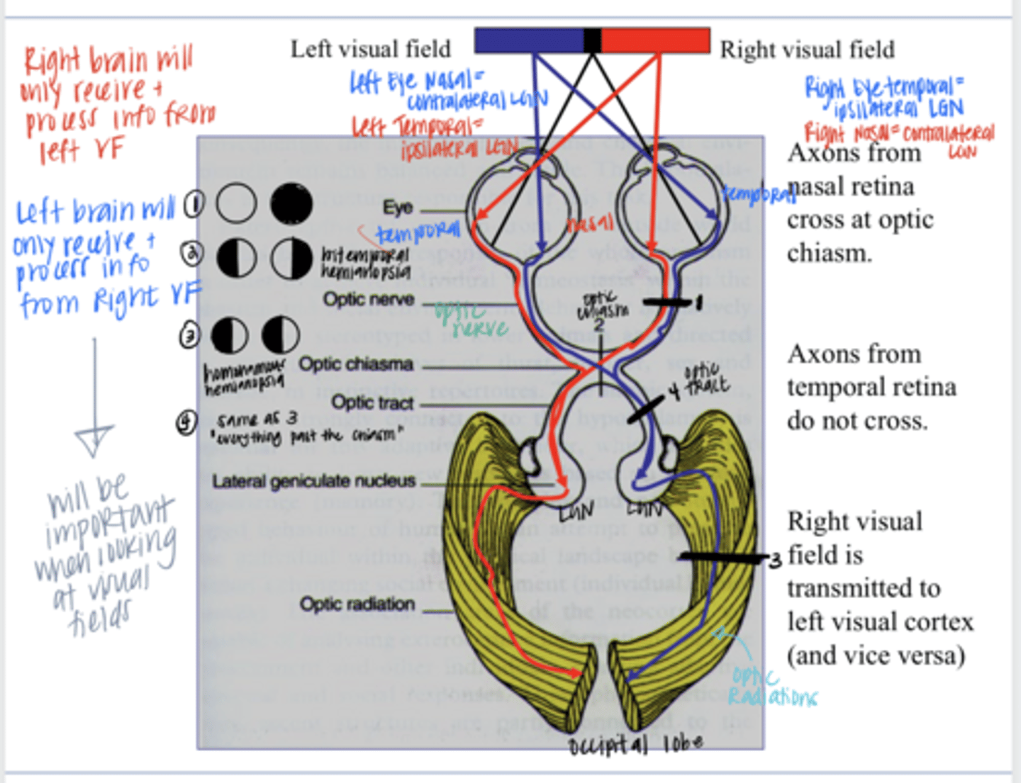
above
Optic radiations carrying info from the inferior VF (falls on the superior retina) will take a more direct route to the synapse (above/below) the calcarine fissure of the occipital lobe
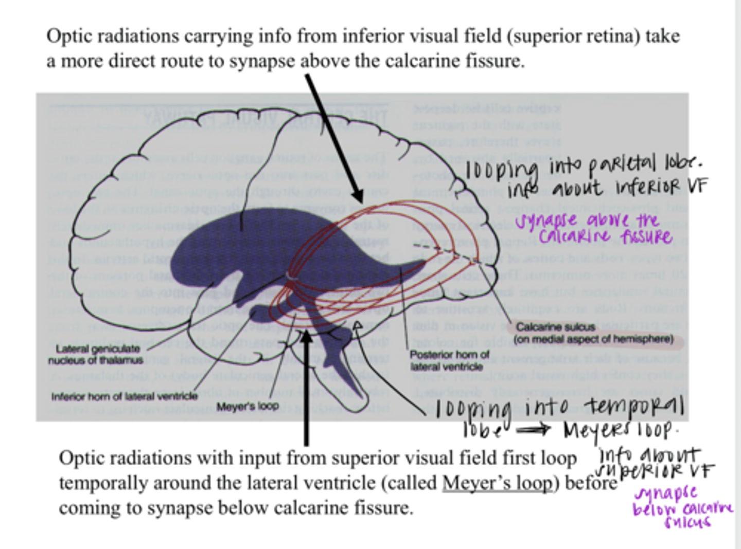
loop through the parietal lobe
Optic radiations carrying info from the inferior VF (falls on the superior retina) will loop through which lobe on their way to the occipital lobe?
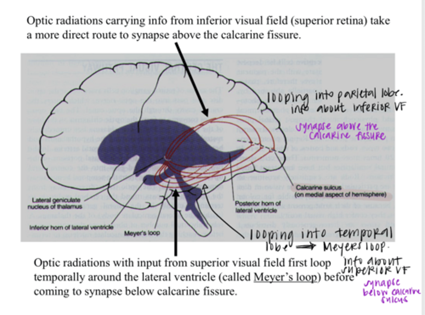
below
Optic radiations carrying info from the superior VF (falls on the inferior retina) will take a more direct route to the synapse (above/below) the calcarine fissure of the occipital lobe
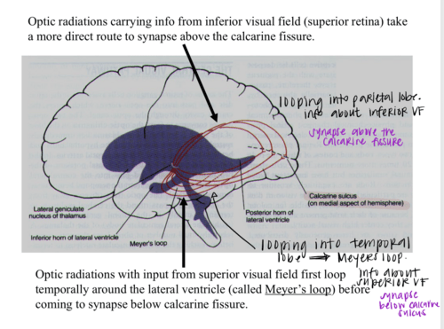
through the temporal lobe
Optic radiations carrying info from the superior VF (falls on the inferior retina) will loop through which lobe on their way to the occipital lobe?
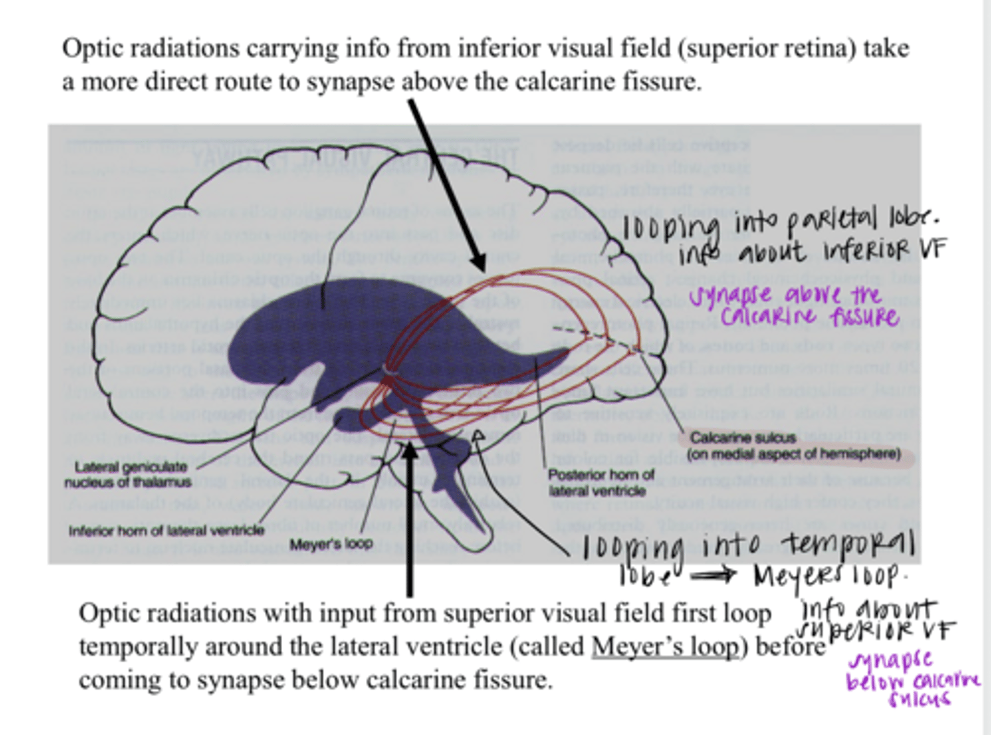
Meyer's loop
Optic radiations carrying info from the superior VF (falls on the inferior retina) will loop through temporal lobe and are called what?
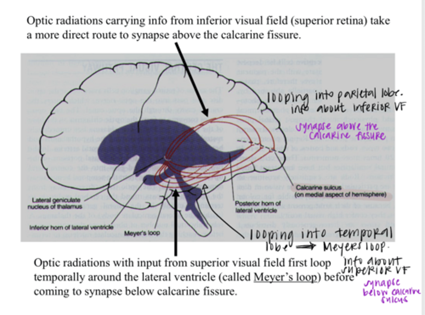
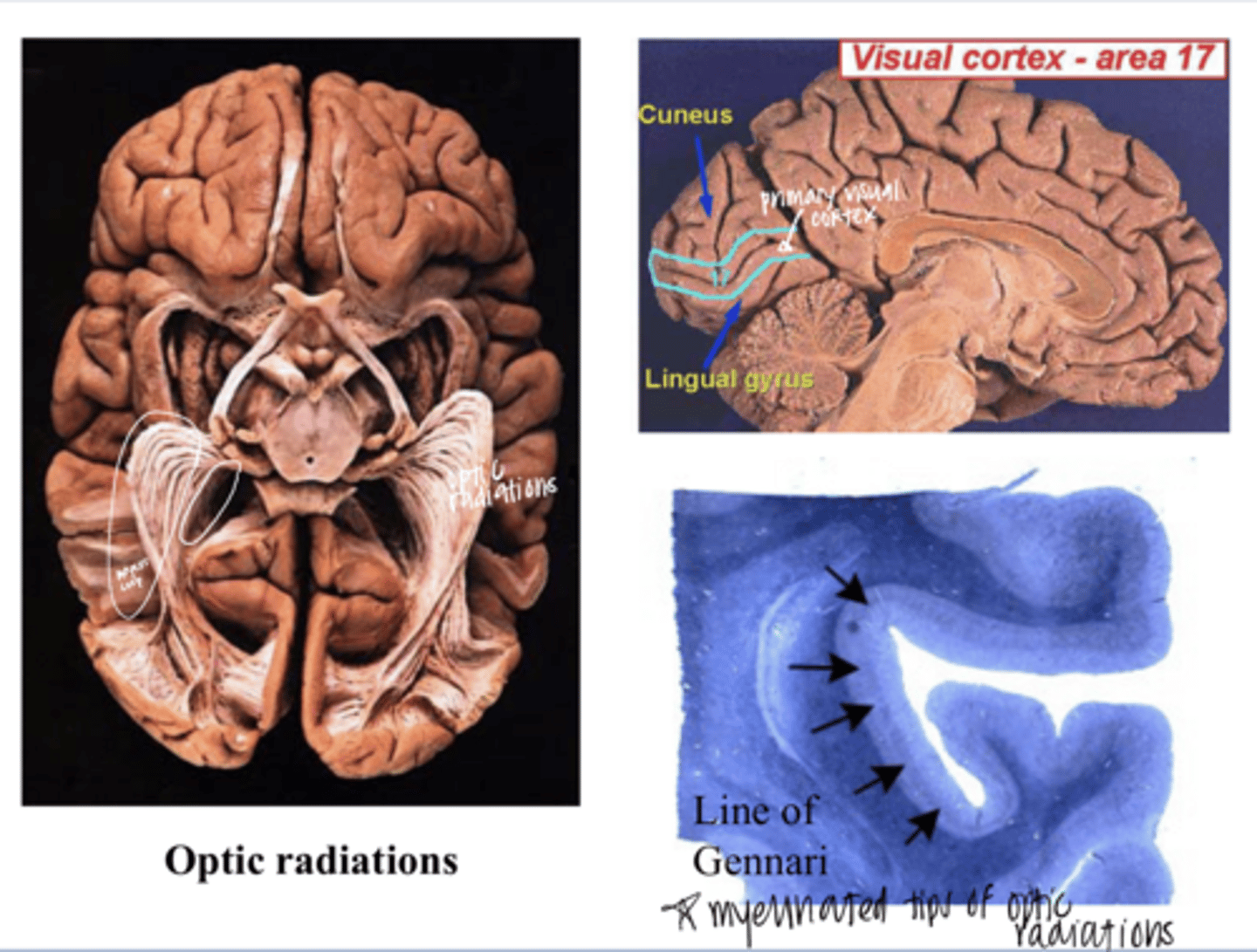
Image of the Superior and Inferior Optic Radiations
Image of the Superior and Inferior Optic Radiations
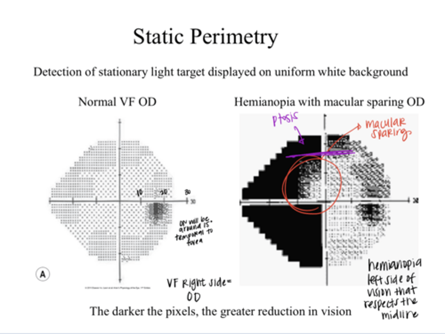
static perimetry
detection of stationary light target displayed on uniform white background
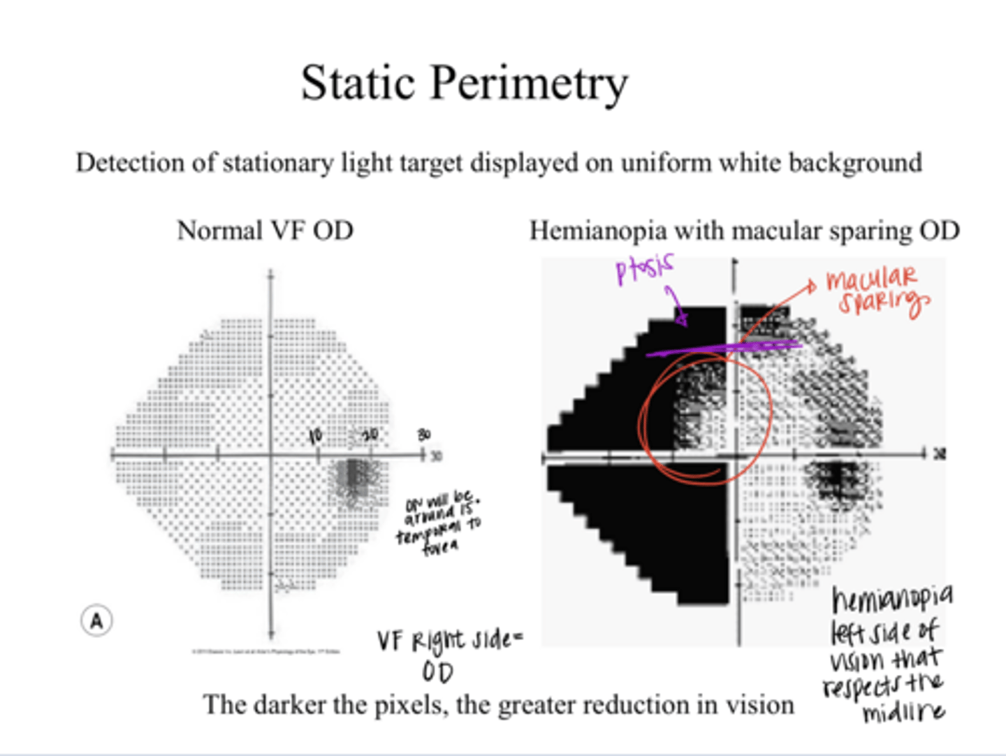
temporal
The ON blind spot (physiological blind spot) will be in the (nasal/temporal) VF of each eye
15
The ON blind spot (physiological blind spot) will be around ______º from the fovea
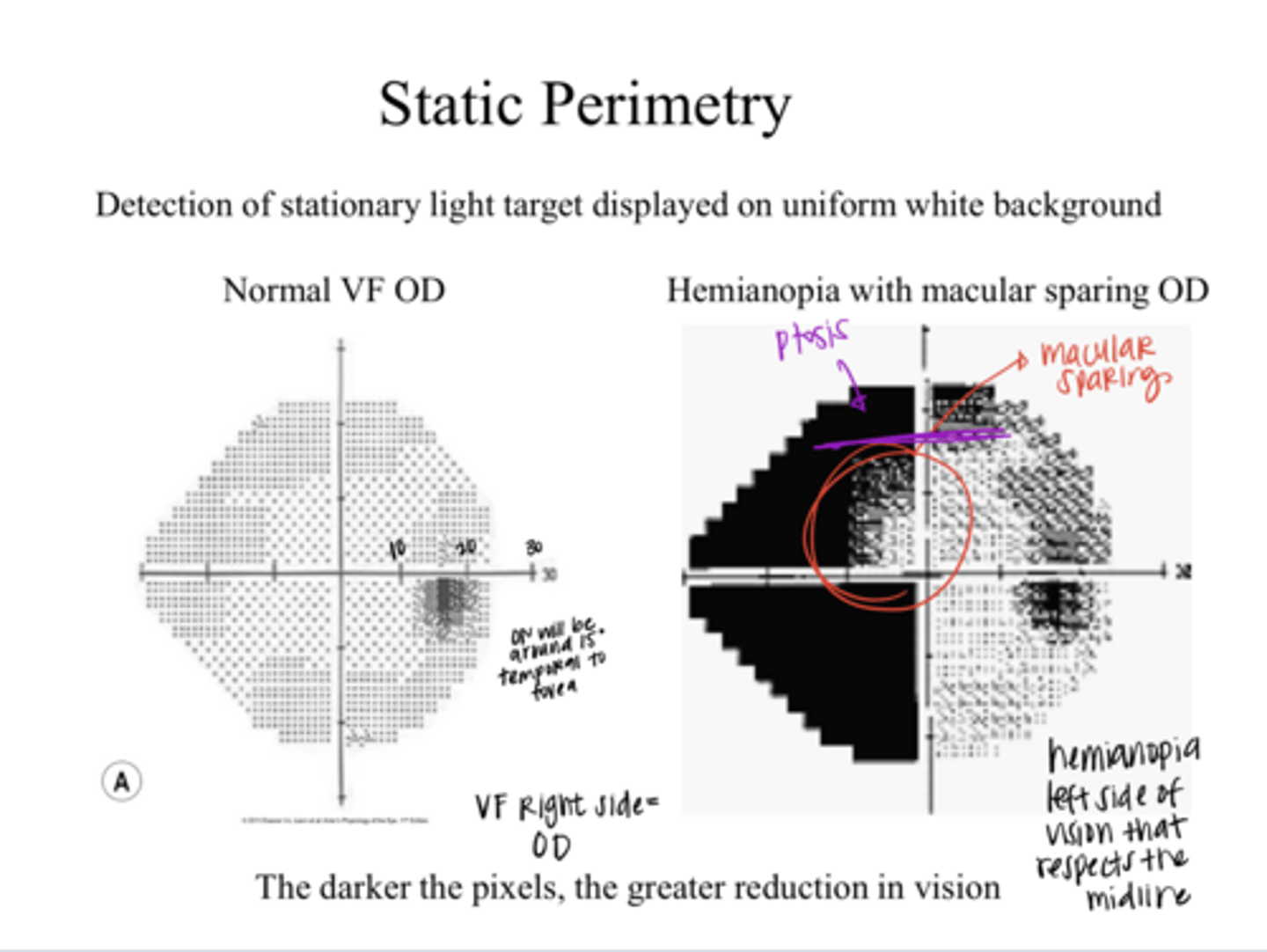
The darker the pixels in a VF printout, the (lesser/greater) the reduction in vision
greater
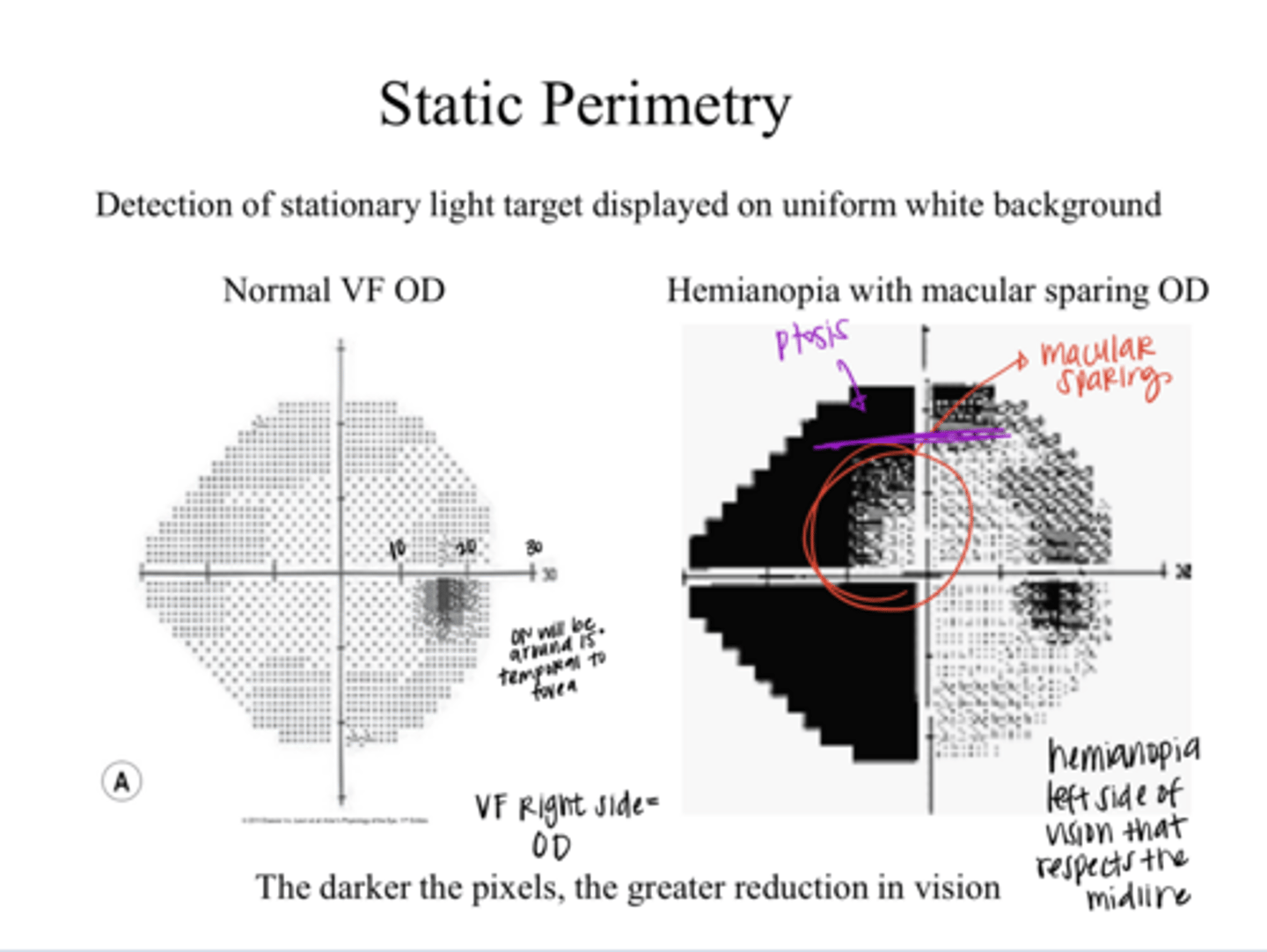
Lesion is relatively close to the optic chiasm and the lesion will clip some of the axons that loop into the contralateral side of the brain = Knee of Von Willebrand
Why is the OD affected in this VF pictured?
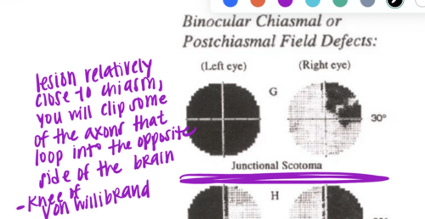
Left homonymous Superior quadrantanopia
**Meyer's loop = superior VF falling on the inferior retina
What would be the VF defect if Meyer's loop through the Right temporal lobe was cut?
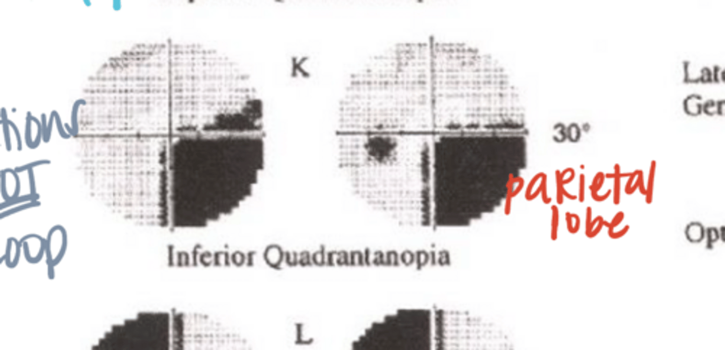
Right homonymous inferior quadrantanopia
**Inferior VF falling on the superior retina
What would be the VF defect if the optic radiations through the left parietal lobe were cut?
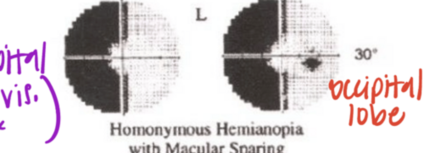
Left homonymous hemianopia with macular sparing
What would a defect of the Right occipital lobe (primary visual cortex) look like on VF?
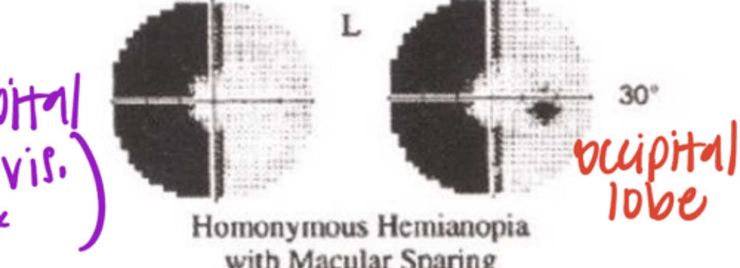
difficult to knock out entire central vision d/t a lot of axons related to macular/foveal vision
Why is macular sparing present with a defect to the occipital lobe?
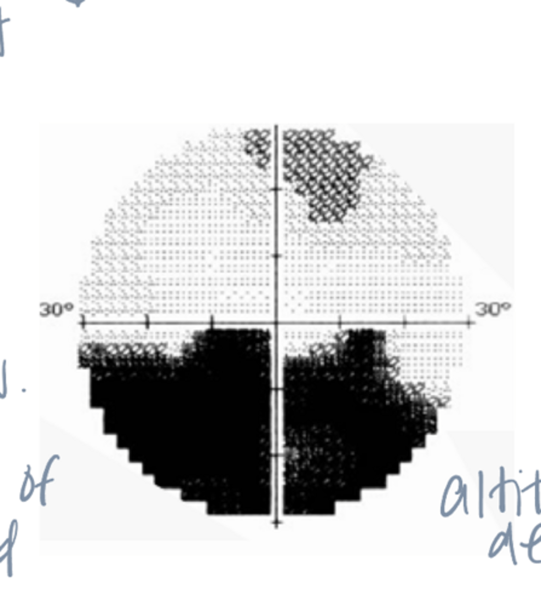
1) Altitudinal defect -- suspect retina or ON problem
2) Look for retinal problem 1st (BRVO, BRAO, retinal detachment)
3) If not retina, look for ON problems (paleness of ONH). Has to be pre-chiasmic
4) Ischemic optic neuropathy is the appropriate dx for this patient given no retinal problems seen.
A 55 year old man reports that when he woke up 10 days ago, he noticed problems "seeing out of the bottom of OD." Your associate measured VA and VF while you finish with another patient.
VA: 20/25 OD; 20/20 OS
Results of Automated VF testing unremarkable OS. OD VF findings shown.
Diagnosis?
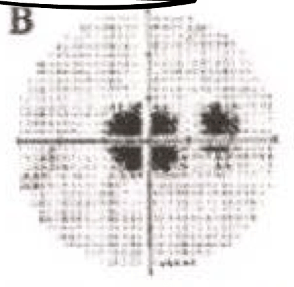
central scotoma
What type of monocular pre-chiasmic VF defect is present in the given image?
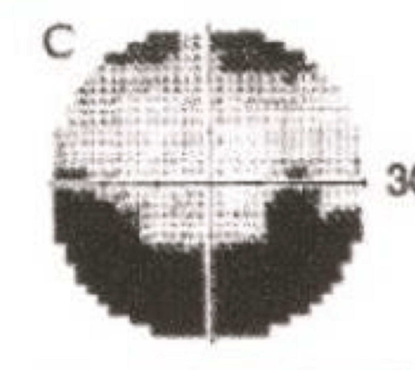
Nerve Fiber Bundle Scotoma
What type of monocular pre-chiasmic VF defect is present in the given image?
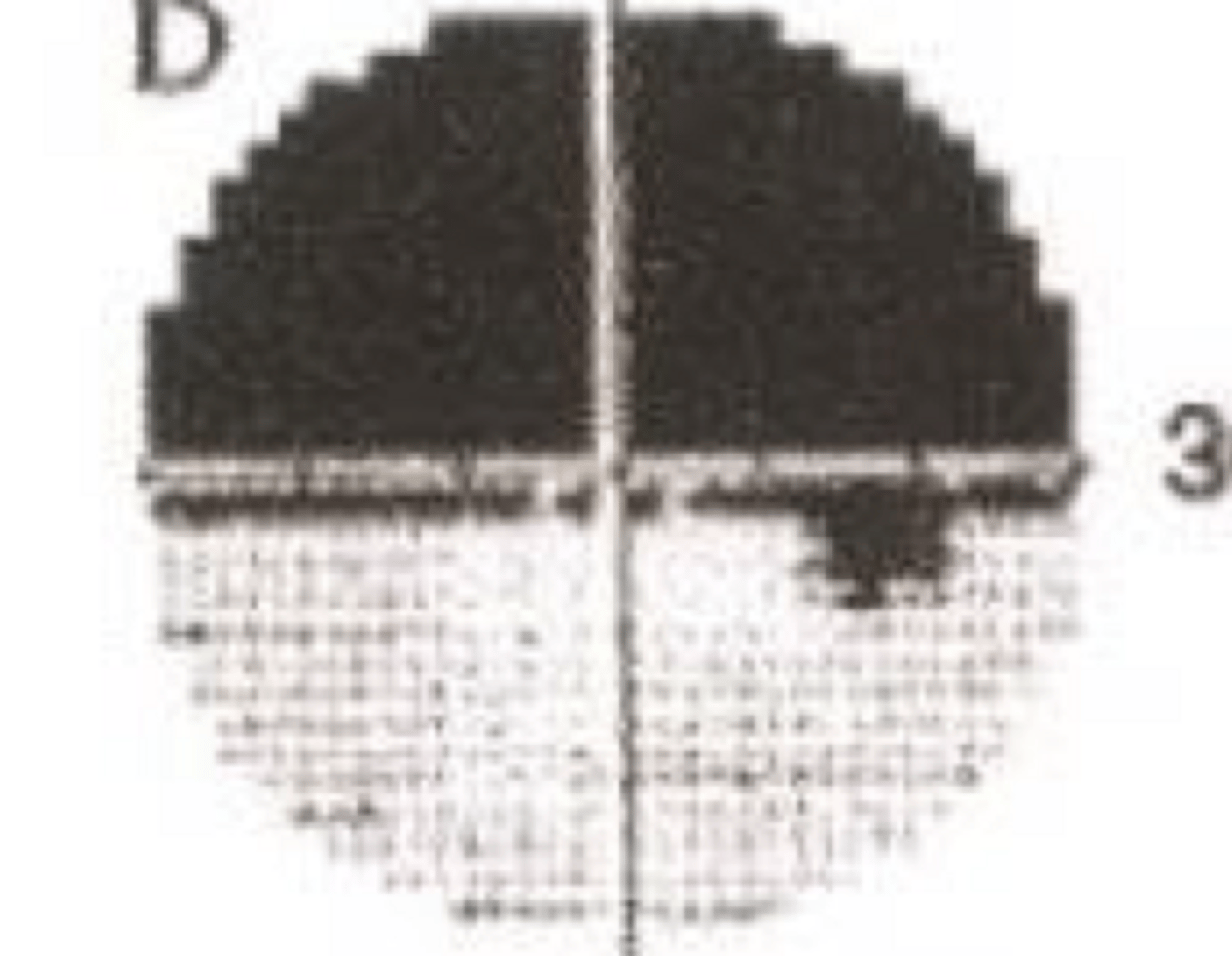
altitudinal scotoma
What type of monocular pre-chiasmic VF defect is present in the given image?
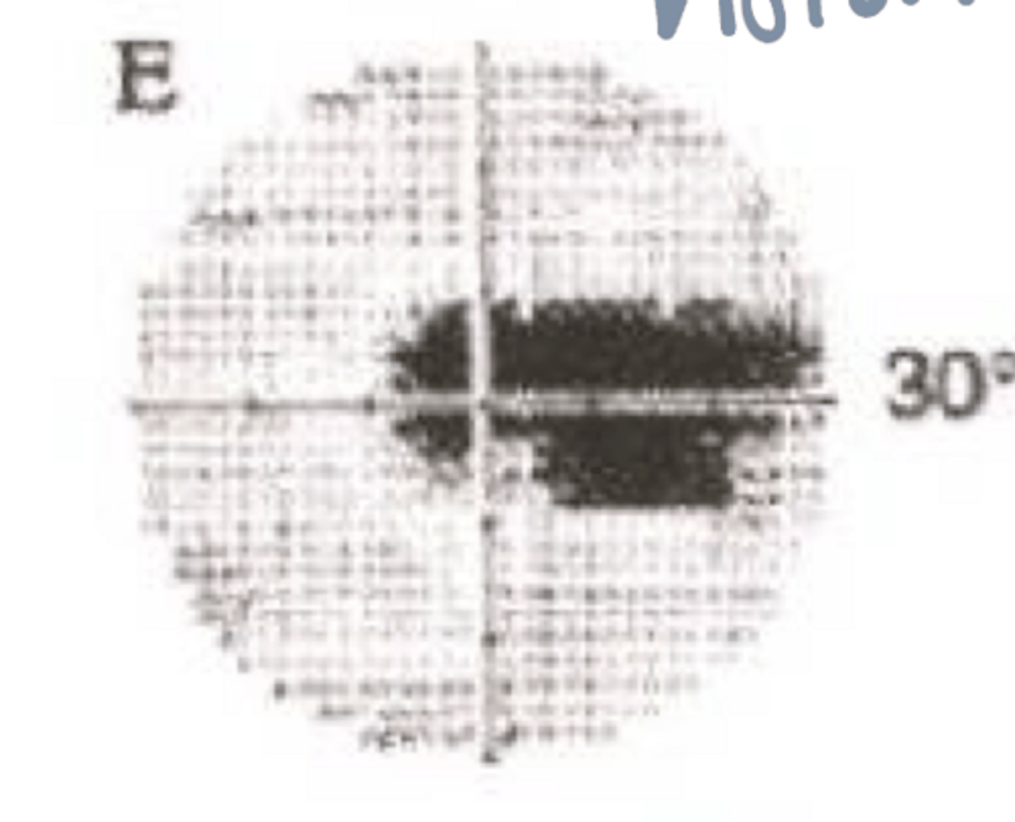
Ceco-central scotoma
What type of monocular pre-chiasmic VF defect is present in the given image?
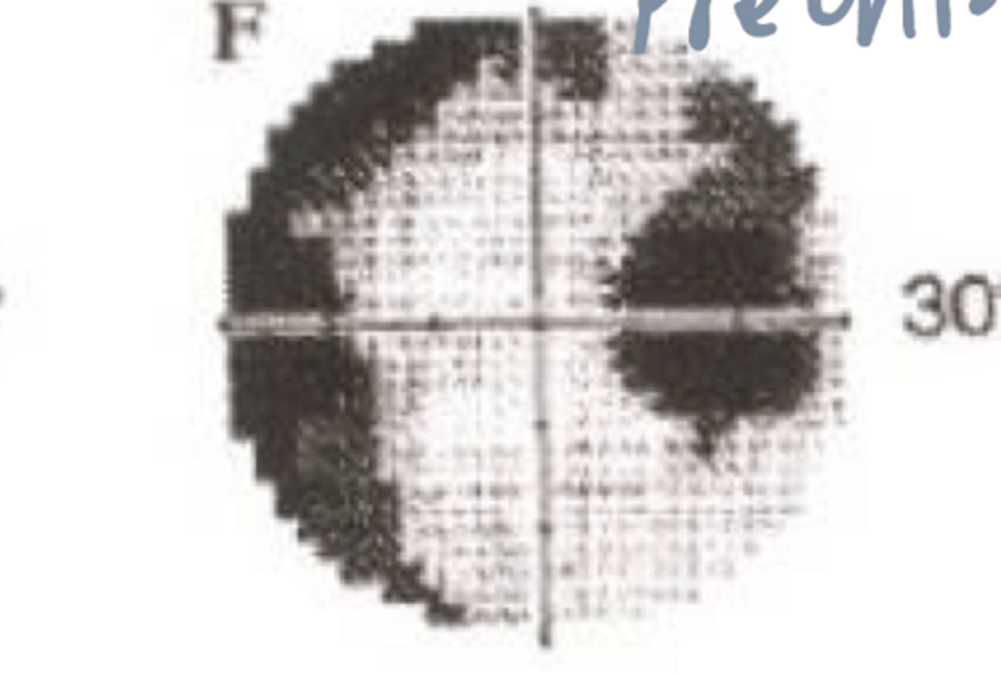
enlarged blind spot with peripheral constriction
What type of monocular pre-chiasmic VF defect is present in the given image?
true
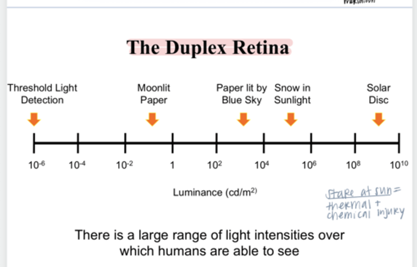
True or False:
There is a large range of light intensities over which humans are able to see
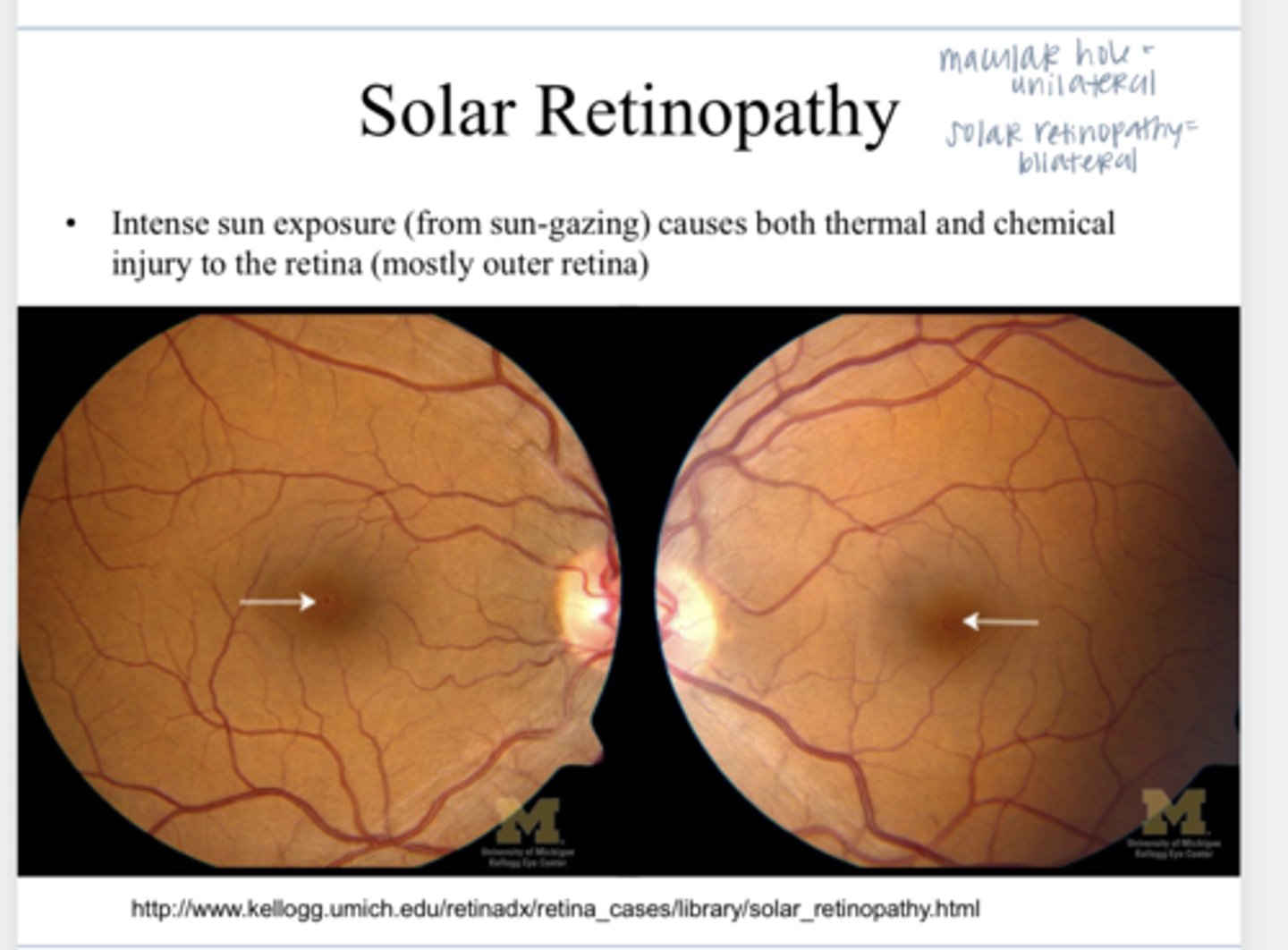
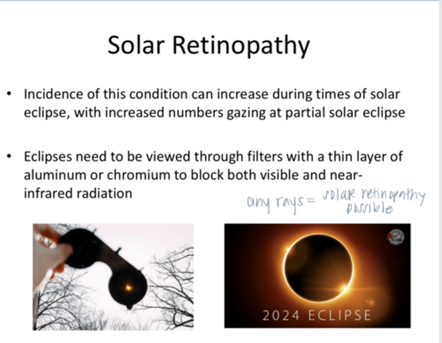
solar retinopathy
Intense sun exposure (from sun-gazing) causes both thermal and chemical injury to the retina (outer)
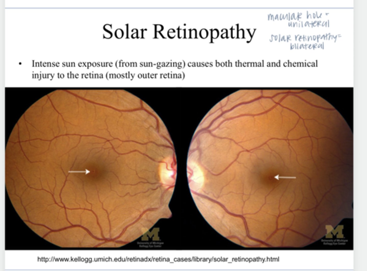
usually bilateral
Is solar retinopathy unilateral or bilateral?
during a solar eclipse, with increased numbers gazing at partial solar eclipse
Incidence of solar retinopathy can increase during what times?
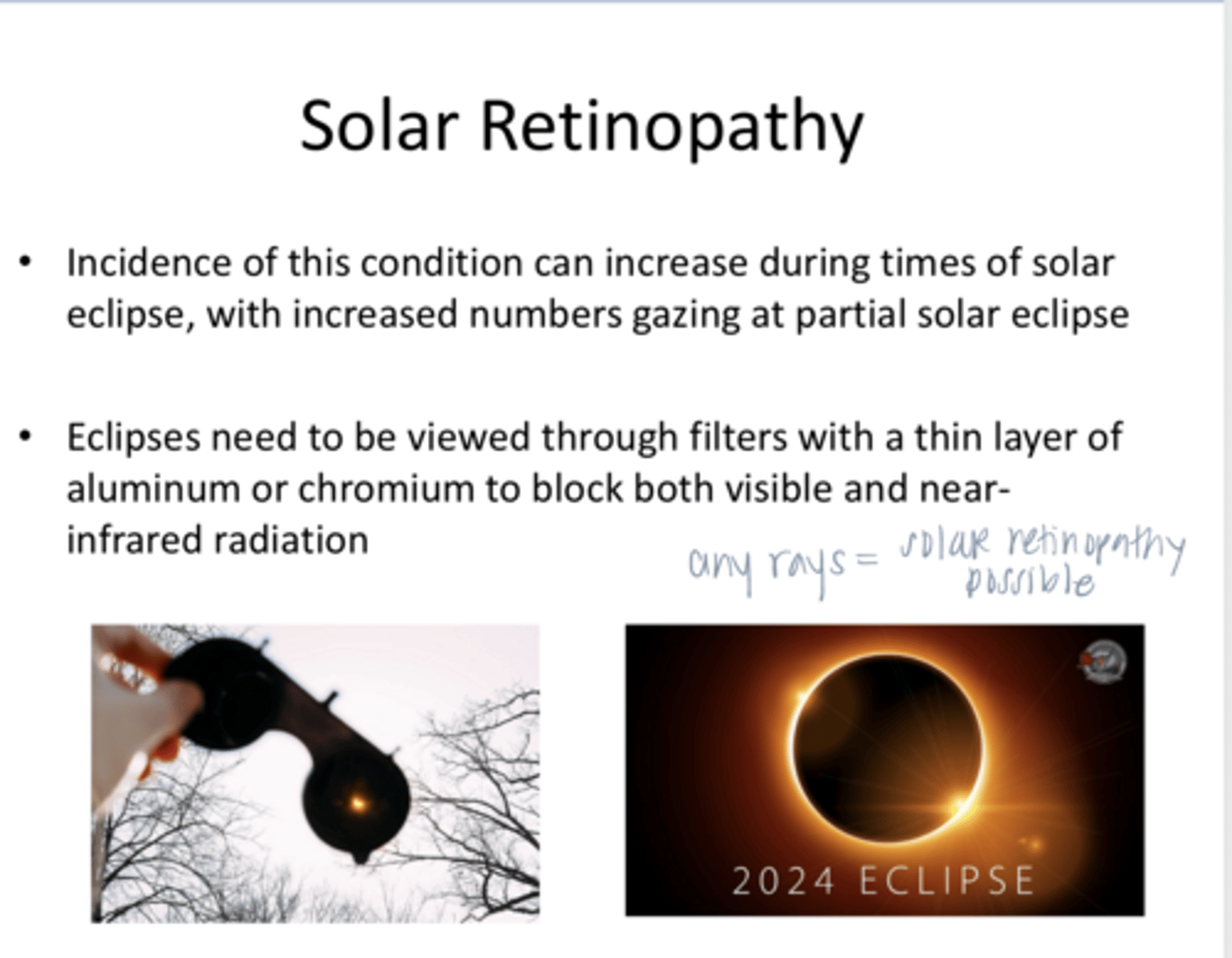
through filters with a thin layer of aluminum or chromium to block both visible and near-infrared radiation
How should eclipses be viewed?
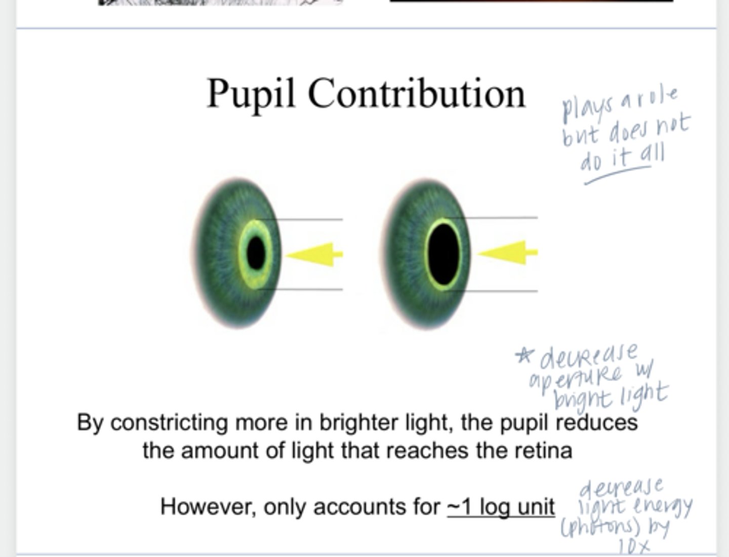
the pupil reduces the amount of light that reaches the retina (~1 log unit)
What is the function of the pupil constricting more in bright light?
2
The retina has ____ different photoreceptor systems
scotopic; photopic
The 2 different photoreceptor systems are optimized for either ____ or _____ conditions
dark
scotopic vision is present in (dark/light) condition
light
Photopic vision is present in (dark/light) condition
rods
Scotopic vision is mediated by what?
single photons can elicit detectable responses (very sensitive to dim light)
Scotopic vision conditions
poor
(poor/good) VA in scotopic visual conditions
no
Is any color discrimination present in scotopic vision conditions?
yes -- no longer respond to increasing levels of light & higher luminance
Is saturation present in scotopic conditions?
cones
Photopic vision is mediated by what?
good
(poor/good) VA in photopic visual conditions