head and neck final ch 1-12
 Call Kai
 Learn
Learn Practice Test
 Spaced Repetition
 Match
Match Flashcards
 Knowt Play
1/188

There's no tags or description
Looks like no tags are added yet.
Name | Mastery | Learn | Test | Matching | Spaced | Call with Kai |
|---|
No analytics yet
Send a link to your students to track their progress
189 Terms
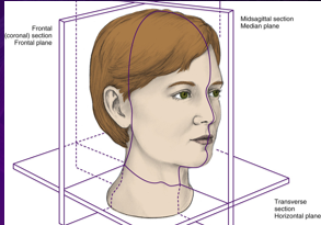
frontal plane/ coronal section
plane related to imaginary line dividing body at any level into anterior and posterior parts
splits body into ventral/ dorsal
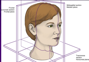
horizontal plane/ transverse section
plane related to imaginary line dividing body at any level into superior and inferior parts
splits body horizontally
splits body into “hamburger” style
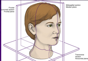
inferior
area facing away from head and toward feet
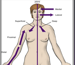
median
structure at median plane
exact midline
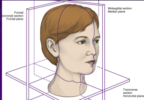
median plane
plane related to imaginary line dividing body into right and left halves
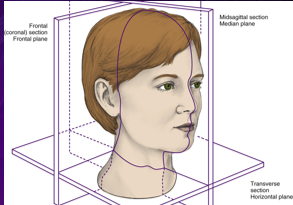
sagittal section/ median plane
section through median plane
section cuts body vertically into exact left and right halves
cuts body “hot dog style”
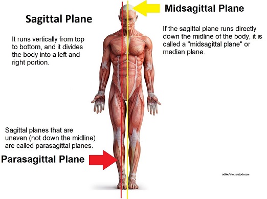
superior
area facing toward head and away from feet
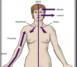
transverse section
section through any horizontal plane
splits the body “hamburger style”
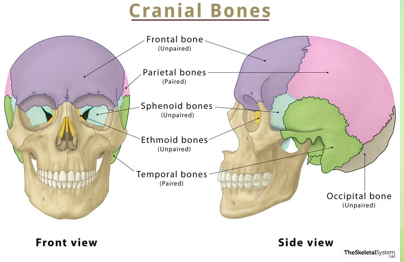
cranium bones
contains the brain
PROTECT THE BRAIN
8 total
occipital
frontal
sphenoid
ethmoid
parietal
temporal
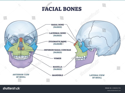
facial bones
support face
serve as a base for dentition
PROTECT THE BRAIN
vomer
lacrimal
nasal
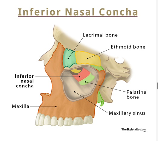
inferior nasal conchae
zygomatic
maxilla
mandible
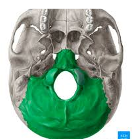
occipital bone
posterior portion of the skull and base of cranium
articulation = parietal, temporal, and sphenoid bone
contains magnum foramen (passage of spinal cord)
condyles are in articulation with the atlas
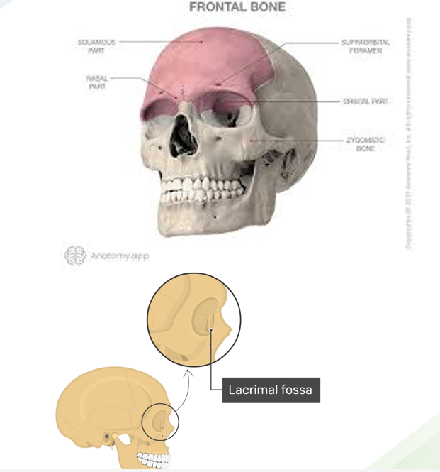
frontal bone
anterior part of skull, superior to eyes and located in frontal region
includes forehead and roof of each orbit
parietal bones
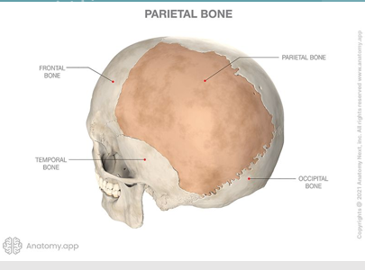
paired
articulate with each other at the sagittal suture
articulate with occipital bone at lambdoidal suture
also articulates with frontal bone, temporal bones, sphenoid bone
temporal bones
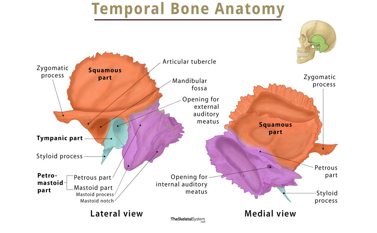
paired
form lateral wall of the skull and part of the base of the skull
articulate with zygomatic bone, parietal bone, occipital bone, sphenoid, mandible
three parts
squamous, tympanic, pterous
sphenoid bone
appearance of a butterfly
goes through midsagittal plane and is located between other bones
forms base of cranium, lateral of skull, floors and walls of orbits
articulates with frontal bone, parietal bones, ethmoid bone, temporal bones, zygomatic bones, palatine bones, comer, occipital bone, maxilla
houses pituitary gland
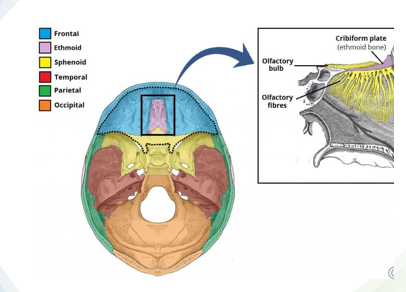
ethmoid bone
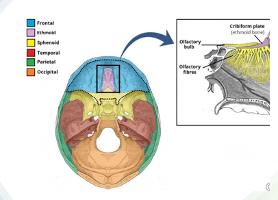
goes through midsagittal plane
located anterior to sphenoid bone
contains cribriform plate
cannot be seen from certain aspect of the skull
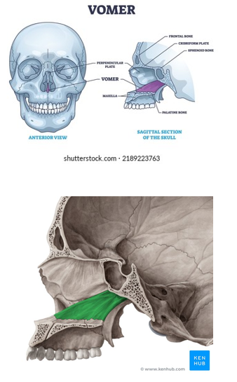
vomer
articulates with sphenoid bone on its superior border
on its inferior border, the vomer articulates with the median palatine suture
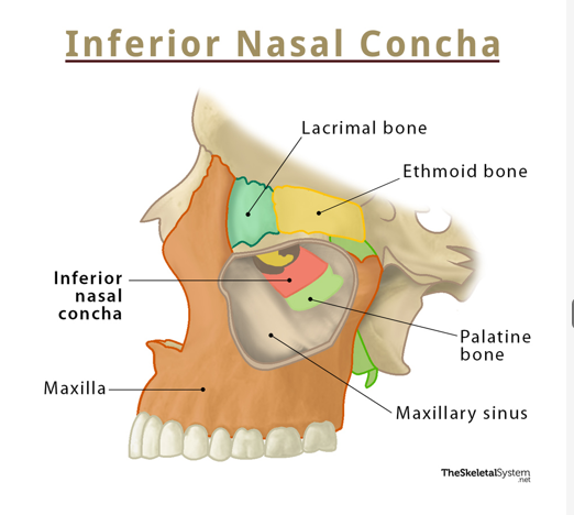
lacrimal bones
2
are the smallest and most fragile of the facial bones
located posterior to the frontal processes of maxillae
each bone articulates with the ethmoid bone and frontal bone as well as maxillae
nasal bones
2
fit between the frontal processes of the maxillae and articulate with the maxillae laterally, and the frontal bone superiorly
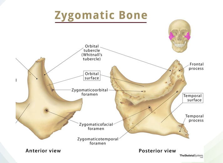
zygomatic bones
form cheek of face
articulates with the frontal bone, temporal bone, and sphenoid bone as well as maxilla
helps to form lateral wall and floor of orbit
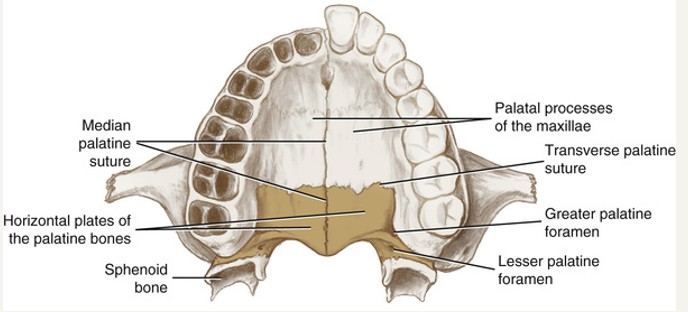
palatine bones
not tehcnically facial bones
L shaped, consists of two horizontal and vertical plates
greater palatine foramen transmits the greater palatine nerve and blood vessels, serving as a landmark for block
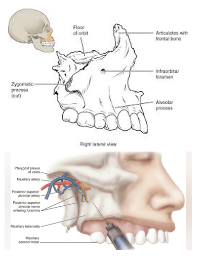
maxilla
2 fused bones at intermaxillary suture
infraorbital foramen
canine fossa (depressions)
canine eminece (raised protrusion)
maxillary tuberosity
posterior superior alveolar foramina
incisive foramen
forms the floor of the nasal cavity
contains maxillary (upper) teeth
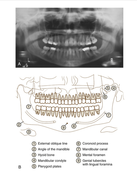
mandible
single facial bone that forms lower jaw
only freely moveable bone of the skull
contains the mandibular foramen, mental foramen, and coronoid notch, along with teeth of lower dentition
origin
end of muscle attached to least movable structure
insertion
end of the muscle attached to more moveable structure
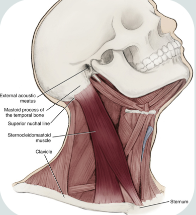
cervical muscles
sternocleidomastoid
trapezius
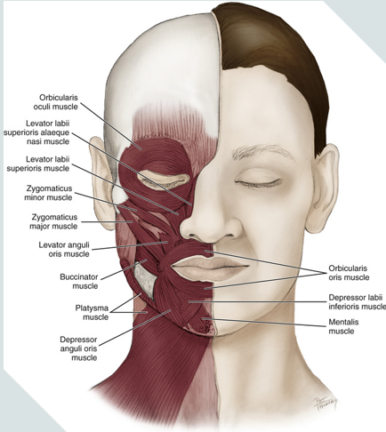
muscles of facial expression
responsible for expression, lip movement, smiling, and frowning
epicranial
frontalis
occipital belly
orbicularis oculi
orbicularis oris
depressor anguli oris
mentalis
platysma
risorius
buccinator
corrugator supercilli
zygomaticus minor
zygomaticus major
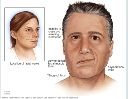
bells palsy
causes paralysis or weakness in one side of the face
paralysis of facial nerve
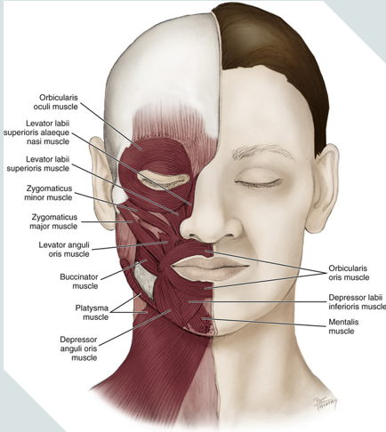
zygomaticus major facial expression
smiling (draws corners of the mouth up)
elevates corners of mouth
zygomaticus major, levator anguli oris, risorius facial expressions
all of these contract when smiling
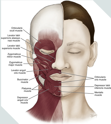
temporalis muscle
a fan shaped muscle
main function is to elevate and retract mandible
originates from the temporal fossa and inserts at the coronoid process of the mandible
its fibers pass deep to zygomatic arch to attach to mandible
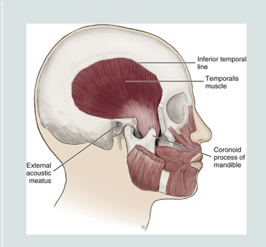
muscles of mastication
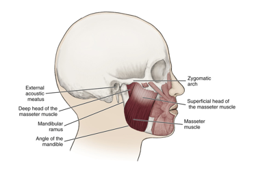
masseter
temporalis
medial pterygoid
lateral pterygoid
elevate, depress, protrude, retrude, and laterally deviate the mandible
innervated by the mandibular division of trigeminal nerve
motor fibers that contract these muscle travel in the trigeminal nerve
MONSTOR TRUCKS MAKE LOUDNESS
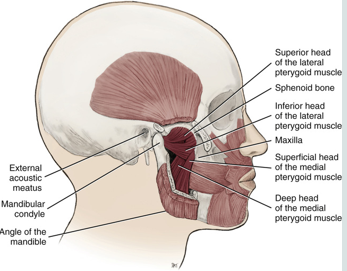
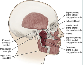
lateral pterygoid muscle
protrudes and laterally shifts mandible
a muscle of mastication
lies superior to the medial pterygoid muscle
a thick and short triangular shaped muscle located in the infratemporal fossa of the skull
superior and inferior heads
causes the power stroke ( moving side to side)
bilateral contraction:
mainly protrusion of mandible with mandible forward
slight depression of mandible during opening of jaws
unilateral contraction
lateral deviation of mandible, shift mandible to contralateral side
trismus
patient cannot open their mouth due to dysfunction in lateral pterygoid muscle
limited opening due to muscle or joint restriction
medial pterygoid muscle
elevates mandible
attaches to the mandible and to the medial surface of the lateral pterygoid plate
bilateral contraction
elevation of mandible during closing of jaws
aids in elevating the mandible while closing the jaw, protruding the mandible, and mastication
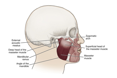
masseter muscle
a muscle of mastication
attaches to the zygomatic arch
when contracted it elevates the mandible
most superficial and is the strongest muscle of mastication, primary chewing muscle
bilateral contraction:
elevation of mandible during closing of jaw
bruxism
bilateral enlargement of the masseter muscle from trauma (grinding)
grinding of teeth
alters facial dimensions
causes muscular pain in masseter and temporalis
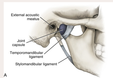
temporomandibular joint
formed by and articulation between the mandibular condyle and the temporal bones
the muscles involved are attached to the cranium and the mandible
allows for the mandible to be elevated and depressed
contains the mandibular condyle, temporal fossa, and articular disc
joint disc contains synovial fluid
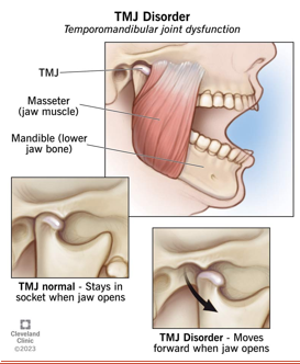
TMD
temporomandibular disorder
joint tenderness, neck/shoulder pain from tension, chronic headaches
vascular system of the head and neck..
consists of an arterial blood supply
a capillary network
venous drainage
artery
carry oxygenated blood away from the heart
strong, muscular blood vessels that carry oxygen-rich blood from the heart to the body
handle a large amount of force and pressure from your blood flow but dont carry a large volume of blood
bacteremia
bacteria traveling within vascular system
vascular plexus
large network of blood vessels, usually veins within the system
vein
deoxygenated blood vessel traveling to the heart carrying blood
carry oxygen-poor blood
hold most of the blood in the body
venous sinus
blood-filled space between two layers of tissue
venule
smaller vein draining capillaries and then joins larger veins
the veins of the head and neck start out as these and become larger as they near the base of the neck on their way to the heart
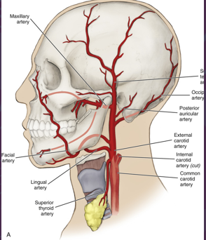
major arterial blood supply of the head and neck
major arteries that supply the head and neck
common carotid artery
subclavian arteries
different origins depending on the side of the body
unique for head and neck arteries because most are symmetrical
common carotid artery
left side of the body: originate directly from aorta
right side of body: branch off brachiocephalic artery
branchless and travels along neck in lateral position to trachea and larynx
travels in carotid sheath deep to SCM
internal and external
key blood supply to head and neck structures
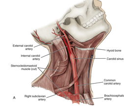
subclavian artery
left side of body: originate directly from aorta
right side of body: branch off brachiocephalic artery
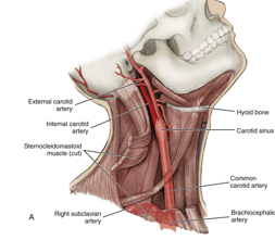
carotid sheath
contains
common carotid artery
internal jugular vein
vagus nerve X
surrounds the vascular compartment of neck
can palpate common carotid against the larynx and obtain pulse
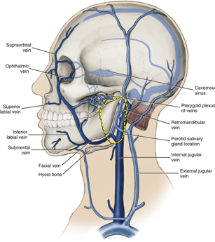
jugular vein
drains deoxygenated blood from the brain and face
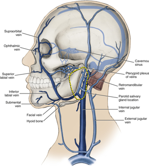
veins in the head and neck start
as small venules and get larger as they approach the heart
pterygoid plexus
network of veins that collects blood from deep areas of face and then flows into maxillary vein
near maxillary tuberosity
helps drain upper and lower jaw
incorrect PSA block can pierce this or maxillary artery
help protect maxillary artery from being squeezed when chewing by adjusting blood volume
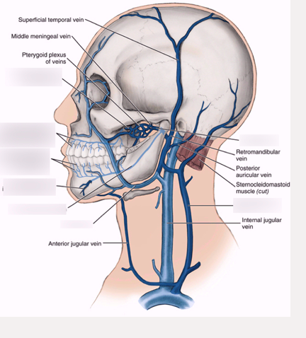
internal jugular vein
drains blood from most of head and neck
exists skull through jugular foramen
runs alongside common carotid artery and vagus nerve in the carotid sheath
travels down neck and joins subclavian vein
duct
passageway to carry secretion from the exocrine gland to set location
exocrine
gland with associated duct serving as passageway
all major and minor salivary glands or sweat glands
only glands that have ducts
endocrine
ductless gland with secretions being poured directly into vascular system
what they make is secreted directly into bloodstream
adrenal glands
parotid gland
the largest salivary gland
is encapsulated
innervated by the 9th cranial nerve
empties around the 2nd maxillary molar which is a site of calculus build-up
another name for parotid duct
stenson duct
percentage of saliva excreted from parotid duct
25%
serous secretion
mumps is a result of this enlarged saliva gland
parotid
submandibular gland
is encapsulated
innervated by the 7th cranial nerve
gland drains into the submandibular lymph nodes
most common site for salivary stones
seen externally as the sublingual caruncle
another name for submandibular gland
Wharton’s duct
“men go to war”
percentage of saliva secreted from submandibular gland
65%
mixed secretions, mostly serous
sublingual gland
located below the tongue
is the only gland not encapsulated
innervated by the 7th and 9th cranial nerve
seen externally as sublingual caruncle
percentage of saliva excreted by sublingual gland
10%
mixed secretions but mostly mucous
minor salivary glands
they are most numerous but generally smaller
located on soft palate, hard palate, and every type of mucosa
all secrete mucous saliva EXCEPT Von Ebner
thyroid gland
2 lobes
is the largest endocrine gland in the body
located below the thyroid cartilage where the larynx meets the trachea
not visible in healthy patients
deals with metabolism and hormones
thyroxine (T4)!!
parathyroid
4 small bean shaped glands on the back of the thyroid gland
cannot be palpated
produces and secretes the parathyroid hormone which regulates calcium and phosphorus levels
lacrimal glands
located within the orbit above the lateral end of the eye
sits in lacrimal fossa
tears flow down to the medial canthus and drain into the inferior nasal meatus
(runny nose when crying)
innervated by the 7th cranial nerve
composed of epithelial and lymphatic tissue
cervical lymph nodes
receive all the lymph from the head and neck
either directly or indirectly via the superficial lymph nodes
these lie along the internal jugular vein
thymus
primarily to produce and mature T-cells
superficial and lateral to the trachea and deep to sternum (cannot palpate)
attached to lobes of thyroid gland
innervated by the 10th cranial nerve
supplied by inferior thyroid and internal thoracic arteries
shrinks after puperty
T-cells
a type of white blood cell that helps the body fight infection and disease
afferent nerve
SENSORY nerve carry info from body periphery TO brain or spinal cord
periphery → brain/spinal cord
efferent nerve
MOTOR nerve carrying info AWAY from brain or spinal cord to periphery
brain/spinal cord→ periphery
dental plexus
network of nerves within both maxillary and mandibular arches
the two components of nervous system
central nervous system
peripheral nervous system
CN I- Olfactory
afferent only
responsible for sense of smell
passes through cribriform plate of ethmoid bone
( cribs smell)
CN II - Optic
afferent only
responsible for vision
enters optic canal of sphenoid bone
both R and L nerves join at the optic chiasma where the fibers cross to contralateral sides before entering the brain
CN III - oculomotor
efferent only
controls most eye muscles and pupil constriction
passes through superior orbital fissure of sphenoid bone
CN IV - Trochlear
efferent only
controls superior oblique muscles of the eye
exits skull through superior orbital fissure of sphenoid bone
CN V - Trigeminal
both sensory and motor roots
afferent- teeth, tongue, oral cavity, lacrimal gland, nasal cavities, and skin of face
sensory to teeth, gingiva, oral mucosa
motor to muscles of mastication
efferent - muscle of mastication
three divisions
opthalmic (V1)
maxillary (V2)
mandibular (V3)
opthalmic division of trigeminal nerve
sensory
enters at superior orbital fissure
maxillary branch of trigeminal nerve
sensory
enters at foramen rotundum
mandibular branch of trigeminal nerve
motor AND sensory
enters at foramen ovale
CN VI - Abducens
efferent only
supplies muscles for eye movement and innervates lateral rectus muscle of eye
exits through superior orbital fissure of sphenoid bone
1st nerve affected by serious sinus infections
CN VII - Facial
both afferent and efferent
efferent - facial expression muscles
afferent - taste anterior 2/3 of the tongue
passes through stylomastoid foramen
gives motor innervation to stylomastoid muscles
chorda tympani
branch of facial nerve
CN VIII -Vestibulocochlear
afferent only
helps with hearing and balance
passes through internal acoustic meatus
damage causes vertigo, hearing loss, tinnitus
CN IX- Glossopharyngeal
both motor and sensory
efferent - stylopharyngeus muscle, parotid gland
afferent - taste and sensation to posterior 1/3 of tongue
includes gag reflex
passes through jugular foramen
CN X- Vagus
both motor and sensory
efferent - soft palate, pharynx, larynx, thoracic organs
afferent - taste sensation for epiglottis
passes through jugular foramen
helps with speech and swallowing
CN XI - Accessory
motor only
provides innervation for trapezius and sternocleidomastoid muscles, muscle of soft palate, and muscles of pharynx
passes through jugular foramen
damage may cause shoulder droop and difficulty turning head
CN XII- Hypoglossal
motor only
controls tongue movements
aids in speech
passes through hypoglossal canal of occipital bone
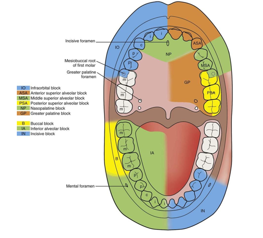
branches we block for trigeminal nerve
V2 Maxillary
V3 Mandibular
maxillary arch local anethetic blocks
PSA
MSA
ASA
NP
GP
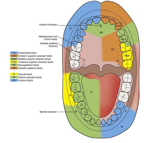
Mandibular arch local anethetic blocks
IA
Long buccal nerve block
mental/ incisive nerve block
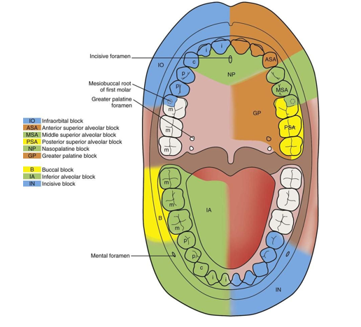
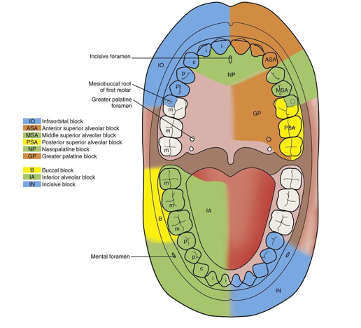
posterior superior alveolar nerve block (PSA)
insertion: mucobuccal fold above the DB root of the 2nd molar
target is maxillary tuberosity
can cause hematoma if injected into pterygoid plexus
teeth anesthetized: maxillary molars except the MB root of the 1st molar
soft tissue anethetized: buccal gingiva of the maxillary molars
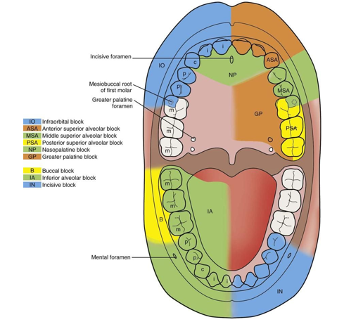
middle superior alveolar nerve block (MSA)
insertion: mucobuccal fold above 2nd premolar
teeth anesthetized: maxillary premolars and MB root of the 1sst molar
soft tissue anethetized: buccal gingiva of maxillary premolars
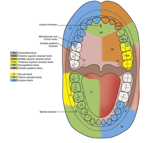
anterior superior alveolar nerve block (ASA)
insertion: height of the mucobuccal fold near mesial aspect of maxillary canine
teeth anesthetized: maxillary incisors/cuspids and MSA teeth
soft tissue anesthetized: maxillary incisors and cuspid gingiva
Nasopalatine nerve block (NP)
insertion: palatal mucosa just lateral to the incisive papilla
soft tissue anethetized: lingual gingiva of the maxillary incisor/cuspids