S26 Anatomy 3-1 Liver and Bile Ducts
1/38
Earn XP
Description and Tags
S26 Anatomy Practical #3
Name | Mastery | Learn | Test | Matching | Spaced | Call with Kai |
|---|
No analytics yet
Send a link to your students to track their progress
39 Terms
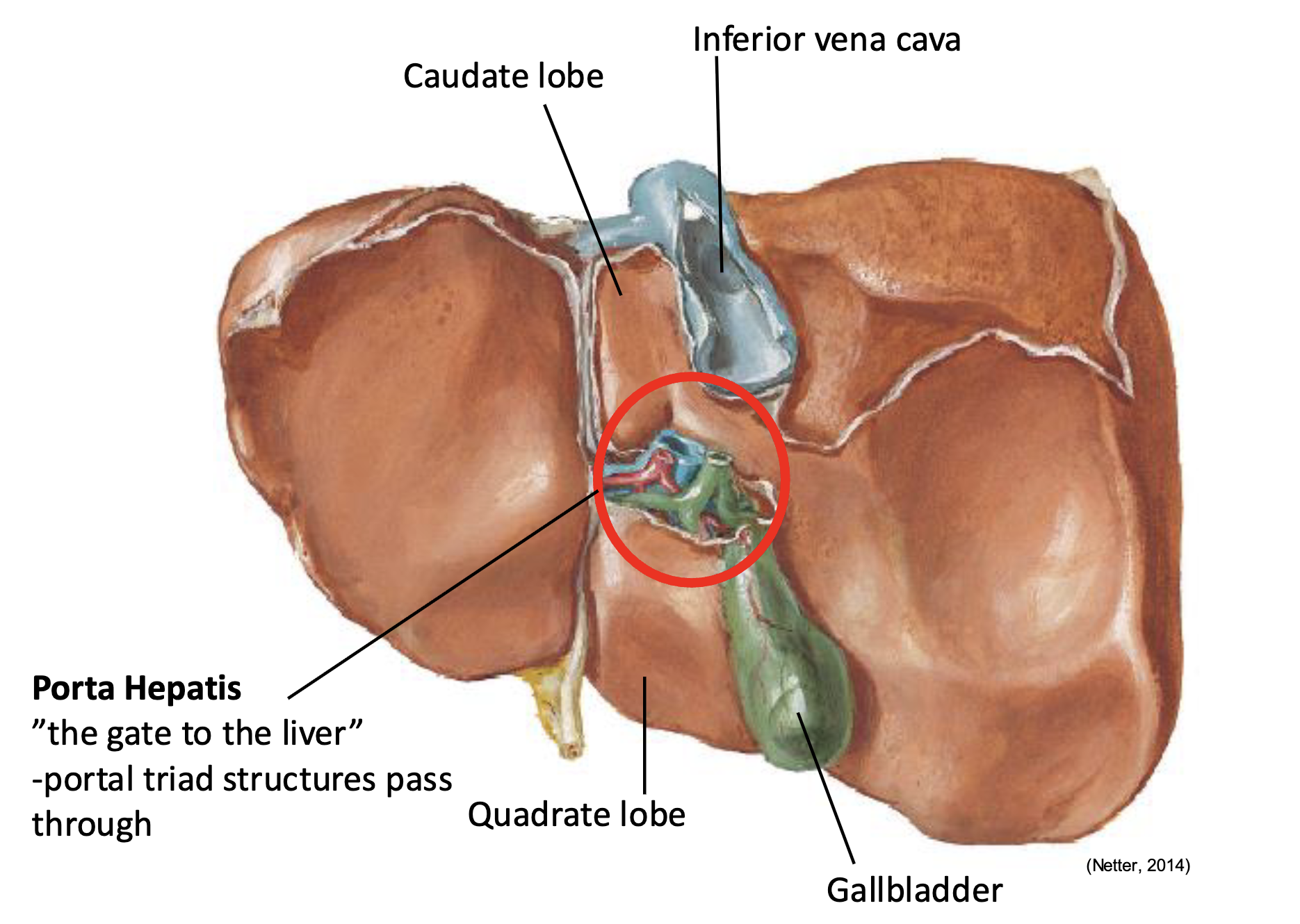
What structure separates the right and left lobes of the liver?
Falciform ligament
Quadrate lobe is found near what landmark?
near the gallbladder
The Caudate Lobe is found near what anatomical landmark?
Caudate lobe near IVC
What travels between the Caudate Lobe and Quadrate Lobe
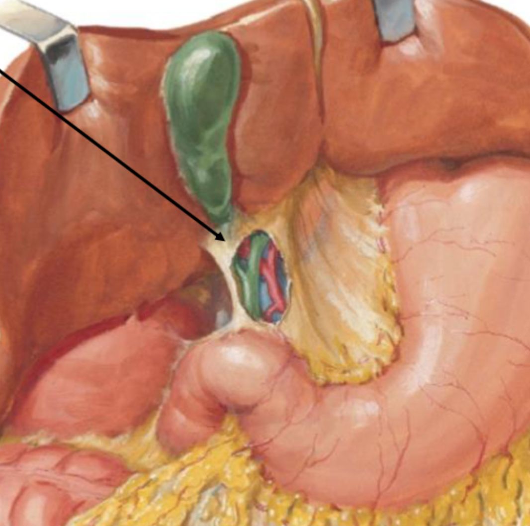
Porta hepatis
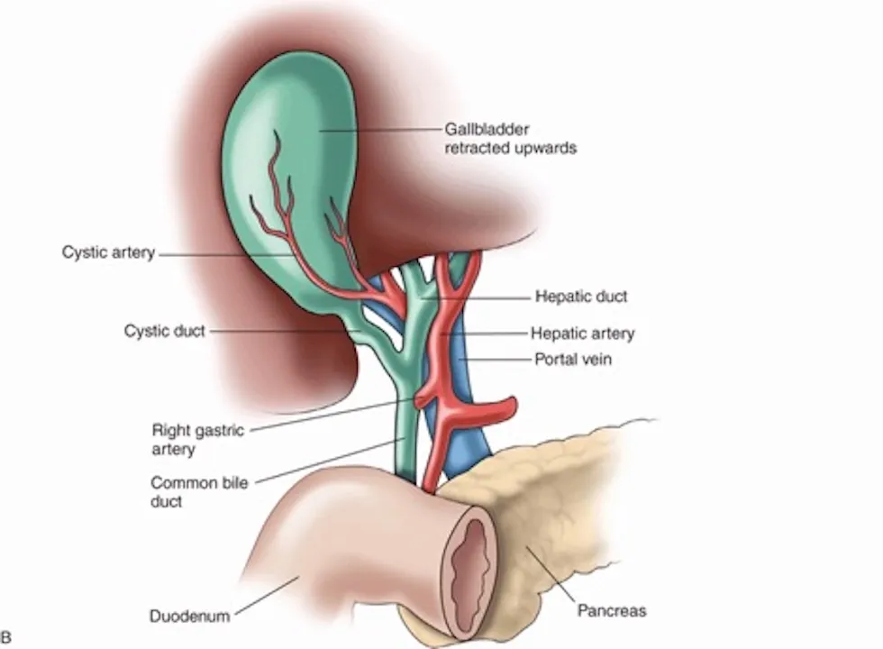
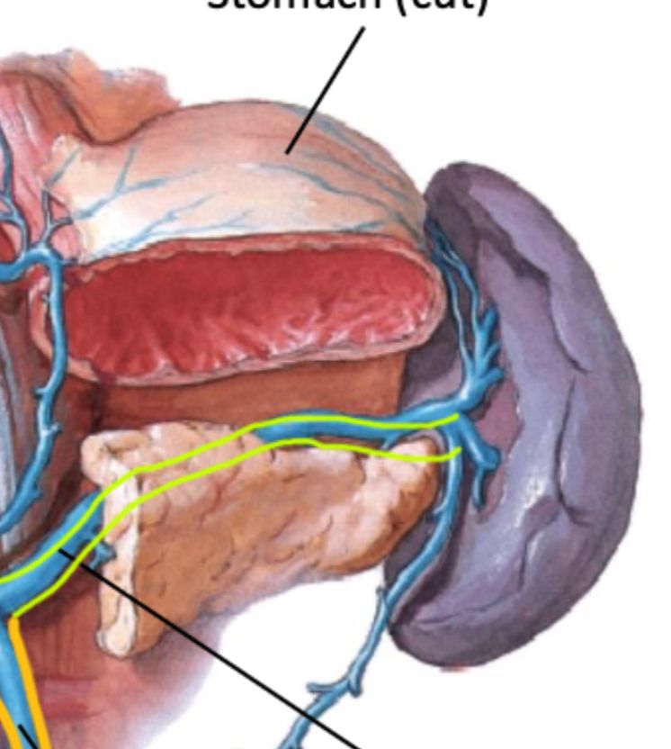
What are the contents of the portal triad?
Hepatic portal vein (deepest), hepatic artery proper, common bile duct (Right*)
The hepatic portal vein is formed by the union of what two veins?
Splenic vein and superior mesenteric vein
Of the portal triad structures which is the deepest?
Hepatic portal vein
Of the portal triad structures which is most towards the right?
Common bile duct

Of the portal triad structures which is most towards the left?
Hepatic artery proper
The cystic duct joins what duct to form the common bile duct?
Common hepatic duct
The spleen sits in what quadrant of the abdomen?
Left upper
The spleen is supplied by what artery?
Splenic artery
The inferior vena cava is located where in relation to the abdominal aorta and vertebral column?
To the right of the abdominal aorta and vertebral column
What do hepatic veins drain into?
Inferior vena cava
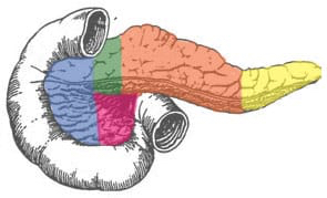
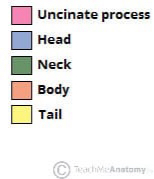
The uncinate process of the pancreas sits directly in which organ?
2nd part of the duodenum
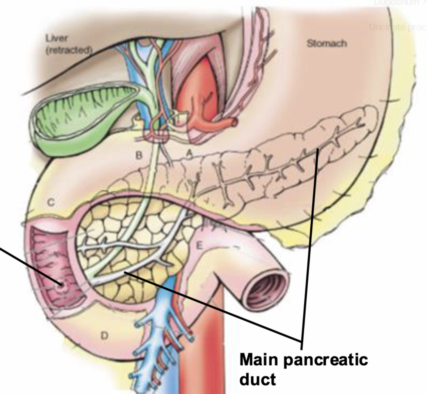
The tail of the pancreas extends to touch which organ?
Spleen
What nerves innervate the pancreas?
Greater thoracic splanchnic (sympathetic) and Vagus nerve (parasympathetic)
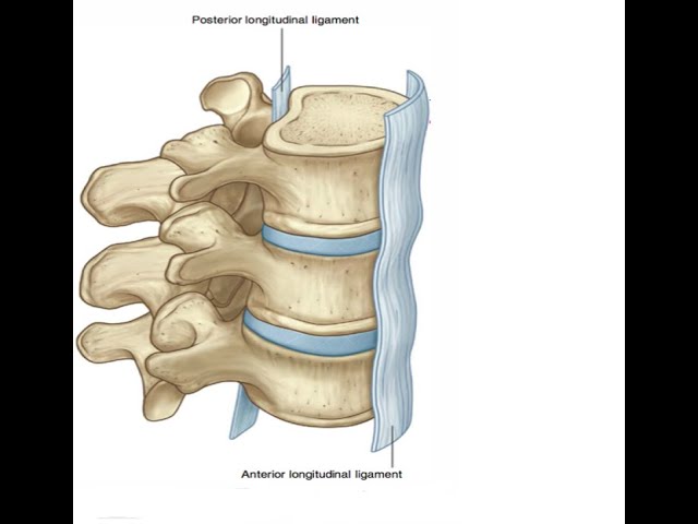
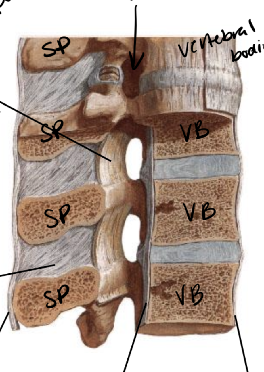
Describe the location and vertebral levels of the Anterior longitudinal ligament.
Anterior and lateral surfaces of all vertebral bodies and intervertebral discs from C2-sacrum
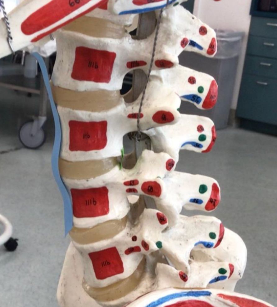
blue tape - anterior longitudinal ligament
green tape - posterior longitudinal ligament
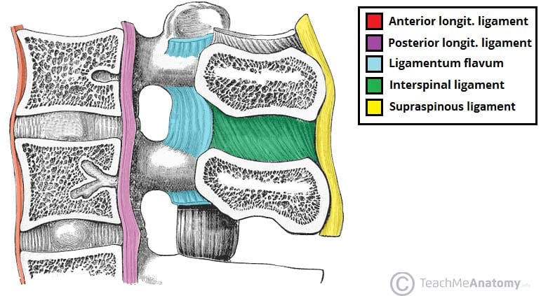
Describe the location and vertebral levels of the Posterior longitudinal ligament.
Posterior surfaces of all vertebral bodies from C2-sacrum
Describe the location and vertebral levels of the Ligamentum Flavum.
Connects the lamina above and below together from C2-S1
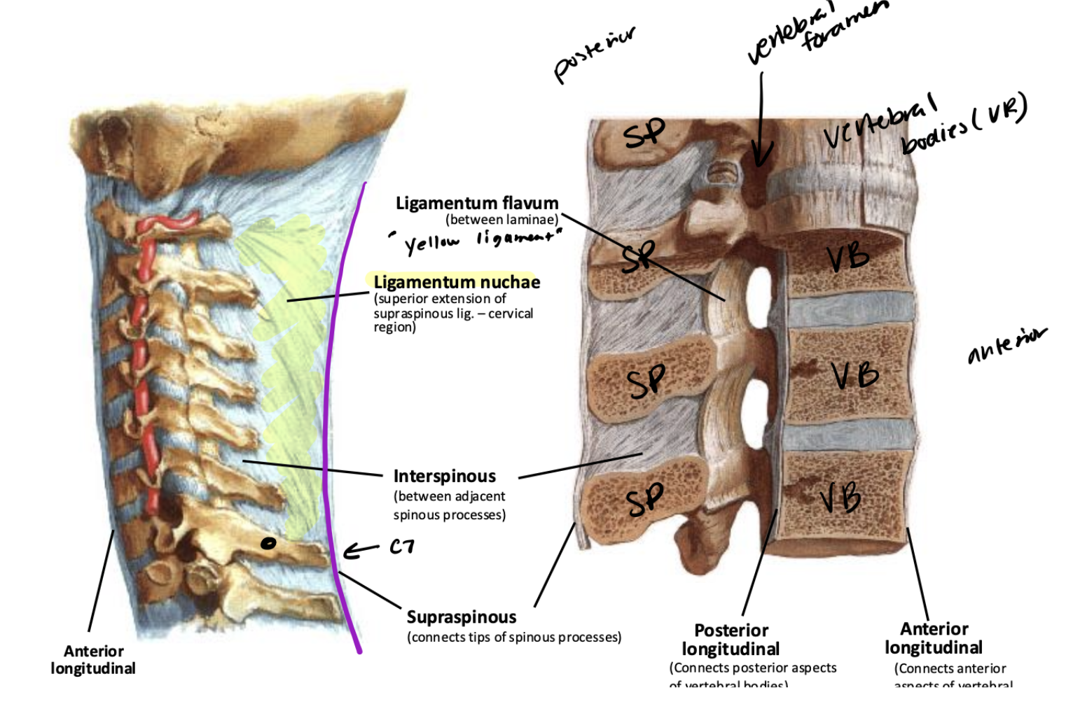
Describe the location and vertebral levels of the Supraspinous ligament.
Connects the tips of all spinous processes from C7 to the sacrum
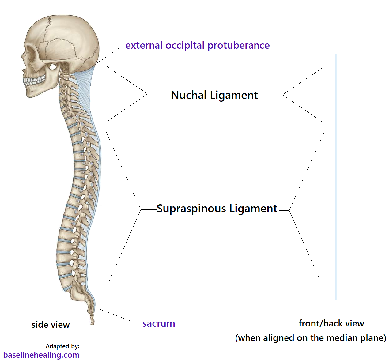
Describe the location and vertebral levels of the Ligamentum nuchae.
Superior continuation of supraspinous ligament, above C7 to midsagittal line on occipital bone from foramen magnum to external occipital protuberance

Describe the location and vertebral levels of the Interspinous ligament.
Connects all the spinous processes together

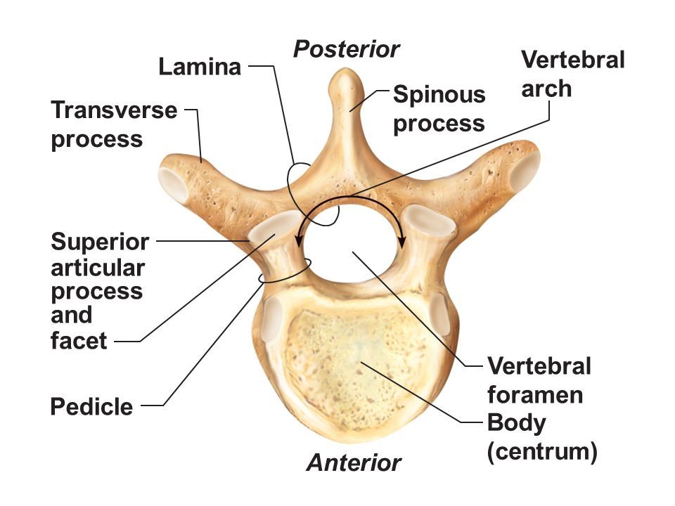
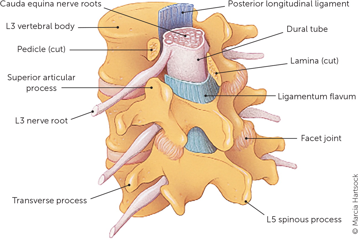
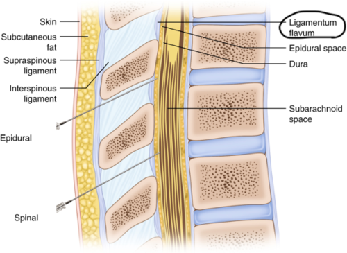
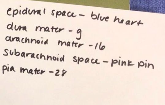
If below L3 vertebral level, posterior and in the midline, what are the structures from superficial to deep ending in the subarachnoid space?
Skin, subcutaneous fat, supraspinous ligament, interspinous ligament, ligamentum flavum, epidural space, dura mater, subdural space, arachnoid mater, subarachnoid space
Explain the arterial supply to the spinal cord and where these arteries branch from?
Anterior spinal artery (from vertebral a.) and 2 posterior spinal arteries (cerebellar arteries)
Subarachnoid space is between which two meningeal layers?
Arachnoid mater and pia mater

What is found in the subarachnoid space?
Cerebrospinal fluid
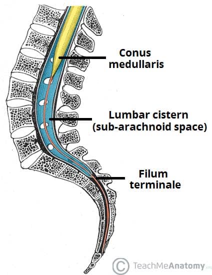
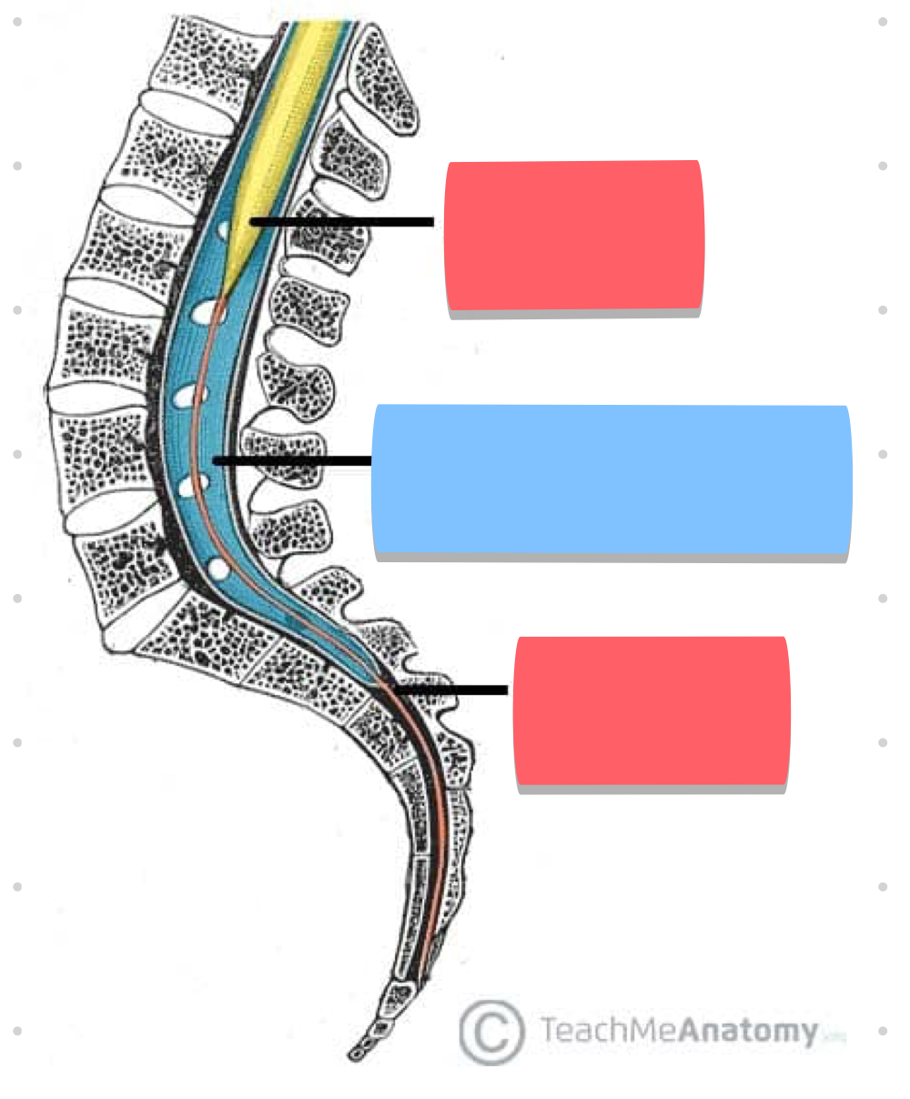
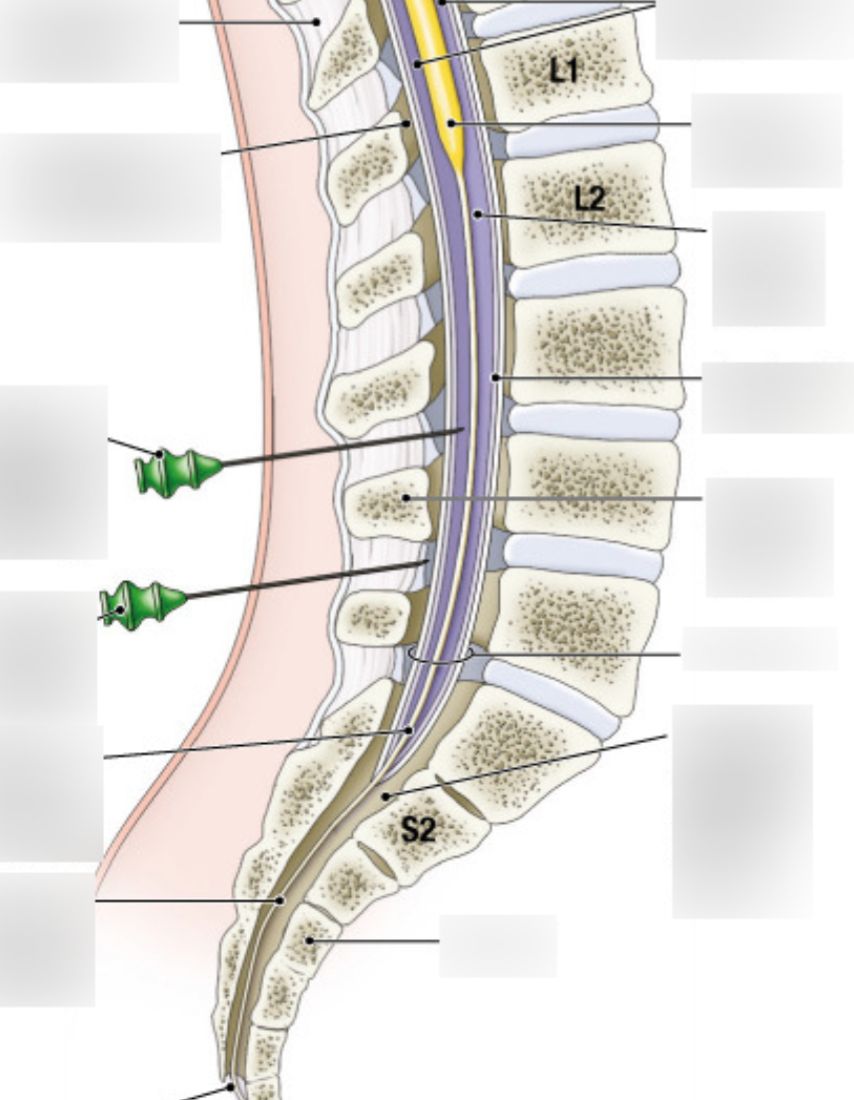
Lumbar cistern is found between what vertebral levels and this is the location of what clinical procedures?
L2 and S2; location for clinical procedures

What is found in the epidural space in the vertebral column?
Adipose tissue, blood vessels, venous plexus
The dural sac ends at what vertebral level?
S2
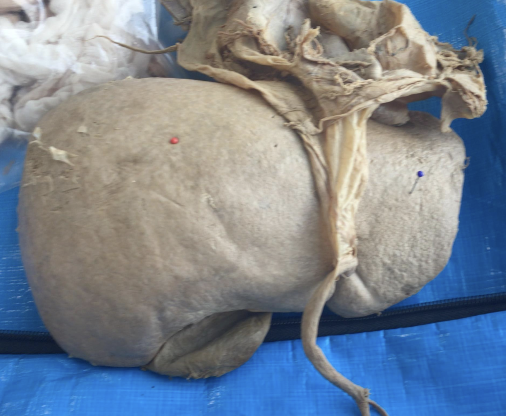
Red - Right Lobe
Navy Blue - Left Lobe
Green - Caudate Lobe (Near IVC)
Yellow Bear - Quadrate Lobe (near gallbladder - GQ)
black - IVC
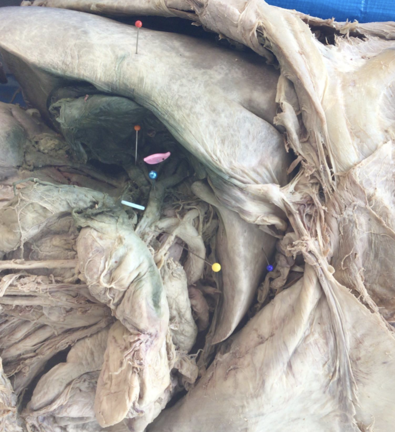
Pink Bird: Common Hepatic Duct
Orange: Cystic Duct
Blue Button: Common Bile Duct
Yellow: Hepatic Portal Vein
Pearly Blue: Proper Hepatic Artery
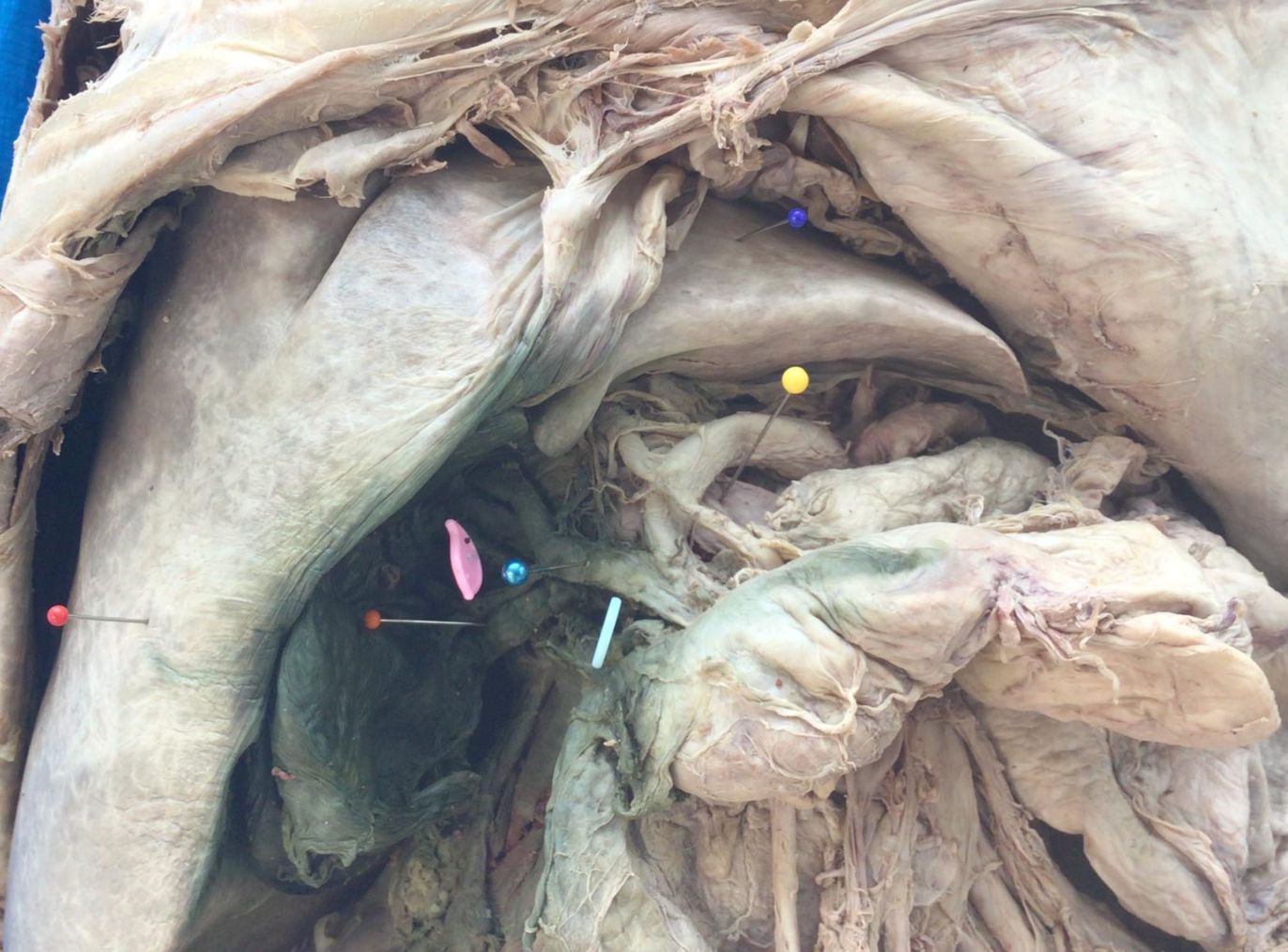
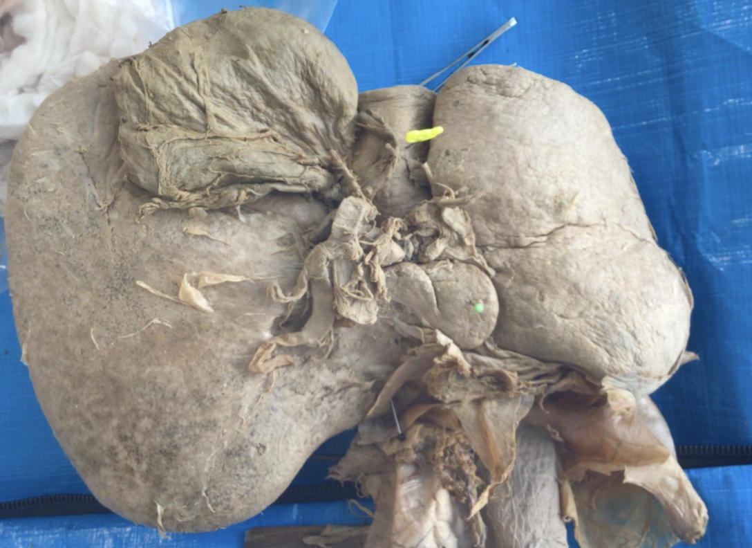
blue - left lobe
red - right lobe
orange - cystic duct
pink - common hepatic duct
seafoam - common bile duct
pearl blue - proper hepatic artery
yellow - hepatic portal vein