Lens I
1/92
There's no tags or description
Looks like no tags are added yet.
Name | Mastery | Learn | Test | Matching | Spaced | Call with Kai |
|---|
No analytics yet
Send a link to your students to track their progress
93 Terms
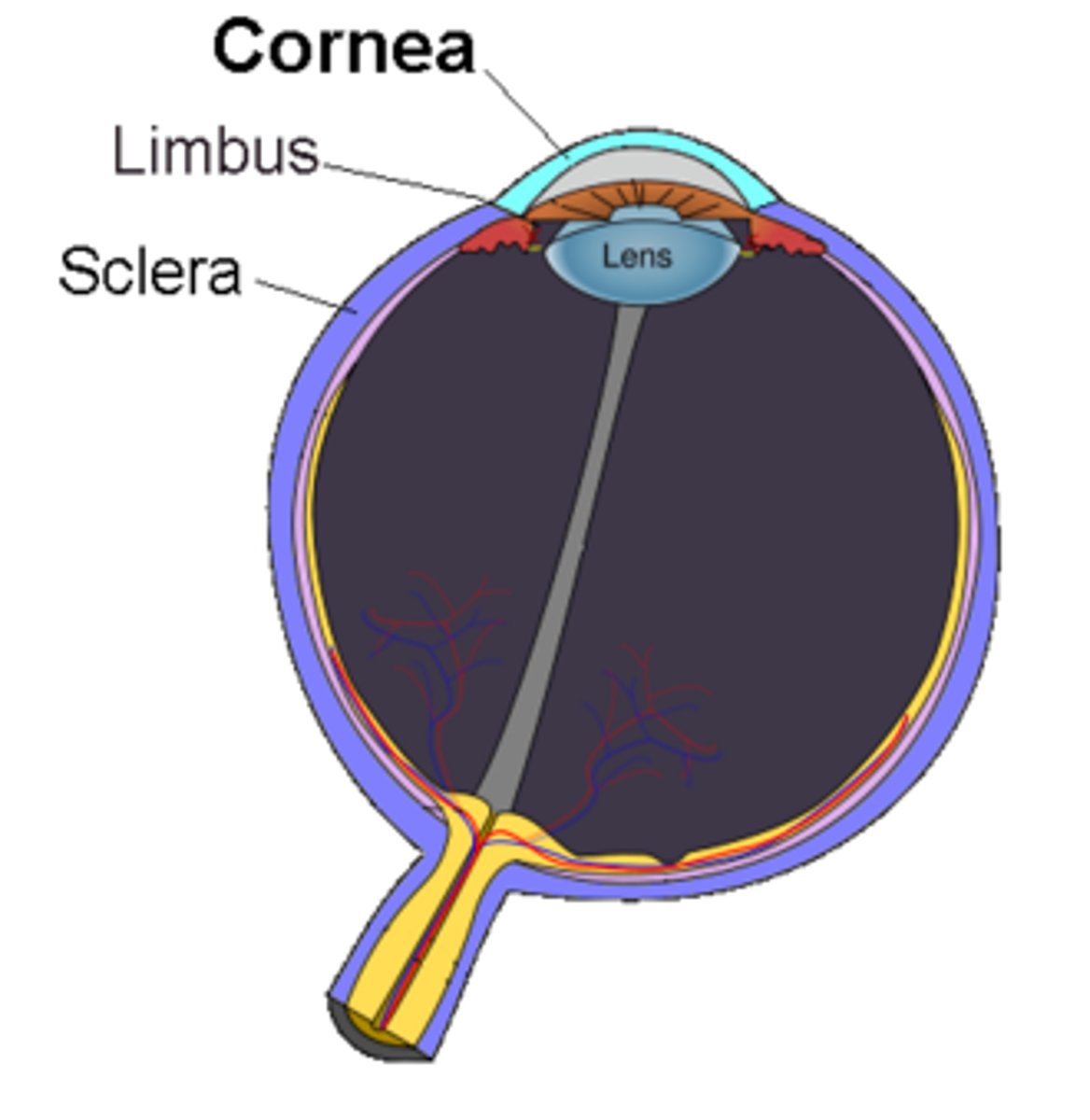
Greek root word for lens?
Phak-
Posteriorly, the lens rests in the ________ _____
Patellar fossa
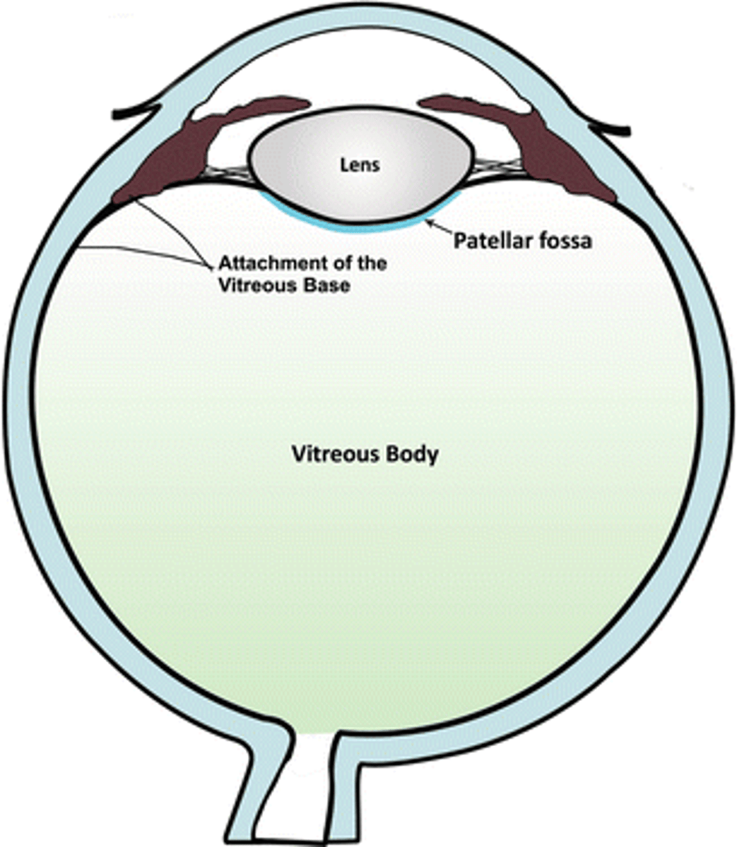
There is about a ____ mm gap between the edge of the lens and the pars plicata
0.5 mm
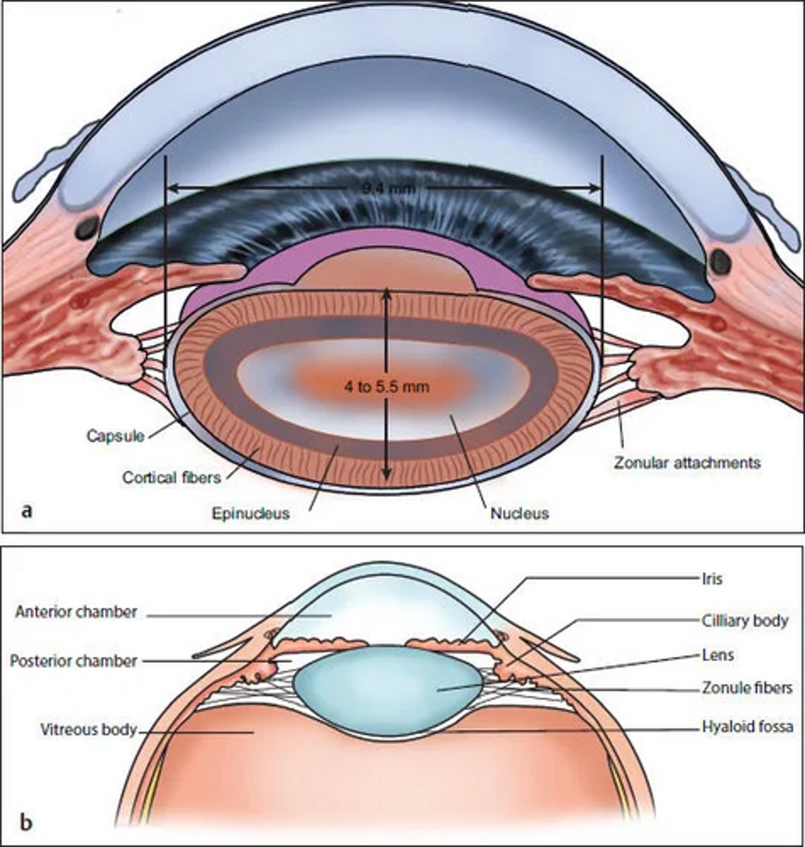
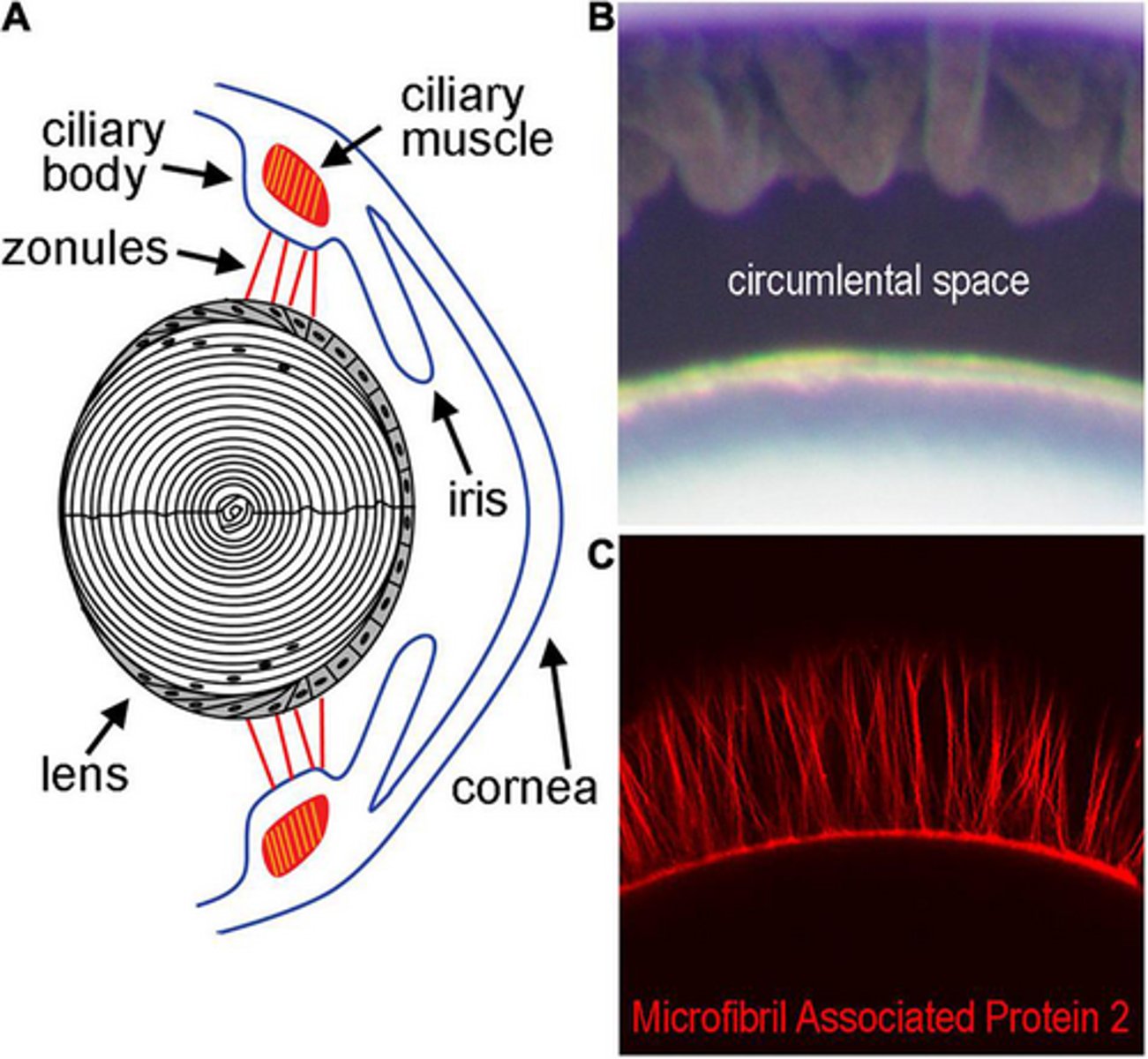
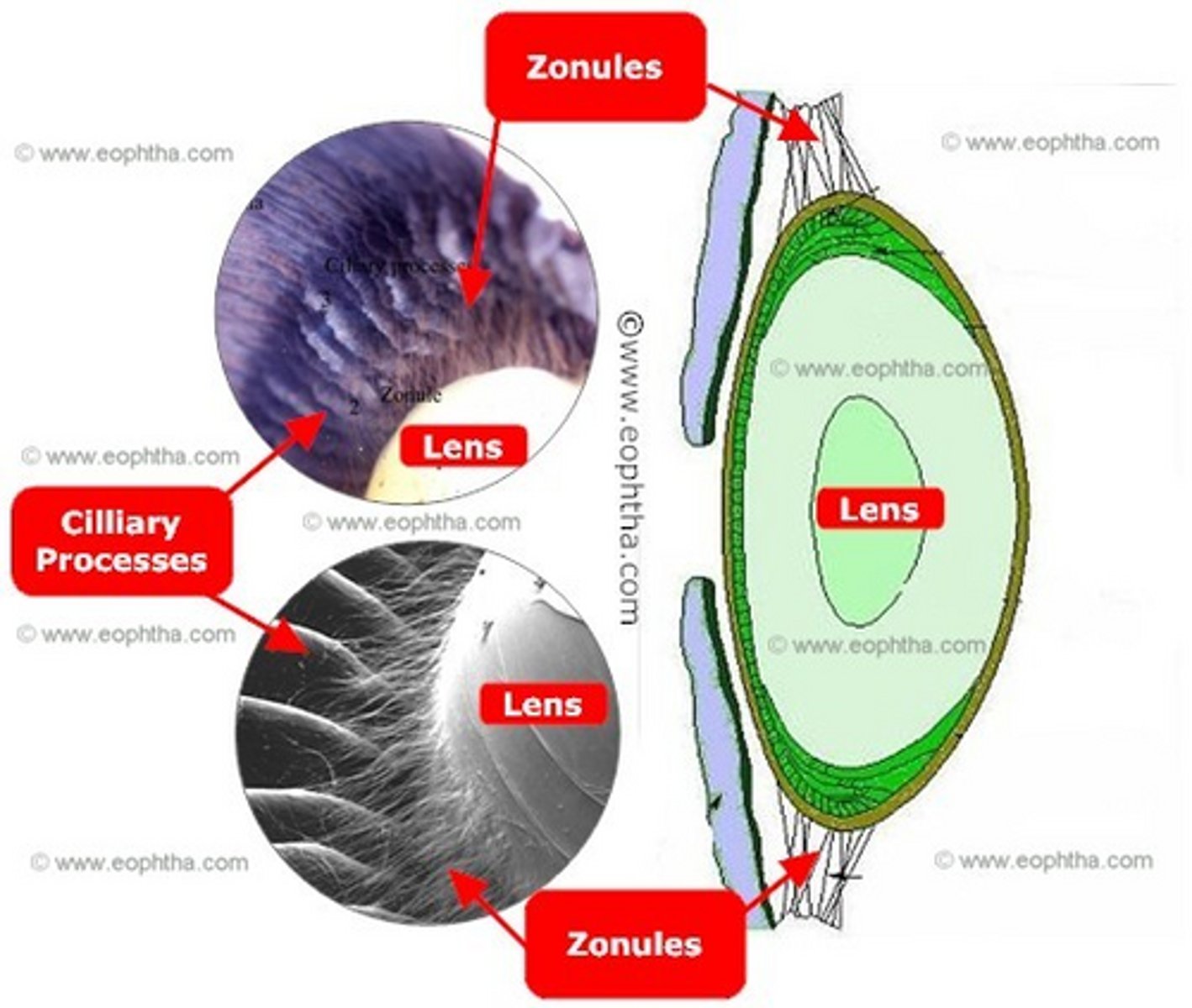
The lens is held in place by _______ ______
Zonular fibers
A thickening of vitreous that acts as a ligament around the space of Berger?
Hyaloid-capsular ligament (Wieger's)

Potential space in front of patellar fossa?
Space of Berger
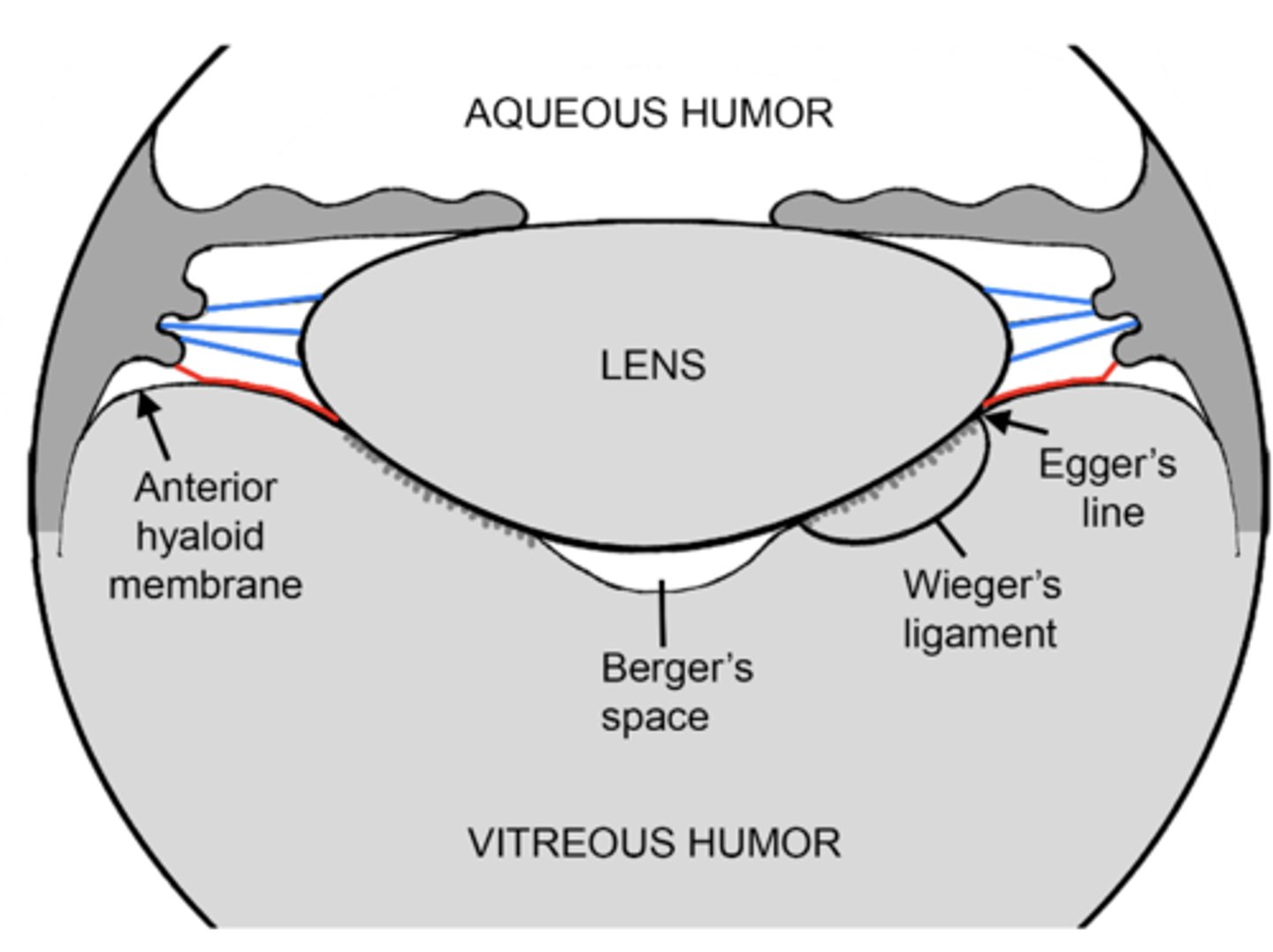
Where zonules attach to the lens capsule?
Zonular girdle
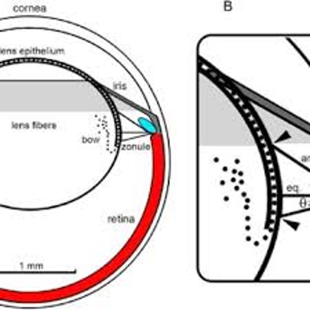
Aqueous space around lens equator?
Circumlental space
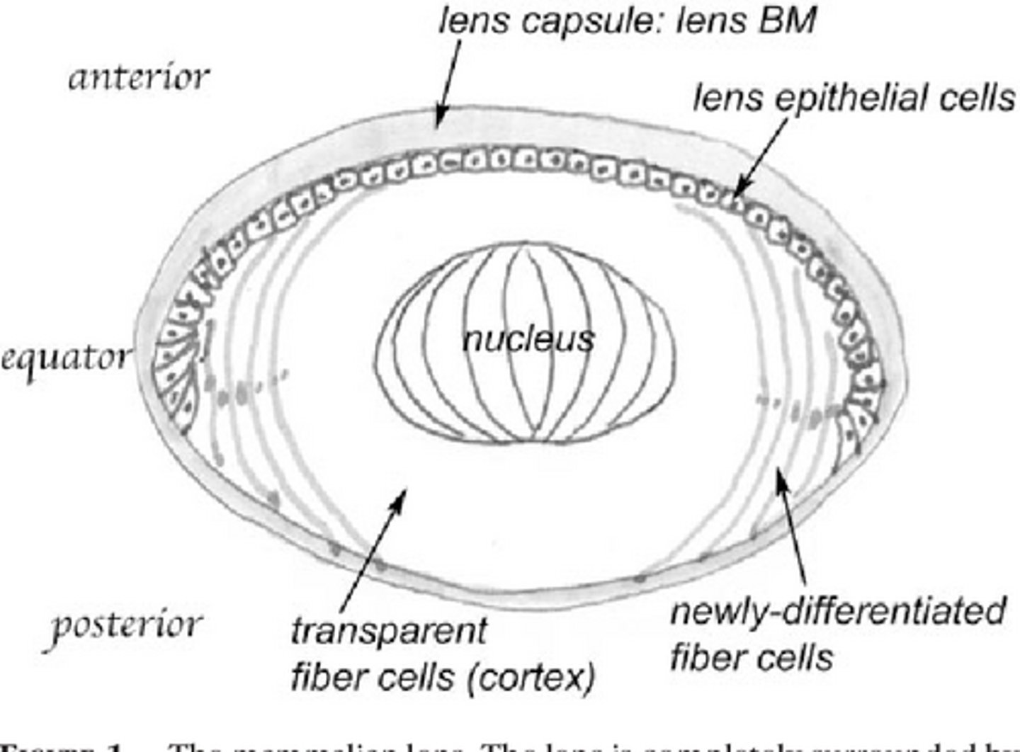
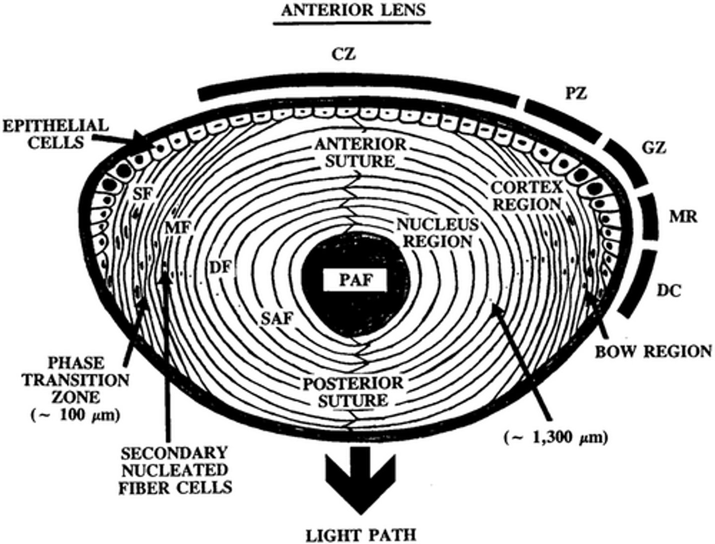
Three parts of the lens:
Capsule
Epithelium
Lens fibers
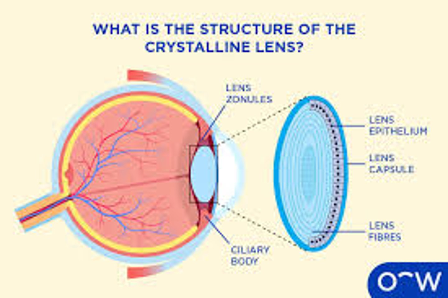
The lens fibers are formed from the _______________ cells
Epithelial
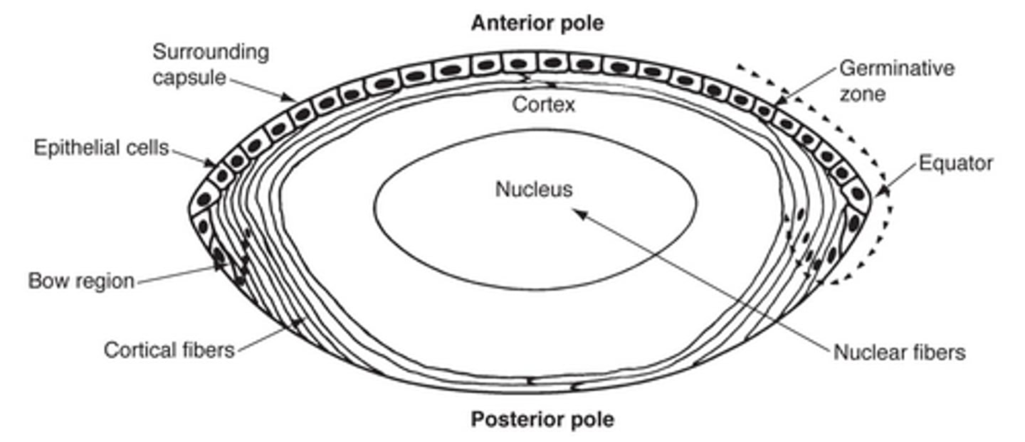
The fibers in the center of the lens?
Nucleus

Fibers outside of the center of the lens?
Cortex

The lens is (vascular/avascular)
Avascular (after birth)
Does the lens stop growing?
No
Is the lens flexible?
Yes, until presbyopia
Lens placode forms from thickened _______ _________
Surface ectoderm
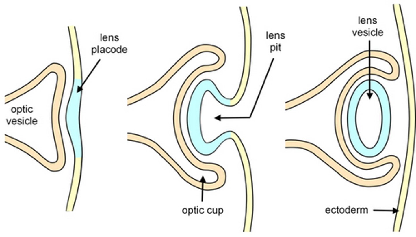
Lens pit forms as placode invaginates along with _____ ____
Optic cup

(posterior/anterior) epithelium elongate and become primary lens fibers during development.
Posterior
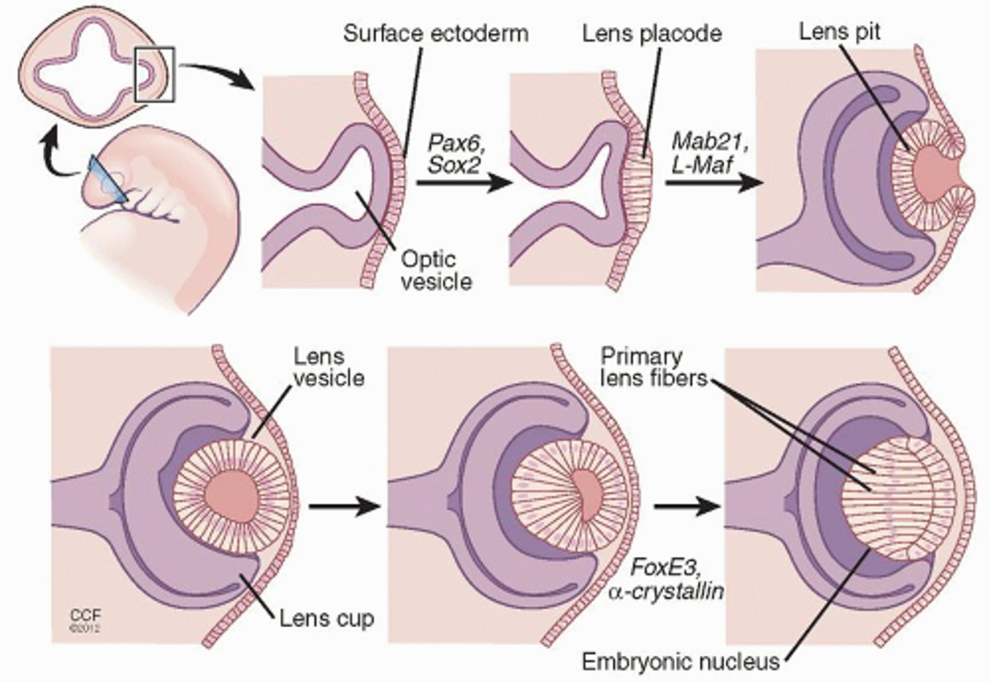
Do primary lens fibers have organelles?
NO
Does the lens have an anterior epithelium?
Yes

Does the lens have a posterior epithelium? (after development)
No
_____ ______ surrounds the lens vesicle during development.
Basal Lamina
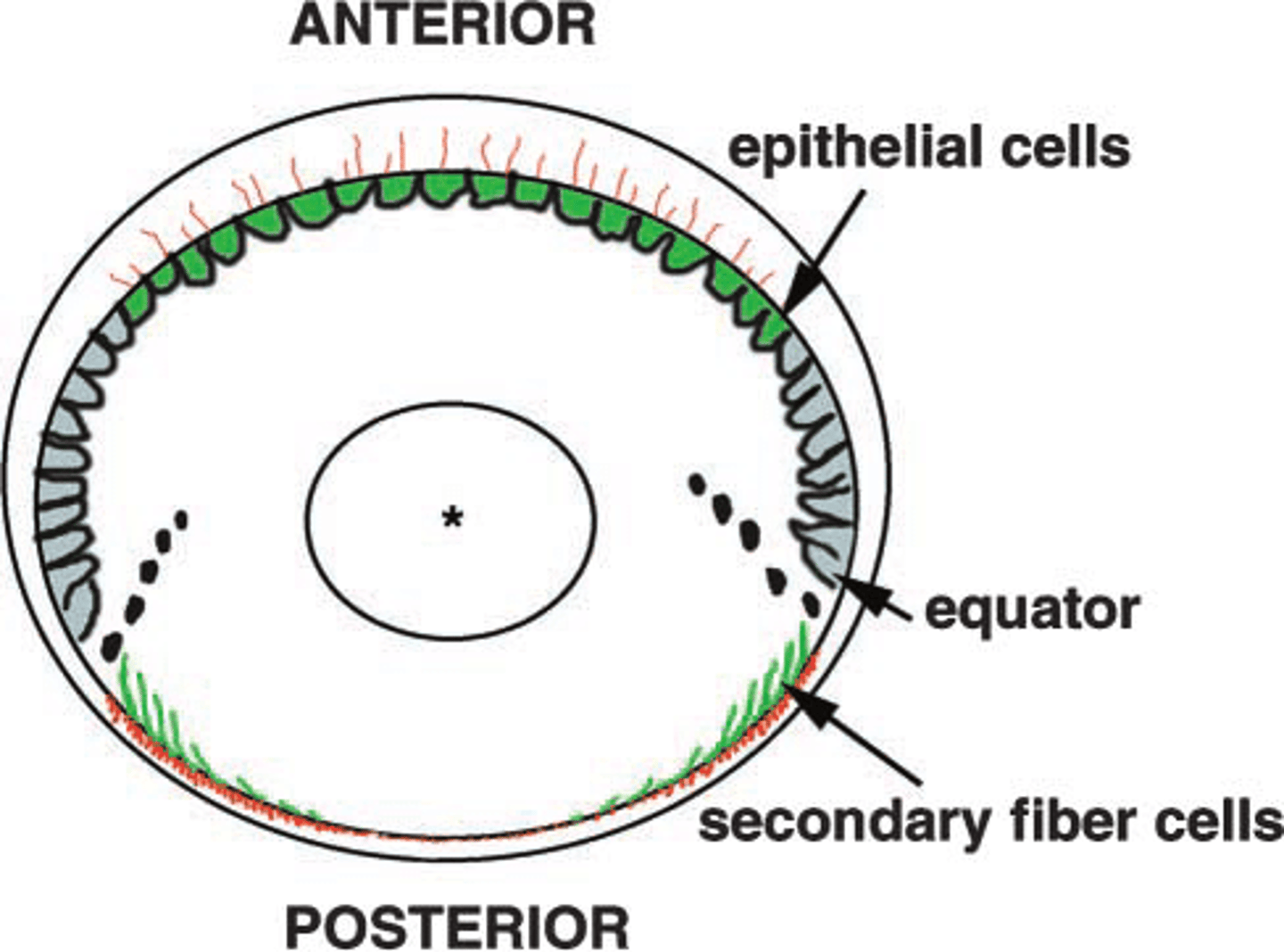
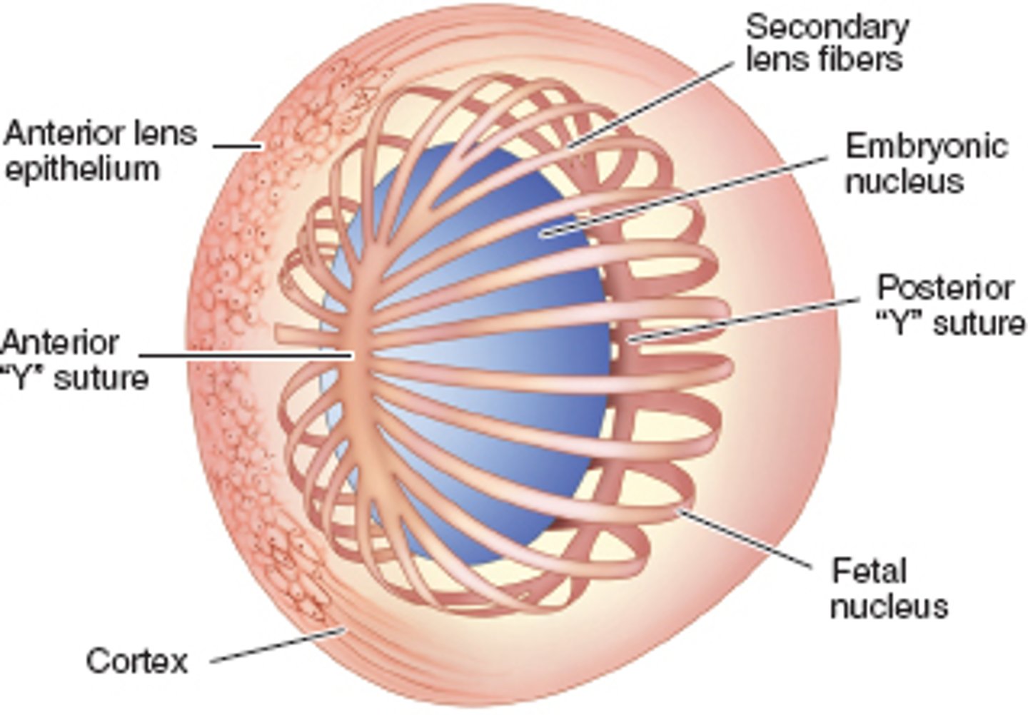
After lens development, where do lens secondary fibers come from?
Anterior epithelium
Lens vascular supply during development? (2)
Anterior pupillary membrane
Posterior tunica vasculosa lentis (from hyaloid artery)
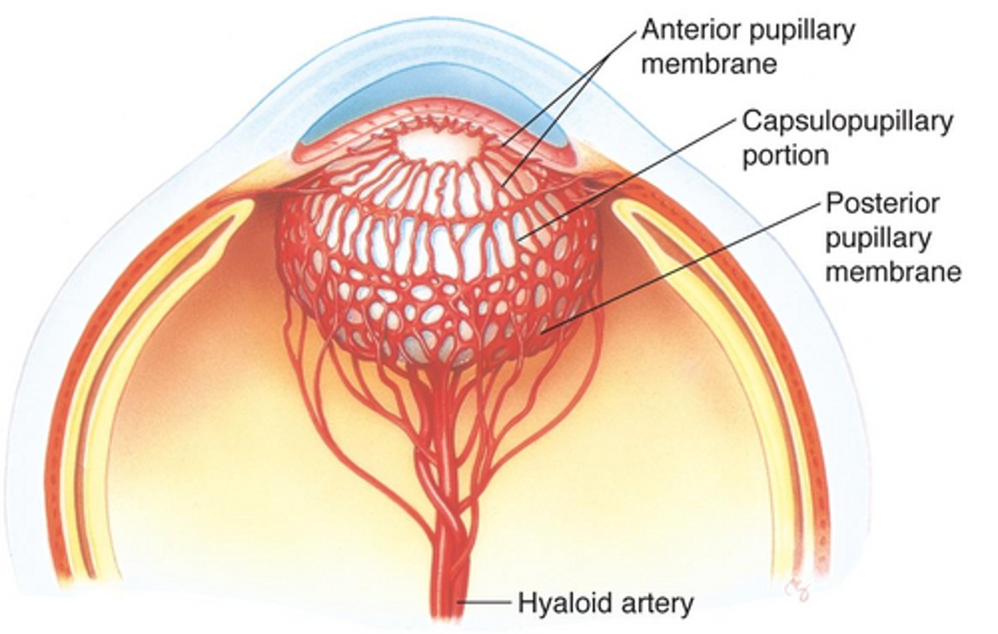
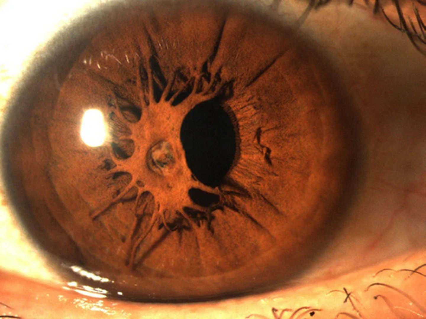
What is remanent of the anterior pupillary membrane called?
persistent pupillary membrane
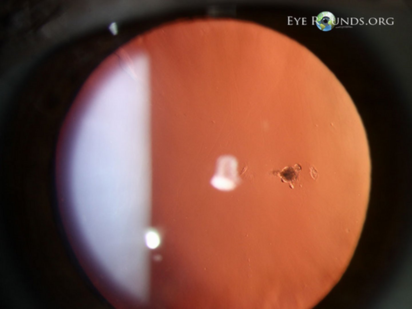
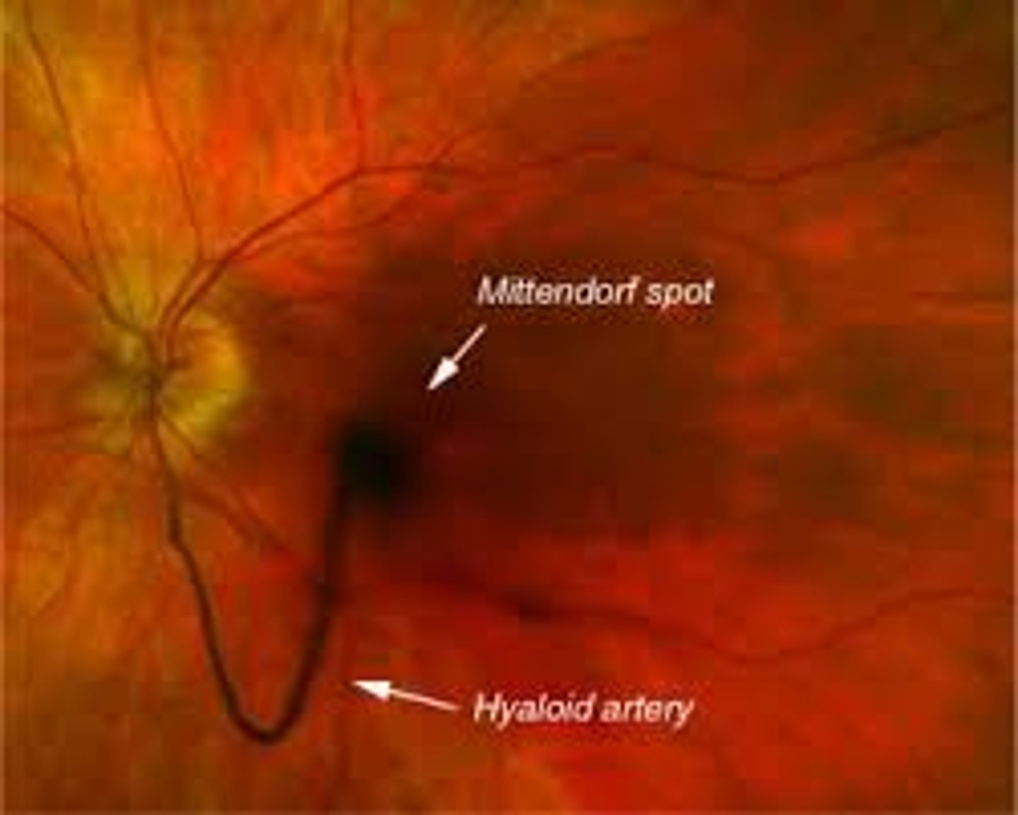
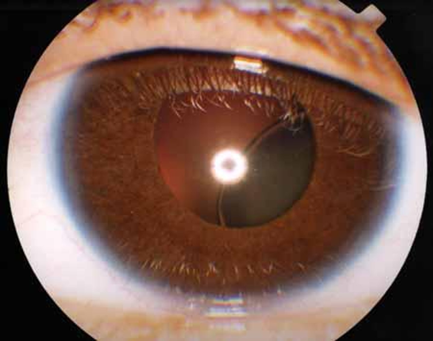
What is a remanent on the hyaloid artery on the lens that looks just like a dot?
Mittendorf dot
What is a complete remanent of the hyaloid artery in the vitreal segment of the eye?
persistent hyaloid
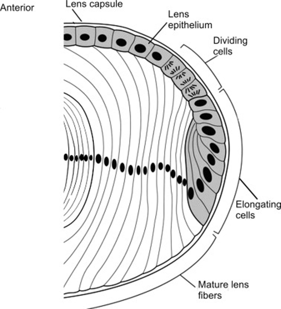
Secondary lens fibers elongate in the ___________ region.
Equatorial

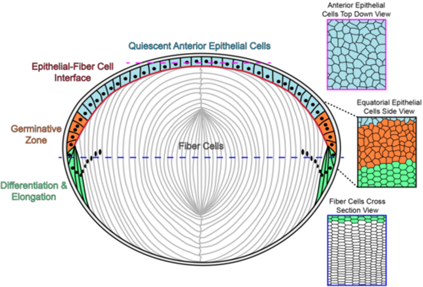
Three zones of epithelium?
Central
Intermediate
Equatorial
Central cells of lens epithelium are more (cuboidal/columnar)
Cuboidal
What do central cells of lens epithelium do?
Not much! Only undergo mitosis in disease or injury
Shape of intermediate zone lens epithelium cells?
Low columnar

Which zone of lens epithelium is home to the germinal zone?
Intermediate zone
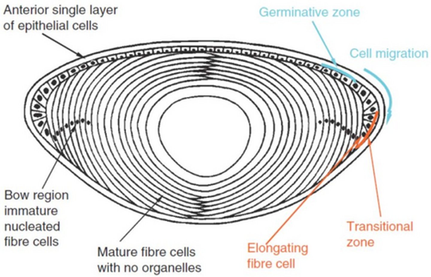
Cells are differentiating as they approach the _______
Equator
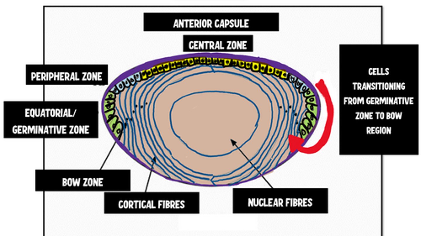
At the equatorial zone, cells begin to produce ___________
Crystallins
What triggers lens epithelium cell differentiation?
Vitreous growth factors
As cells continue to elongate, (apical/basal) processes progress anteriorly underneath the epithelium
Apical
As cells continue to elongate, (apical/basal) processes move posterior underneath the capsule
Basal

As fibers form, they eventually lose what two things?
Nuclei
Attachment to basement membrane
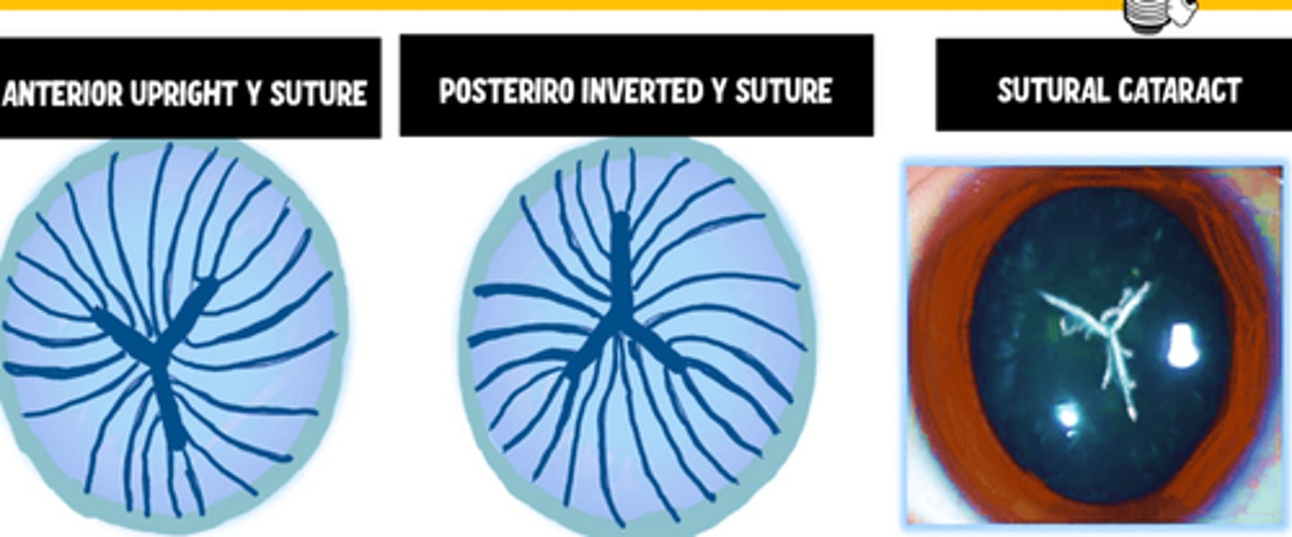
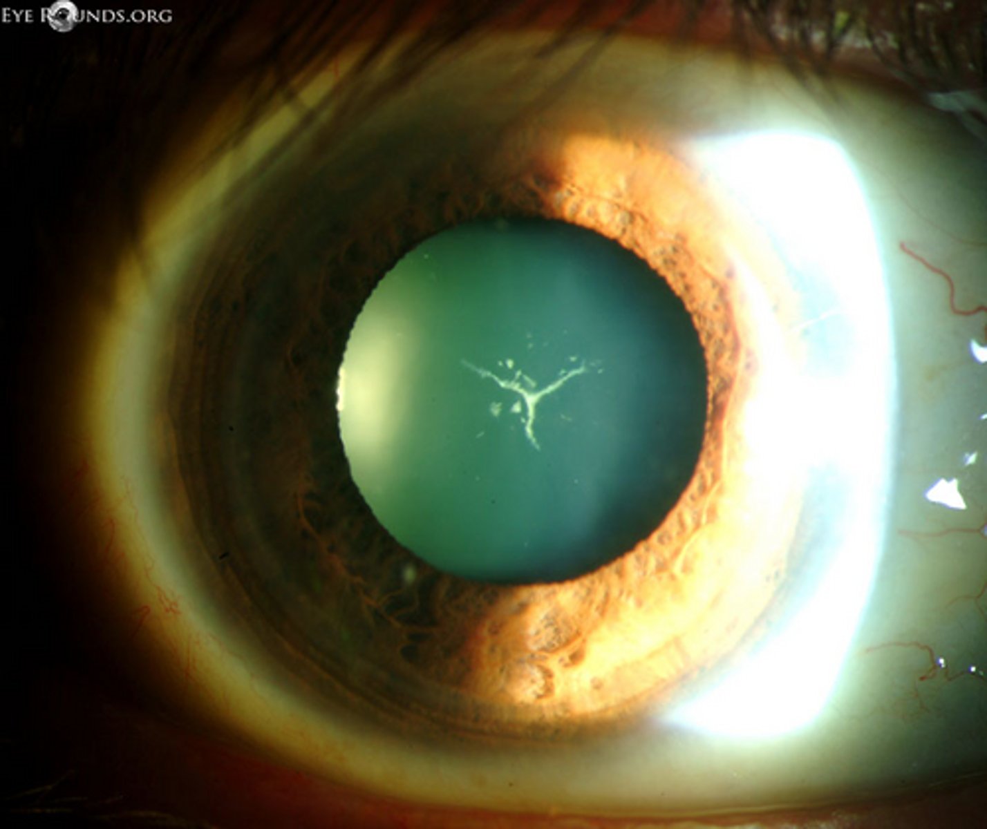
What lens sutures are formed during embryogenesis? (2)
Upright Y anteriorly
Inverted Y posteriorly
Is there a suture in an embryonic nucleus?
no (only fetal)
Why do sutures form in the lens?
Lens fibers meet
Can we ever see Y sutures in the slit lamp?
Yes, although often subtle
Do lens fibers run the full circumference of the lens?
No
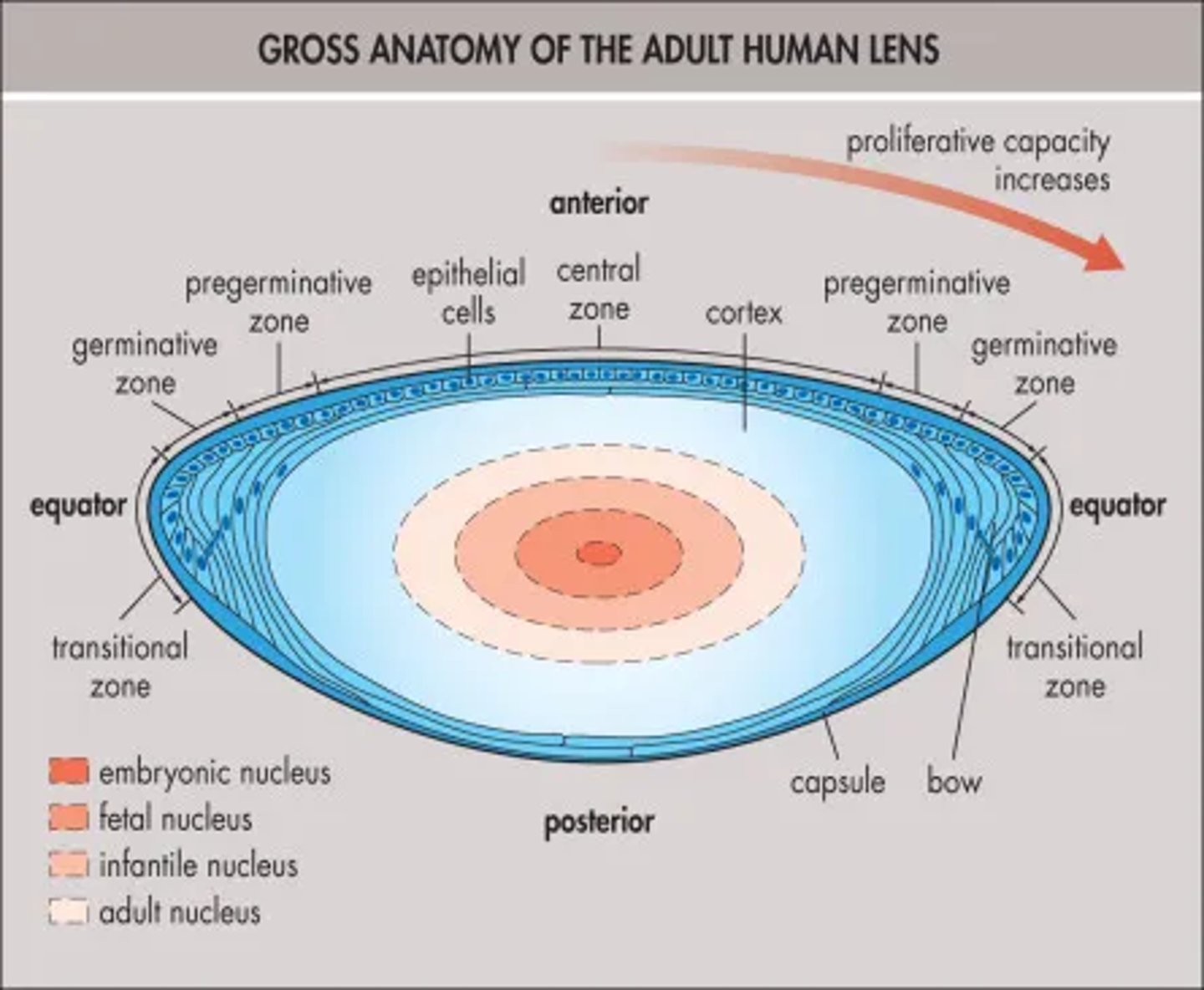
When is the embryonic lens nucleus formed?
During first trimester by primary fibers
When is the fetal lens nucleus formed?
Before birth, includes sutures
When is the adult nucleus formed?
Before sexual maturity
Two layers of the lens capsule?
Inner cuticular layer
External zonular layer
Is the lens capsule consistently thick?
No, varbiable
Where is the lens capsule thickest?
Ring zone 6mm from pupillary axis on anterior side
Where is the lens capsule thinnest?
Posterior pole
The inner cuticular layer is secreted by the __________ and ___________ _____ _____
Epithelium
Superficial fiber cells
The inner cuticular layer is composted of (one/multiple) lamina
Multiple
The inner cuticular layer is secreted from the (inside/outside)
Inside, pushes older capsule out
The inner cuticular layer is mostly made of type __ collagen.
IV
Does the inner cuticular layer contain elastins?
No, but arrangement of fibers makes it elastic-y
Where is the outer zonular layer found?
3-4 mm zone around equator

What is the function of the outer zonular layer of the lens capsule?
Insertion point for zonular fibers.
Cell shape in lens epithelium varies based on if cells are _______ or not.
Mitiotic
(Mitotic/non-mitotic) cells are hexagonal in shape.
Non-mitotic
(Mitotic/non-mitotic) cells have many lateral interdigitations.
Non-mitotic
Do epithelial cells have basal infoldings?
YES, numerous
What is the function of basal infoldings?
Increase surface area
Lateral features of lens epithelial cells? (3)
Gap Junctions
Zonula occludens
Desmosomes
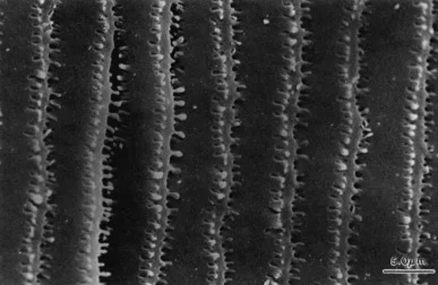
Lens fibers are roughly __________ in shape.
Hexagonal
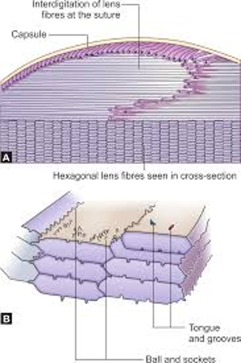
Lens fibers have many ________________.
Interdigitations (ball-and-socket)
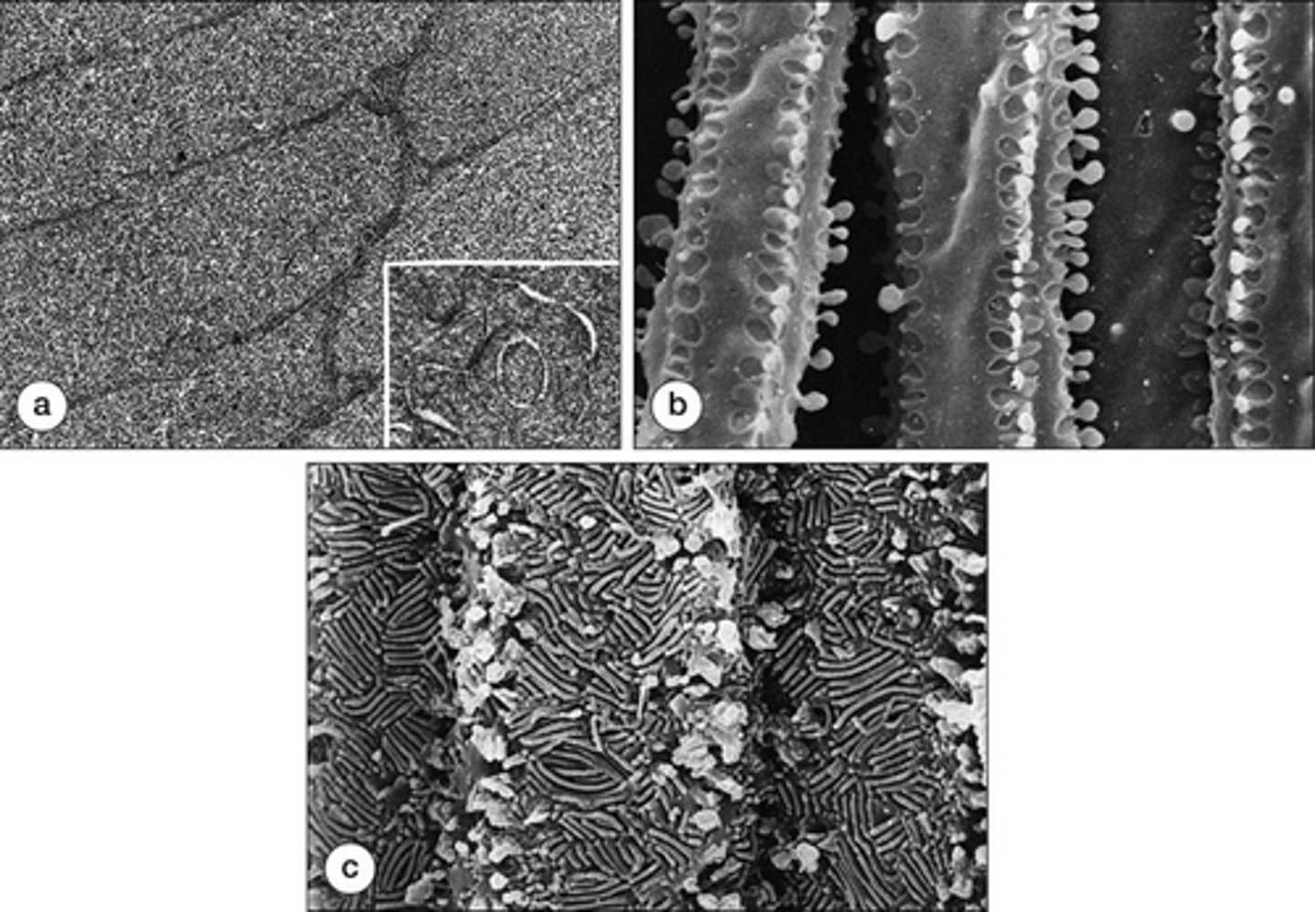
What is the function of interdigitations?
Less interstitial fluid between fibers, maintaining clarity and consistent refractive index
What cytoplasmic proteins make up 35-40% of the lens weight?
Crystallins
Which region of the lens has the highest distribution of crystallins?
Nucleus
Uneven distribution of crystallins creates a _______ refractive index of the lens
Gradient (1.38-1.41)
Benefits of a gradient refractive index in the lens?
Reduce spherical aberration
Features of lens fiber cell membranes? (4)
High proportion of cholesterol
Major intrinsic polypeptide (insoluble protein)
Gap junction plaques
Actin cytoskeleton
Lens develops a ______ _____ as we age.
Yellow tinge (from pigment in ground substance)
Does the lens grow taller throughout the life?
No, stops at about 9mm at age 4
Does the lens grow wider throughout the life?
Yes, can be over 5mm in old age
Weight change in lens throughout life?
60-65 mg at birht, 200mg in old age
What happens when a lens fiber reaches the suture? (3)
Adheres to fiber from across the way
Detaches from capsule
Loses membrane bound organelles
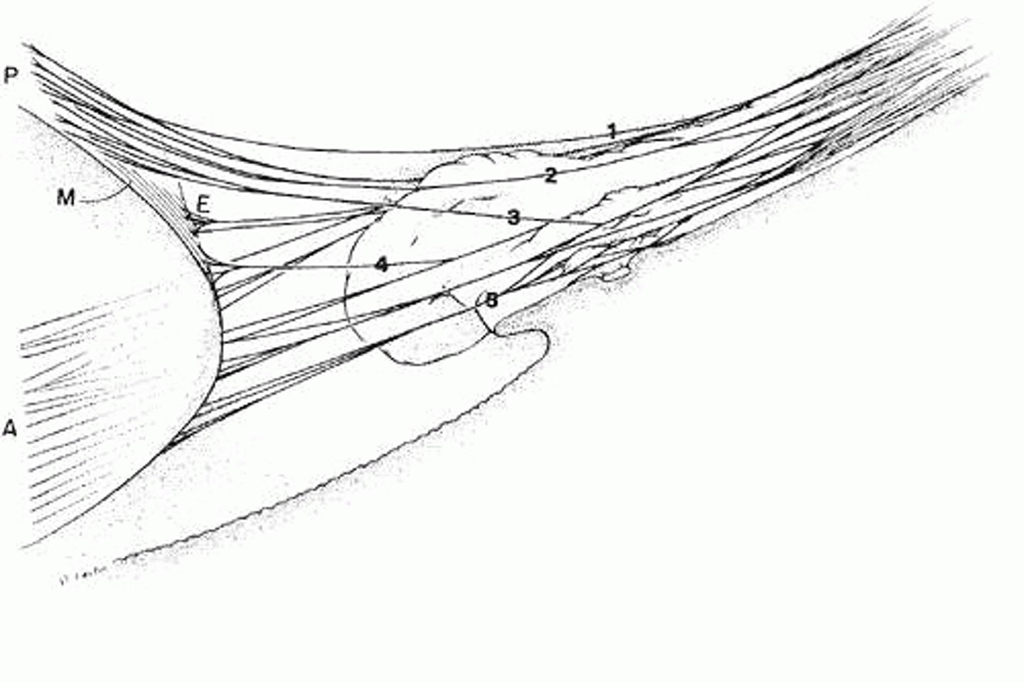
Zonules insert into lens capsule and what else?
Basal lamina of non-pigmented ciliary epithelium
Main structural protein in zonules?
Fibrillin
How are zonules names?
By where they attach
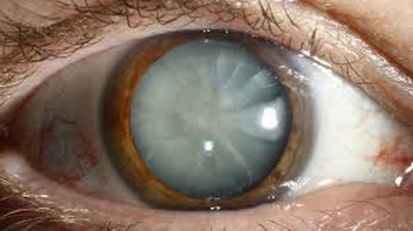
Which condition is a mutation to fibrillin gene resulting in lens dislocation?
Marfans
4 kinds of zonules?
Ubiculo-anterocapsular
Obiculo-posterocapsular
Auxillary/secondary
Equatorial
Zonules insert more (anteriorly/posteriorly) over time
Anteriorly (this may contribute to presbyopia)
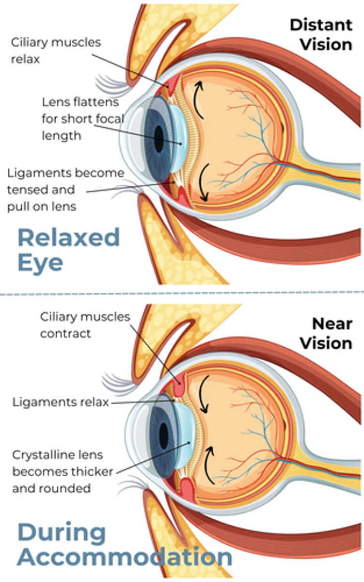
Accommodation occurs via the _______ ______.
Ciliary muscle
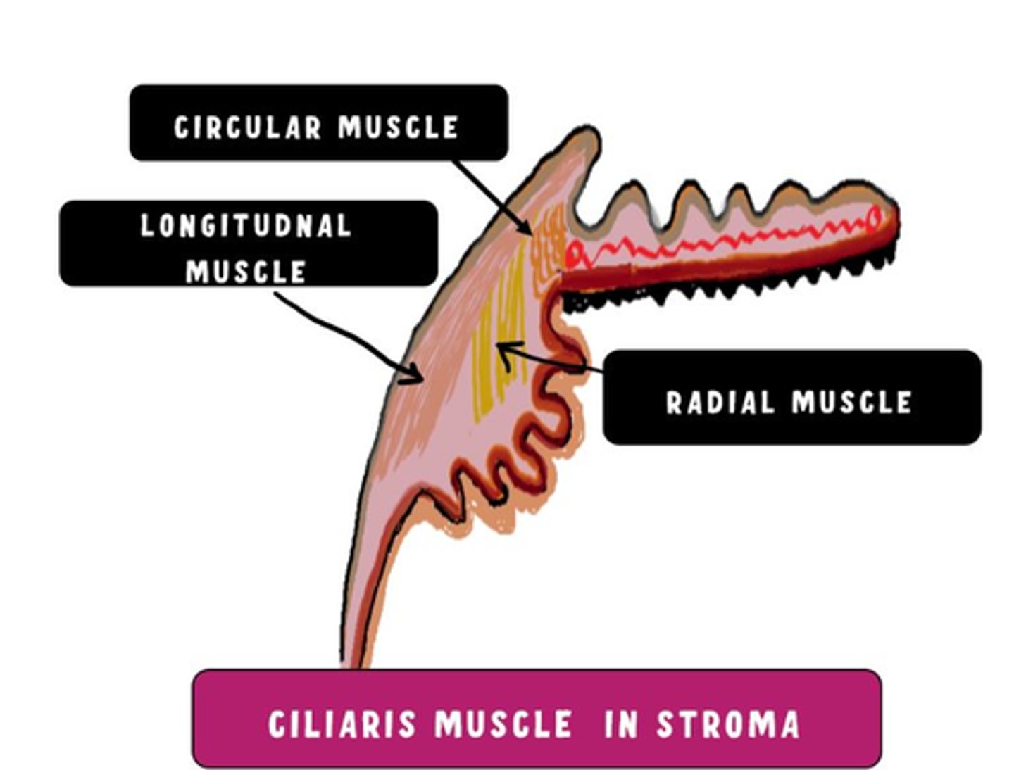
Three regions of ciliary muscle?
Longitudinal
Radial
Circular
If you removed your lens from your eye, it would be (accommodated/unaccommodatied) shape
Accommodated
Does the vitreous play a role in accommodation?
Some, axial length decreases when accommodating
What age does loss of accommodative amplitude begin?
about 18
Factors that likely contribute to presbyopia? (5)
Aging changes to zonules
Lens capsule becomes rigid
Lens fibers become rigid
Index of refraction of lens changes
Less elasticity at ciliary muscle insertion
A loss of accommodation can also result in a change in ________.
Vergence
Unaccommodated lens radii of curvature? (2)
Anterior 10mm
Posterior 6mm
Accommodated lens radii of curvature?
Both about 5.33 mm
What is the major refractive surface of the eye?
Cornea