glycoconjugates
 Call Kai
 Learn
Learn Practice Test
 Spaced Repetition
 Match
Match Flashcards
 Knowt Play
1/9

There's no tags or description
Looks like no tags are added yet.
Name | Mastery | Learn | Test | Matching | Spaced | Call with Kai | Chat |
|---|
No analytics yet
Send a link to your students to track their progress
10 Terms
peptidoglycan
covalently linked short peptides and polysaccharides forming a linked network in the bacterial cell wall
proteoglycan
core protein with covalently attached glycosainoglycans, found on cell surface and extracellular matrix
glycoprotein
glycoylated proteins of other types; generally, focus is on the protein and oligosaccharide is considered an addition to the protein seauence
glycolipid
glycosylated lipids, or lipidated polysaccharides
glycostransferases
they catalyze the transfer of sugars onto proteins, lipids, or other molecules, forming glycosidic bonds that build complex carbohydrates.
proteoglycan aggrecan. Q: these aggrecan glycosaminoglycans are prevalent in cartilage. ur joins have effective circulaton of nutrients without blood, due to mechanical motion, processed through simple biomechanical/biophysical properties of these aggrecans in water on compression/release. how?
Think of aggrecan as a “bottlebrush”: a protein core with so many glycosaminoglycans chains sticking out, each packed with (-) charges.
Holds water: Those dense negative charges attract ions (like Na+) which osmotically pulls in lots of water
Compression: When you load the joint, water is squeezed out of this hydrated network.
Limit to compression: the negatively GAG chains repel each other → this sets a mechanical limit and gives cartilage its stiffness
Expansion: When the water is removed, the electrostatic repulsion + osmotic pressure draws water back in, re-expanding the structure
Net effect: aggrecan behaves like a water-filled, charge-stabilized spring, enabling cartilage to withstand compression and then recover while moving fluid in and out. This means circulation with no blood!
Lectins
proteins that bind specific carbohydrate (sugar) ligands, and they do this using the same kinds of noncovalent forces that govern all protein-ligand interactions (hydrogen bonds, electrostatic interactions)
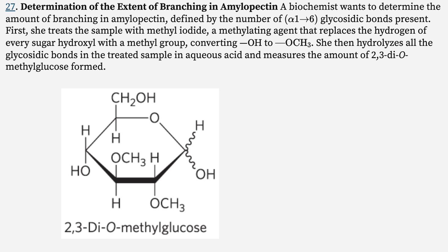
In this representation of 2,3-di-O-methylglucose, the wavy bonds at C-1 indicate that the structure represents both anomers (α and β).
Explain the basis of this procedure for determining the number of (α1→6) branch points in amylopectin. What happens to the unbranched glucose residues in amylopectin during the methylation and hydrolysis procedure?
Only branch-point glucose residues produce 2,3-di-O-methylglucose because their C6 position was involved in an α(1→6) linkage and couldn’t be methylated.
Unbranched: C6 is free → methylated → tri-methyl product
Branched: C6 is involved → NOT methylated → di-methyl product
exoglycosidases
enzymes that cleave off very specific monosaccharides from the nonreducing end of the oligosaccharide
endoglycosidases
cleave glycosidic bonds between interior sugar residues