Pathology: C-spine & soft tissue neck
1/14
There's no tags or description
Looks like no tags are added yet.
Name | Mastery | Learn | Test | Matching | Spaced | Call with Kai |
|---|
No analytics yet
Send a link to your students to track their progress
15 Terms
Clay Shoveler’s Fx
avulsion fx of spinous process in the lower cervical & upper thoracic region
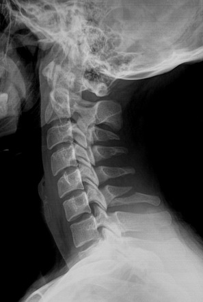
clay shoveler’s fx- radiographic appearance
best seen on the lateral image as lucency through the spinous process
tech factors for this test
no change- for all
jefferson fx
a comminuted fracture of the ring of the atlas; involves both the anterior & posterior arches & causes displacement of the fragments (comminuted)
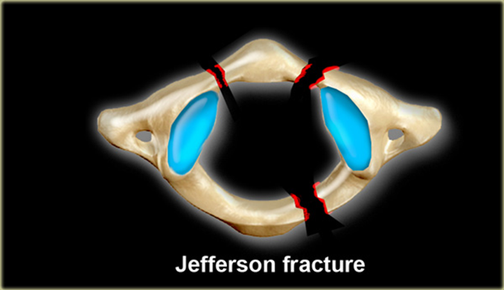
jefferson fx- radiographic appearance
Lateral displacement of lateral masses of C1 bilaterally; Best visualized on odontoid view
hangman’s fx
A fracture of the arch of C2 (axis)
(ex- head forced back by impact)
hangman’s fx radiographic appearance
Best visualized on lateral view
herniated nucleus pulposus (HNP)
Condition in which part or all the soft, gelatinous central portion of an intervertebral disk is forced through a weakened part of the disk (like a gusher- squeezes out)
herniated nucleus pulposus (HNP) radiographic appearance
narrowing of disc spaces
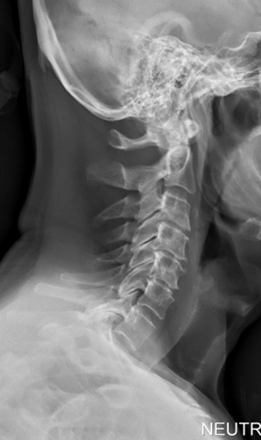
hyperextension injury
Abnormal motion or force applied to the neck that causes movement beyond the necks normal range of motion
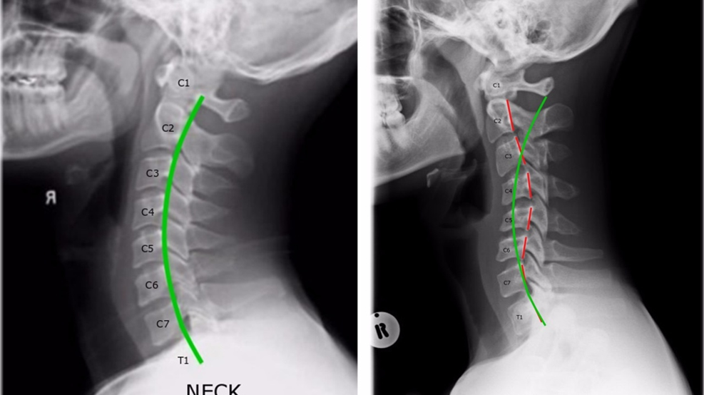
hyperextension injury- radiographic appearance
Straightening of the cervical spine – loss of lordosis
(ex- whiplash)
croup
Viral infection primarily of young children – produces inflammatory obstructive swelling localized to the subglottic portion of trachea
croup- radiographic apperance
(“steeple sign”)
how to determine which bone is C2
biggest bone
how to determine which bone is C7
the long one