Chapter 2-Part 1: Intestinal, Commensal, and Free-living Pathogenic Amebae
1/39
Earn XP
Description and Tags
This is the first part of Chapter 2. Study well.
Name | Mastery | Learn | Test | Matching | Spaced | Call with Kai |
|---|
No analytics yet
Send a link to your students to track their progress
40 Terms

Species Name:
Group:
Pointed in Arrow:
Stage:
Life Cycle Stages of this species:
How many nuclei does it have:
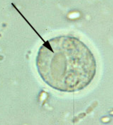
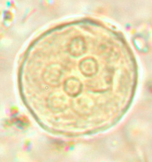
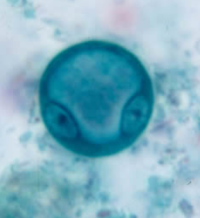
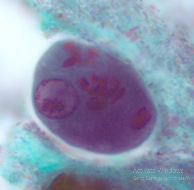
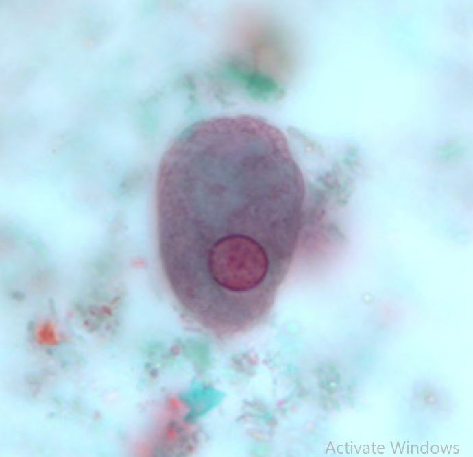
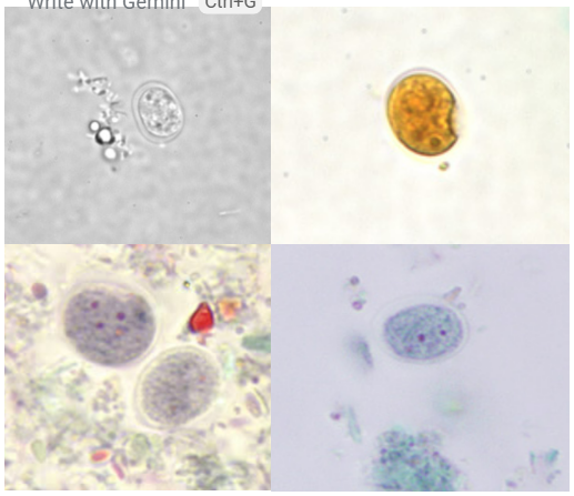
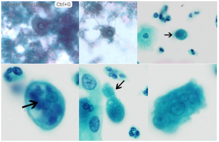
Species Name: E. histolytica
Group: Amoeba (Sarcodina)
Pointed in Arrow: Chromatoid body
Stage: Cyst
Life Cycle Stages of this species: Cyst and Trophozoite
How many nuclei does it have: 4
Species Name:
Pointed in Arrow:
Stage:
Infective and Invasive stage:
Primary Host and Reservoir:
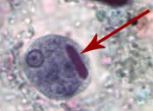
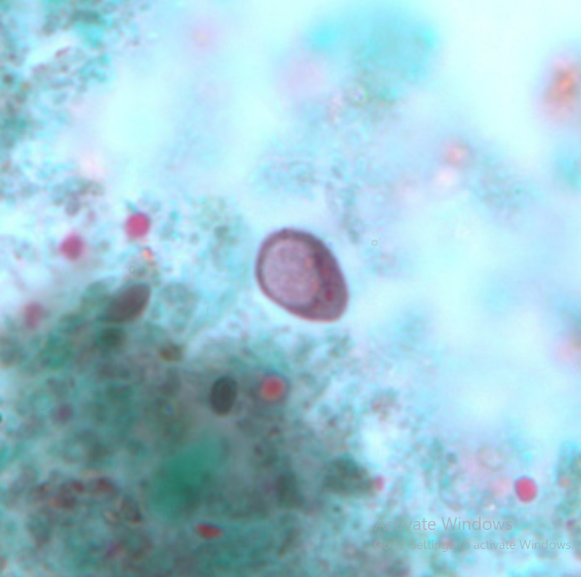
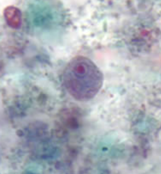
Species Name: E. histolytica
Pointed in Arrow: Chromatoid body
Stage: Cyst
Infective and Invasive stage: Cyst and Trophozoite
Primary Host and Reservoir: Humans

7 Species of Intestinal Amebae in Humans
Pathogenic:
Commensals:
7 Species of Intestinal Amebae in Humans
Pathogenic: Entamoeba histolytica
Commensals: E. dispar, E. moshkovskii, E. hartmanni. E. coli, Endolimax nana, Iodamoeba butschlii
Mnemonic: (HMDCNB - Hugaw Man Diay Ca Na Bata)

Species and what stage:
Infective Stage:
Diagnostic Stage:
Pathognomonic finding:
MOT:
Lab Diagnosis:
Pathogenicity:
Treatment:
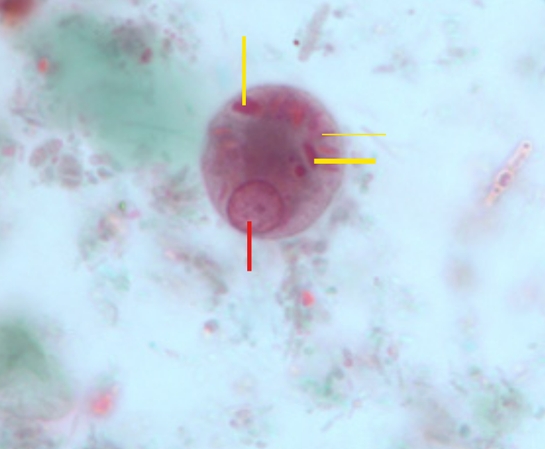
Red Line:
Yellow Line:
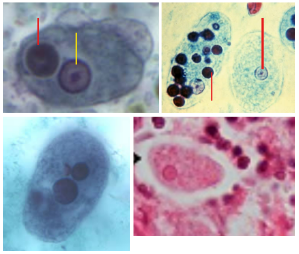
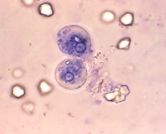
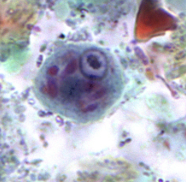
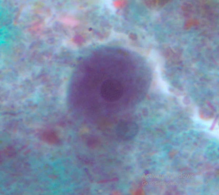
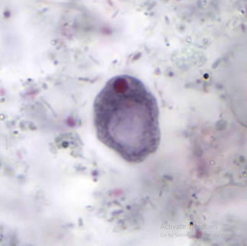
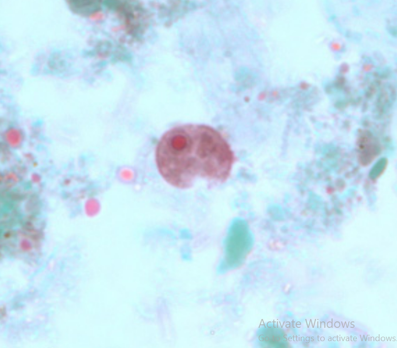
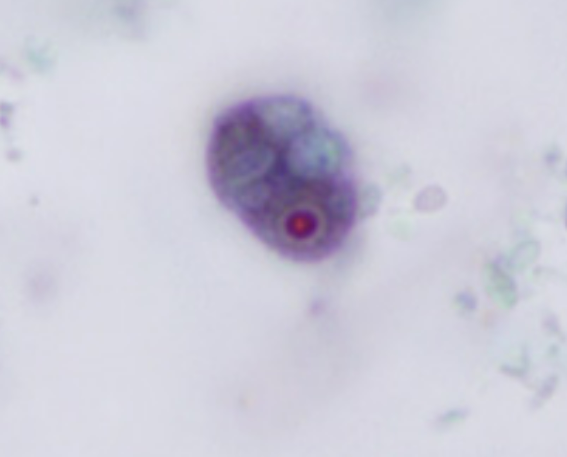
Species: E. histolytica, Trophozoite
Infective Stage: Mature Quadrinucleate Cyst
Diagnostic Stage: Cyst and Trophozoite in Stool
Pathognomonic finding: Trophozoite with ingested RBCs
MOT: Fecal-Oral Route, Mechanical Vectors, Sexual contact
Lab Diagnosis: Stool Microscopy, ELISA, PCR, Isoenzyme analysis
Pathogenicity: Amoebic colitis, Amoebic Liver Abscess (ALA)
Treatment: Metronidazole, Tinidazole
Red Line: Nucleus
Yellow Line: Ingested RBC

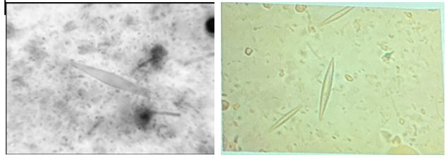
Species and Stage:
Locomotion:
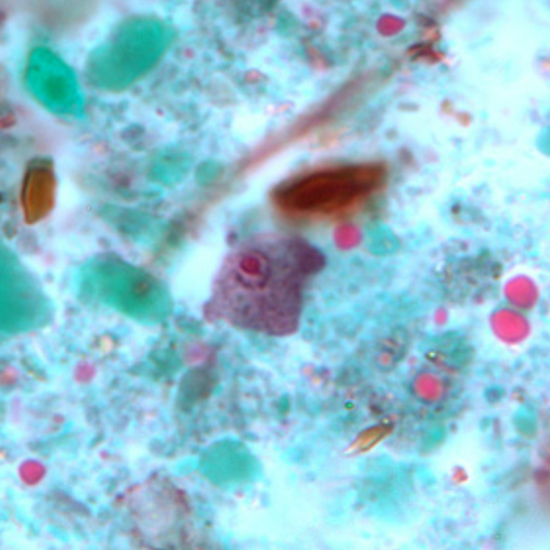
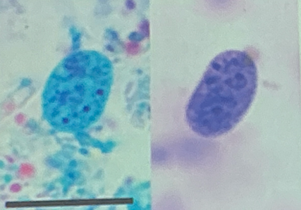
Species and Stage: E. histolytica, Trophozoite
Locomotion: Pseudopodia
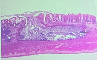
What type of ulcer is this?
What parasite is the cause?
Flask-shaped Ulcer
E. histolytica

What is this?
If you have this, you suffer from?
What species is responsible for this?
Drug of choice. Give the alternative DOC.
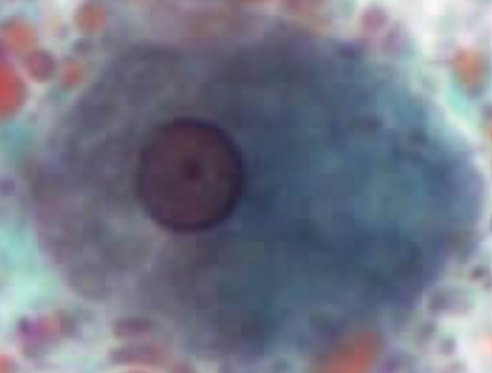
Charcot-Leyden Crystal
Amebiasis
E. histolytica
Metronidazole. Tinidazole, Secnidazole


Species
Describe it’s survival ability.
What species is it morphologically identical with?
Entamoeba moshkovskii
Osmotolerant, Survives 0-41 C
E. histolytica
Species
Stage
Movement
Number of Nuclei
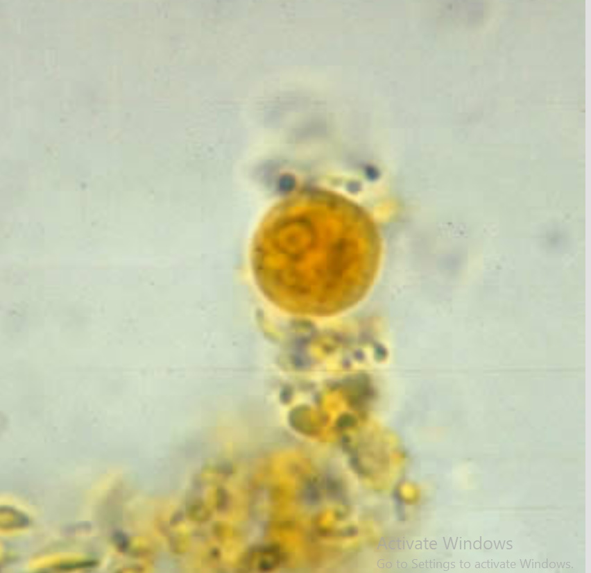
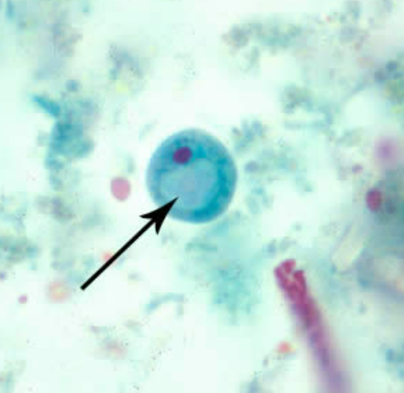
Entamoeba coli
Cyst
Sluggish Movement
8

Species
Stage
Movement
Number of Nuclei
Entamoeba coli
Cyst
Sluggish Movement
8


Species
Stage
Entamoeba coli
Trophozoite

Species
Also known as:
Stage
Entamoeba hartmanni
Small race of E. histolytica
Cyst
Species
Stage
Entamoeba hartmanni
Trophozoite


Species
Stage
Entamoeba hartmanni
Trophozoite

Species
Stage
Entamoeba polecki
Cyst

Species
Stage
Entamoeba polecki
Cyst
Species
Stage
Entamoeba polecki
Cyst


Species
Stage
Red:
Yellow:
Entamoeba polecki
Cyst
Large nucleus with pleomorphic karyosome
Chromatoid bodies
Species
Stage
Entamoeba polecki
Trophozoite

Species
Stage
Entamoeba polecki
Trophozoite


Species
Stage
Entamoeba polecki
Trophozoite

Species
Stage
Distinguishing feature
Source of infection
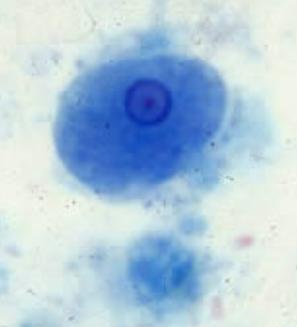
Iodamoeba buetschlii
Cyst
Glycogen vauole
Contaminated hog feces

Species
Stage
Black Arrow
Sources of infection
Iodamoeba buetschlii
Cyst
Glycogen vauole
Contaminated hog feces

Species
Stage
Black Arrow
Source of infection
Iodamoeba buetschlii
Cyst
Glycogen vauole
Contaminated hog feces
Species
Stage
Iodamoeba buetschlii
Trophozoite

Species
Stage
Iodamoeba buetschlii
Trophozoite


Species
Stage
Iodamoeba buetschlii
Trophozoite
Species
Stage
Endolimax nana
Cyst


Species
Stage
Endolimax nana
Trophozoite
Species
Stage
Endolimax nana
Trophozoite

Species
Stage
Endolimax nana
Cyst

Species
Stage
Transmission
Feeds on
Unique Characteristic
Treatment
Entamoeba gingivalis
Trophozoite
Kissing, Droplet spray, shared utensil
Feeds on leukocytes and bacteria
Unique characteristic: No cyst stage
No treatment required (non-pathogenic)


Species
Stage
Causes what diseases:
Prominent Characteristic
Give at least two entry pathway to the body
This is a possible reservoir hosts form medically important bacteria such as… give at least two
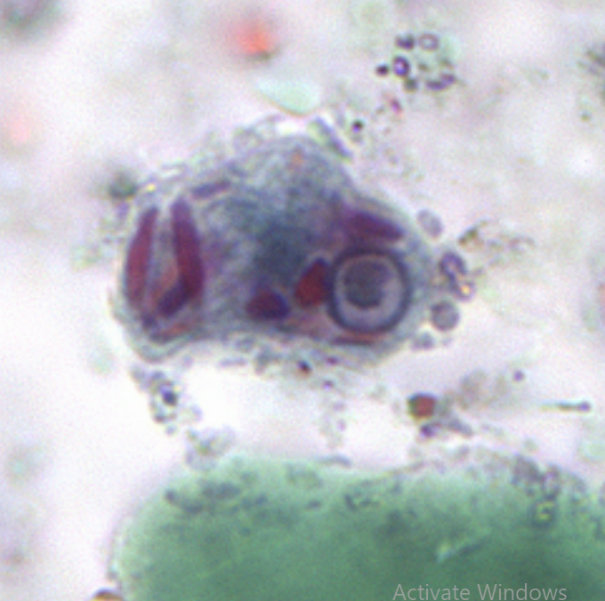
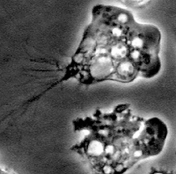
Acanthamoeba spp.
Trophozoite
Acanthamoeba Keratitis (AK), Granulomatous Amebic Encephalitis (GAE)
Acanthopodia (thorn-like projections)
Eye, Nasal Passages, Broken skin
Legionella spp., mycobacteria, E. coli

Species
Stage
Found in… give at least two
Acanthamoeba spp.
Trophozoite
Freshwater and seawater, soil, sewage, hospital equipment, contact lenses and lens solutions
Species that causes Acanthamoeba keratitis
Diagnosis of the disease?
Treatment of the disease?
What medication to avoid?
Acanthamoeba spp.
Epithelial biopsy, corneal scrapings, culture, PCR
Clotrimazole combined with pentamidine, isethionate, neosporin
Topical coticosteroids (retards immune repsonse)
Species that causes Granulomatous Amebic Encephalitis (GAE)
Treatment
Acanthamoeba spp.
Amphotericin B, Penamidine, Sulfadiazine, Flucytosine, Fluconazole, Itraconazole
Species
Stage
Three stages of life cycle
Vegetative forms
This species has two forms of trophozoites
What are these forms, and which one is found in humans.
Environment where they thrive best
Entry pathway to the body
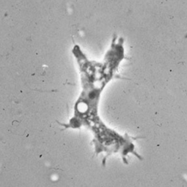
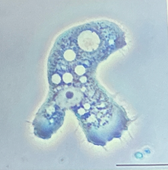
Naegleria spp.
Trophozoites in ameboid
Cysts, trophozoites, flagellated forms.
Ameba (trophozoite form) and flagellate (swimming form)
Naegleria fowleri
Ameboid and Ameboflagellate. Ameboid is found in humans.
Hot springs and other warm environment (thermophilic)
Nasal mucos

Also know as the brain-eating amoeba.
What disease does it cause?
Naegleria fowleri
Primary Amebic Meningoencephalitis (PAM)
Species
Infective stage
Diagnostic stage
Location in this infective stage
Naegleria fowleri
Trophozoite
Trophozoites in CSF and brain tissue
Human tissues and CSF
Species that causes Primary Amebic Meningoencephalitis (PAM)
Diagnosis
Treatment
Naegleria fowleri
Aspirates from suspected infection and introduced into bacteria-seeded agar culture medium
Amphotericin B (Blood-Brain Barrier)