Histology lecture #11: Special Senses the Eye
1/147
There's no tags or description
Looks like no tags are added yet.
Name | Mastery | Learn | Test | Matching | Spaced | Call with Kai |
|---|
No analytics yet
Send a link to your students to track their progress
148 Terms
They capture light stimuli, enabling the sense of sight.
What is the function of photoreceptors in the eye?

By the bony orbits and adipose tissue cushions.
How are the eyes protected within the skull?

1. External fibrous globe (maintains shape)
2. Internal transparent tissues
- A layer of photoreceptors
- Collection of neurons
What are the main structural components of the eye?

Collection, processing, and transmission of visual information to the brain.
What is the overall function of the photoreceptor system?

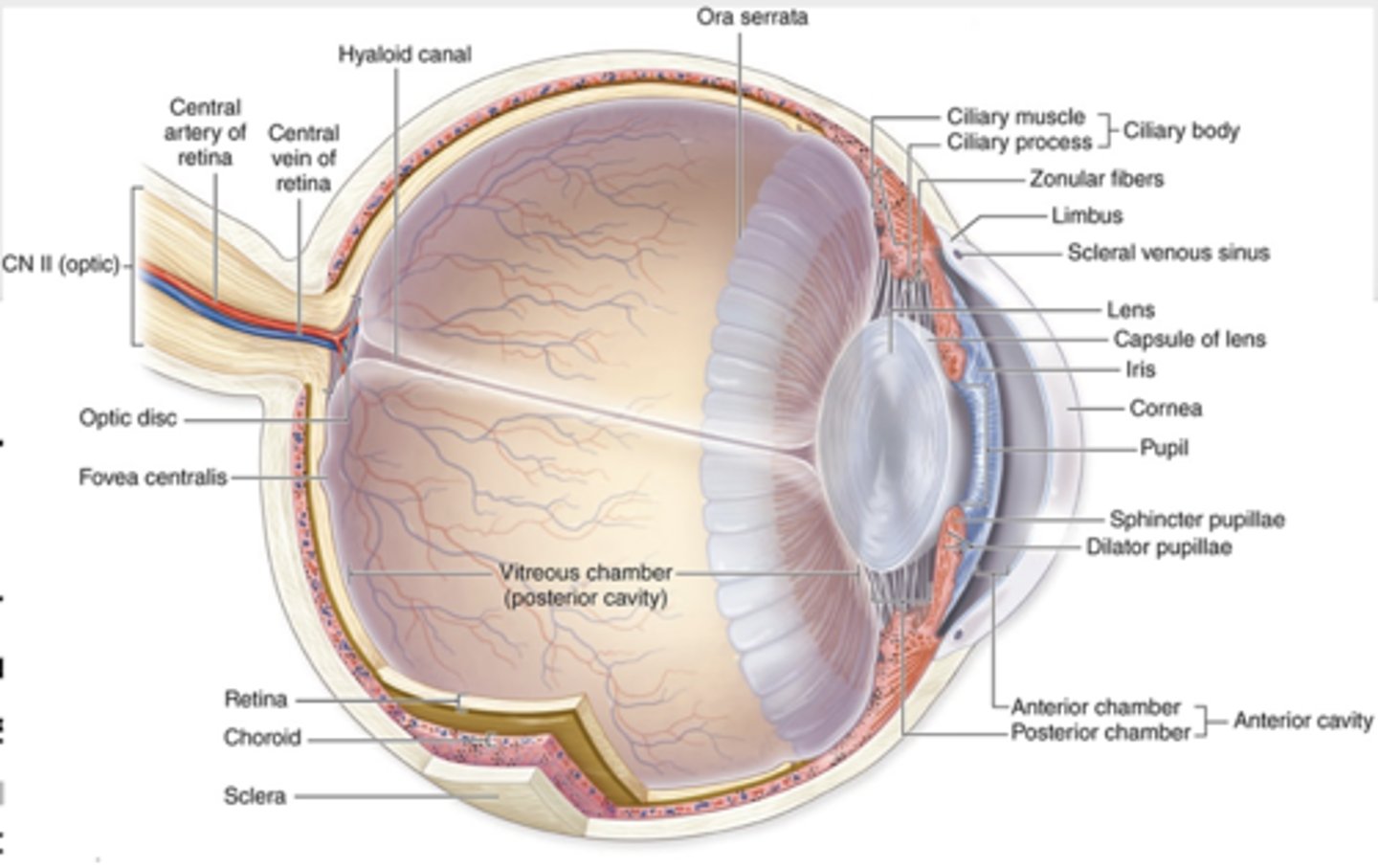
1. Anterior cavity
2. Posterior cavity (Vitreous cavity)
What are the two main cavities of the eye?

1. Anterior chamber
2. Posterior chamber
What are the two chambers within the anterior cavity of the eye?

By the pupil, which allows aqueous humor to flow between them
How are the anterior and posterior chambers connected?

- Surrounded by the retina
- Located behind the lens and zonula fibers
What are the boundaries of the posterior (vitreous) cavity of the eye?

The vitreous body, a connective tissue-like substance that helps maintain eye shape.
What does the posterior (vitreous) cavity contain?
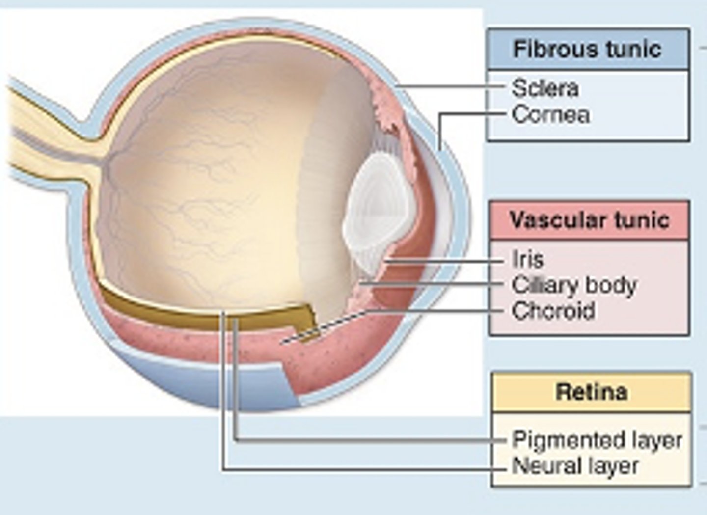
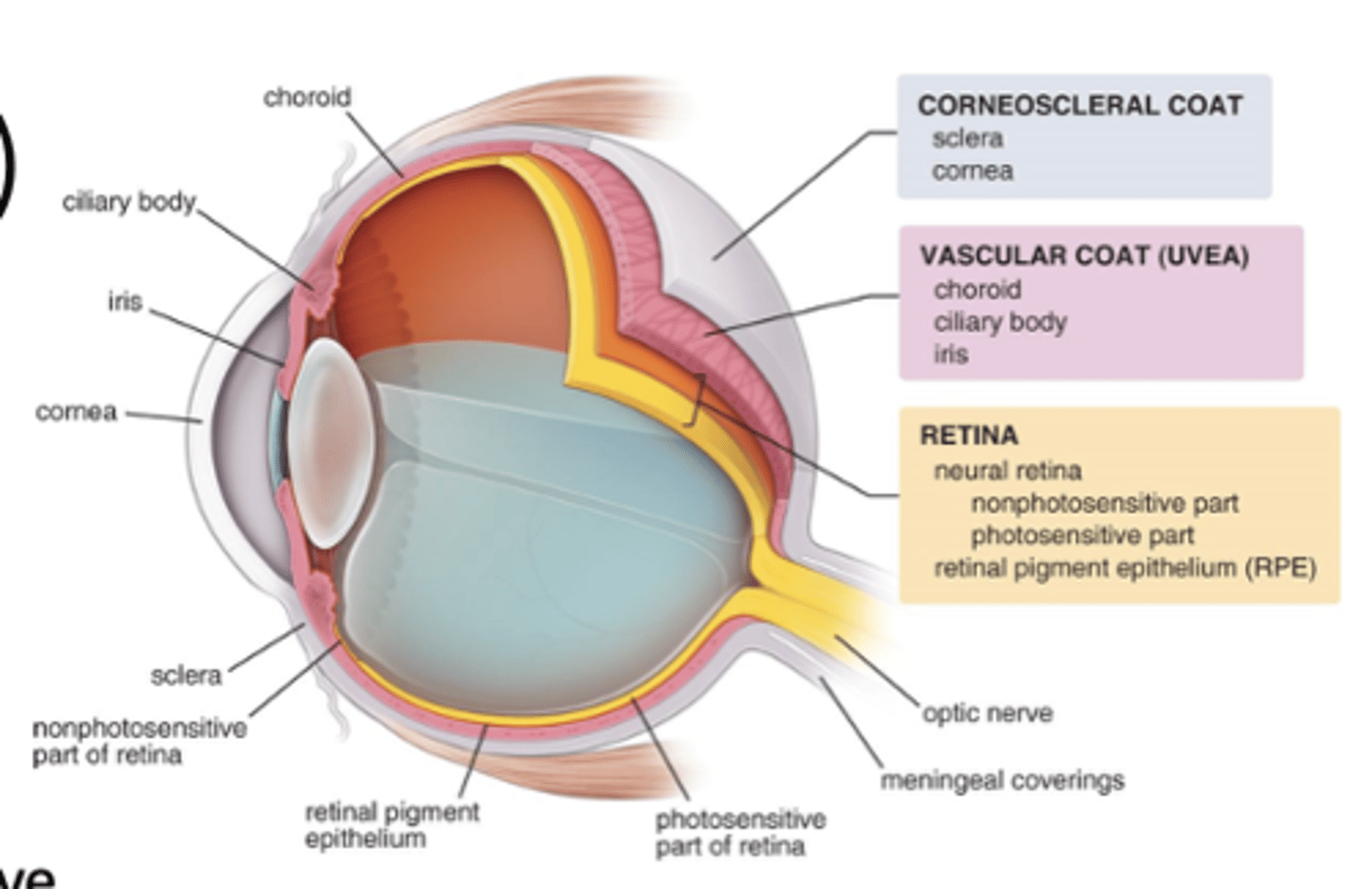
1. Sclera
2. Cornea
What does the Fibrous Tunic of the eye contain? (2)
1. Iris
2. Ciliary body
3. Choroid
What does the Vascular Tunic of the eye contain?
1. Pigmented layer
2. Neural layer
What does the Retina Tunic of the eye contain?
Dense irregular connective tissue
1. Supports the eye shape
2. Extrinsic eye muscle attachment site
What is the component and function of the sclera? (2)

two layers of epithelium with organized CT in between
1. Protects the anterior surface of the eye
2. Refracts (bends) incoming light)
What is the component and function of the Cornea? (2)

Loose (areolar) CT that's highly vascularized
1. Supplies nourishment to the retina
2. Pigments absorb extraneous light
What is the component and function of the Choroid? (2)

Ciliary smooth muscle and ciliary processes; covered with secretory epithelium
1. Hold suspensory ligaments that attach to the lens and change the lens shape for far and near vision
2. Epithelium secretes aqueous humor
What is the component and function of the Ciliary Body? (2)

Two layers of SM (spinchincer papillae and dilator pupillae) and CT, with a central pupil
1. Controls pupil diameter and thus the amount of light entering the eye
What is the component and function of the Iris?

Pigmented epithelial cells
1. Absorbs extraneous light
2. Provides Vitamin A for photoreceptor cells****
What is the component and function of the Pigmented layer? (2)

Photoreceptors, bipolar neurons, ganglion cells, and supporting Muller cells
1. Detects incoming light rays; light rays are converted to nerve signals and transmitted to the brain
What is the component and function of the Neural layer ? (2)
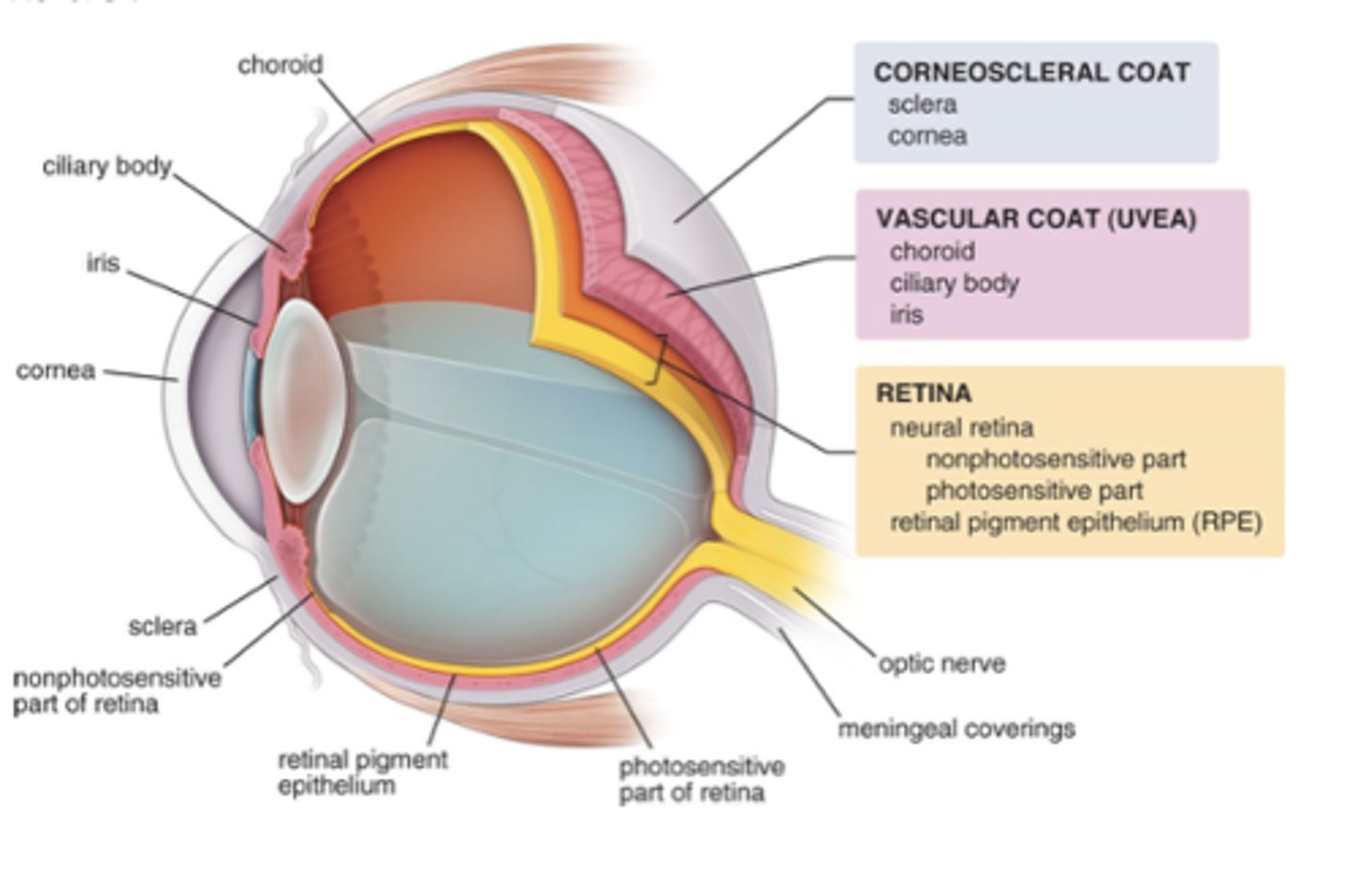
The corneosclera.
What is the fibrous tunic of the eye also called?
1. Posterior sclera
2. Anterior cornea
What are the two regions of the fibrous tunic?

At the limbus
Where do the sclera and cornea meet?

1. Episclera
2. Sclera proper
3. Suprachoroid lamina (lamina fusca)
What are the three layers of the sclera of the fibrous tunic?
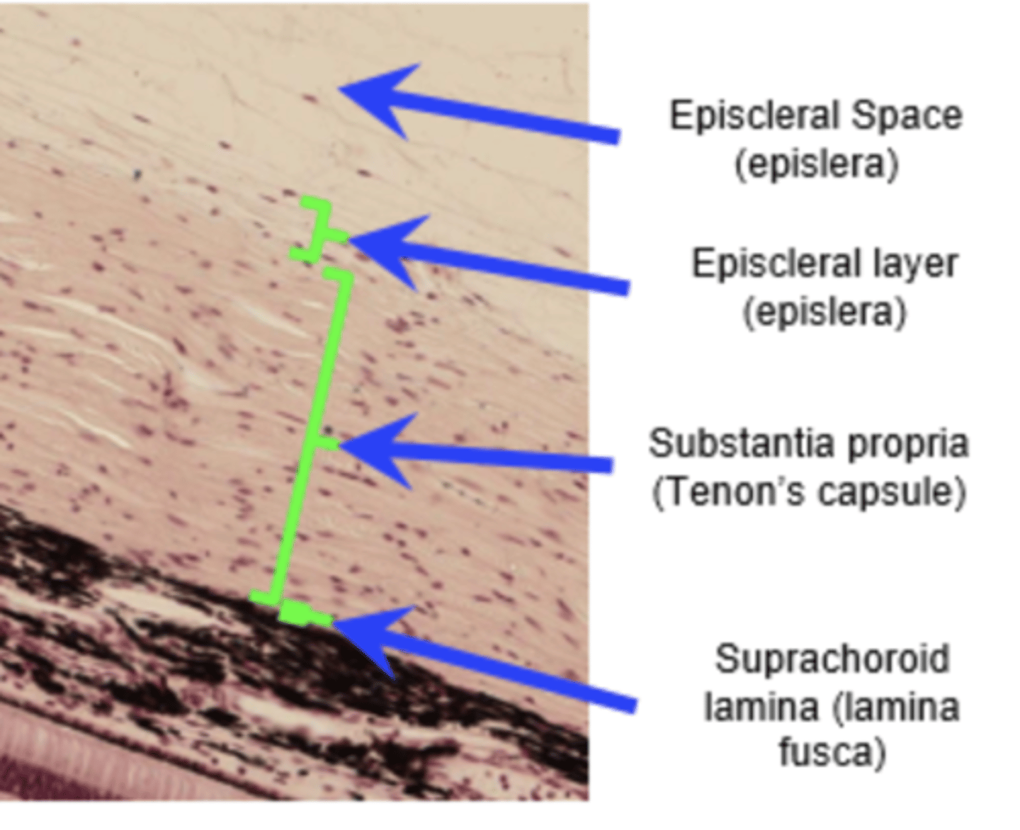
- Thin layer
- Abundant microvasculature
- Loose connective tissue with fibroblasts, macrophages, and lymphocytes
What are the characteristics of the episclera? (3)

- Dense connective tissue
- Dense bundles of type I collagen, parallel to the surface but intersecting in distinct directions
- Avascular****
What are the characteristics of the sclera proper?

- Less collagen
- More fibroblasts
- Contains elastic fibers and melanocytes
What are the characteristics of the suprachoroid lamina (lamina fusca)?

The anterior 1/6 of the eye.
What portion of the eye does the cornea make up?
- Transparent
- Avascular
What are the main characteristics of the cornea?*** (2)
1. Corneal epithelium
2. Bowman’s membrane
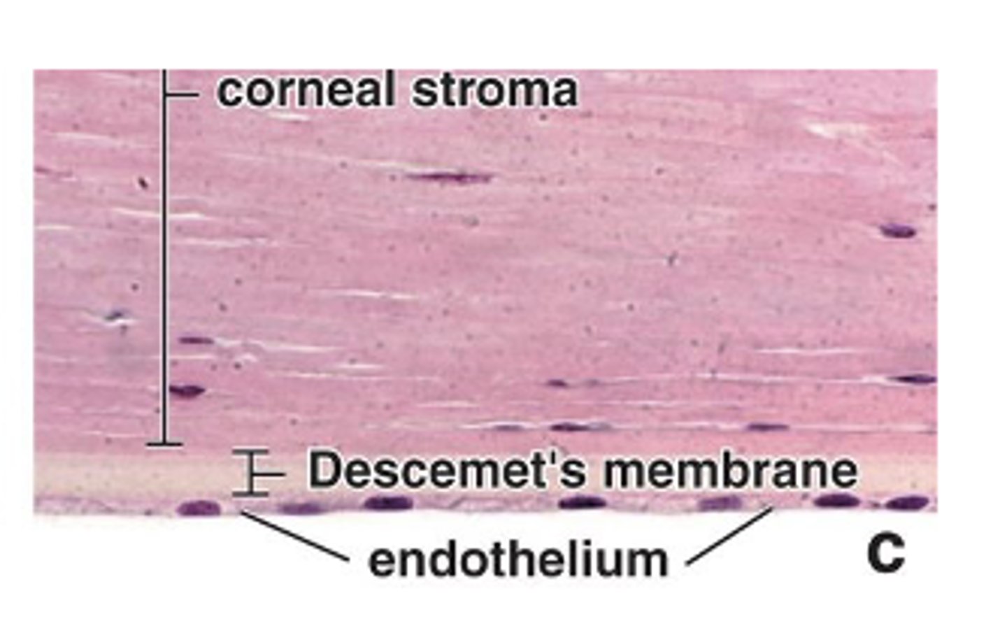
3. Corneal stroma
4. Descemet’s membrane (posterior limiting membrane)
5. Corneal endothelium
"CBCDC"
What are the five layers of the cornea (from outermost to innermost)?****
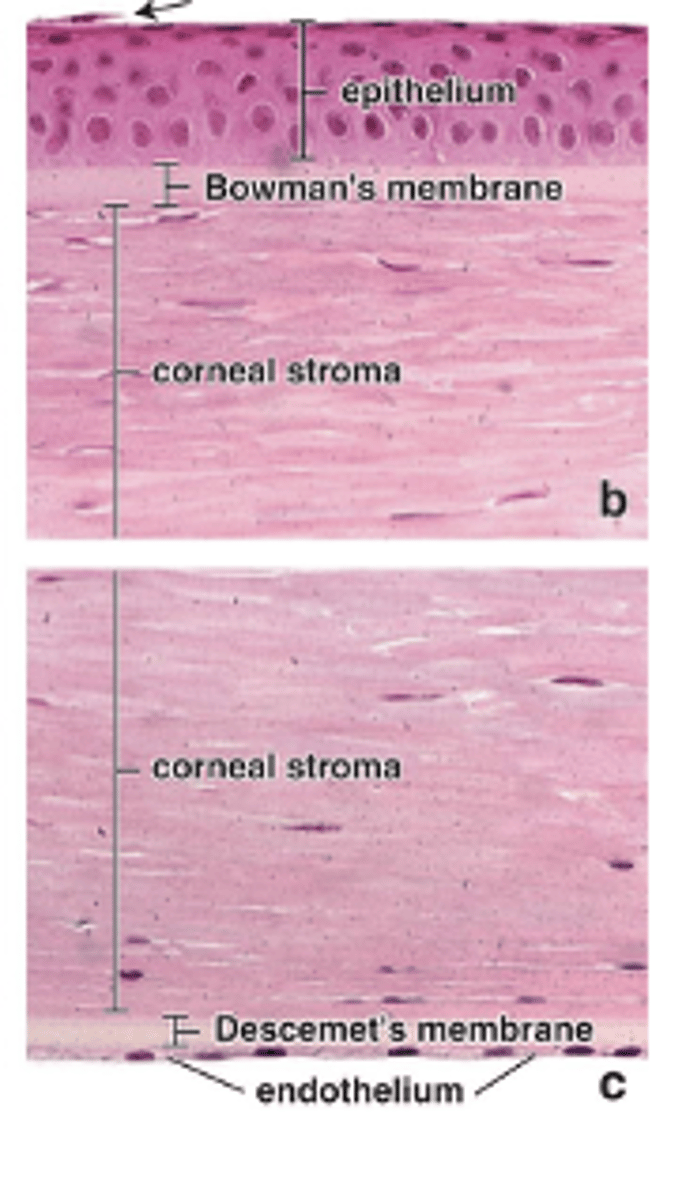
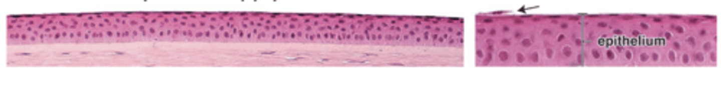
Non-keratinized stratified squamous epithelium.
What type of epithelium is the corneal epithelium?
The conjunctival epithelium.
What is the corneal epithelium continuous with?

5–6 layers of epithelial cells.
How many cell layers make up the corneal epithelium?

- High regenerative capacity
- Emerge from stem cells in the corneoscleral limbus (around the cornea).
What is the regenerative capacity of corneal basal cells, and where do they arise from?

They possess microvilli.
What special feature do squamous superficial cells of the corneal epithelium have?***

A rich sensory nerve supply.
What type of innervation does the corneal epithelium have?

The anterior limiting membrane.
What is another name for Bowman’s membrane?
8–10 μm, making it a very thick basement membrane.
How thick is Bowman’s membrane?
- Provides stability and strength to the cornea
- Protects the stroma against infections
What is the function of Bowman’s membrane in the cornea?(2)
The substantia propria.
What is another name for the corneal stroma?
It is the thickest layer, making up about 90% of corneal thickness.
How much of the cornea does the stroma make up?
- ~60 lamellae with parallel bundles of collagen fibrils
- Collagen fibrils of adjacent lamellae are arranged at right angles.
How many lamellae are present in the corneal stroma, and how are the collagen fibrils arranged?
Flattened fibroblasts (keratocytes).
What cells are found between the lamellae of the corneal stroma?
The posterior limiting membrane.
What is another name for Descemet’s membrane?
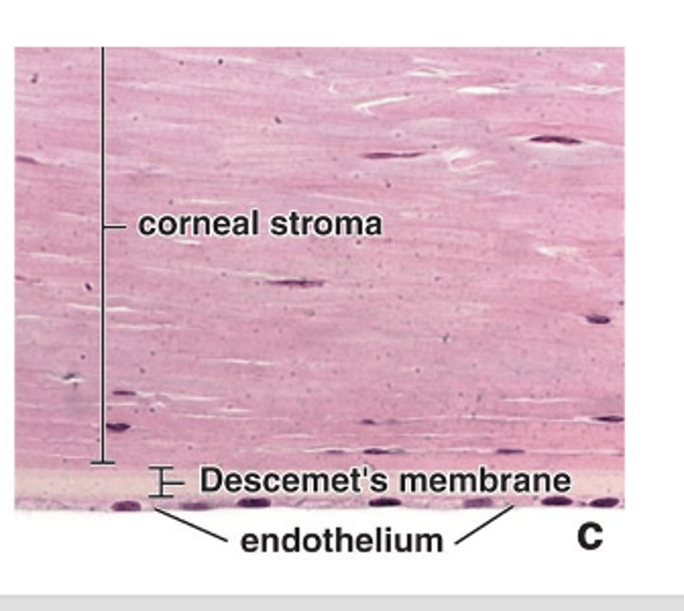
The corneal endothelium.
What is Descemet’s membrane the basement membrane of?
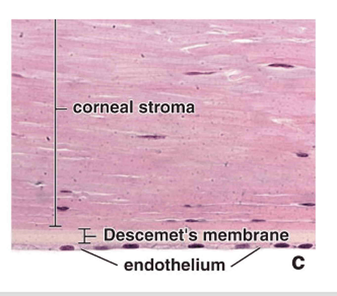
Simple squamous endothelium
What type of cells make up the corneal endothelium?

Descemet’s membrane
What membrane does the corneal endothelium maintain?

Corneal endothelial cells
Which corneal cells are the most metabolically active?

Na⁺/K⁺ ATPase pumps
Which pump is present in the basolateral membranes of corneal endothelial cells?
Regulate hydration of the corneal stroma
What is the main function of corneal endothelial cells in relation to the stroma?

Maintains maximal transparency and optimal light refraction
How does the corneal endothelium contribute to vision?

A transition zone that encircles the cornea where the sclera and cornea meet.
What is the limbus and where is it located?
Bowman’s membrane ends at the limbus; the surface epithelium becomes more stratified and forms the conjunctiva.
Where does Bowman’s membrane end and what happens to the surface epithelium at the limbus?
Extends to the anterior sclera and the internal surface of the eyelids.
To where does the limbus extend?
Epithelial stem cells that give rise to progenitor cells of the corneal epithelium.
What important stem cells are found in the limbus?
It becomes vascular and less organized; collagen fibrils merge with those of the sclera.
How does the stroma of the limbus differ from the corneal stroma?
A system of endothelium-lined channels in the limbus that replaces the corneal endothelium and Descemet’s membrane.
What is the trabecular meshwork and its function?
Penetrates the stroma at the corneoscleral junction.
Where does the trabecular meshwork penetrate?

Allows slow and continuous drainage of aqueous humor.
What is the main function of the trabecular meshwork?
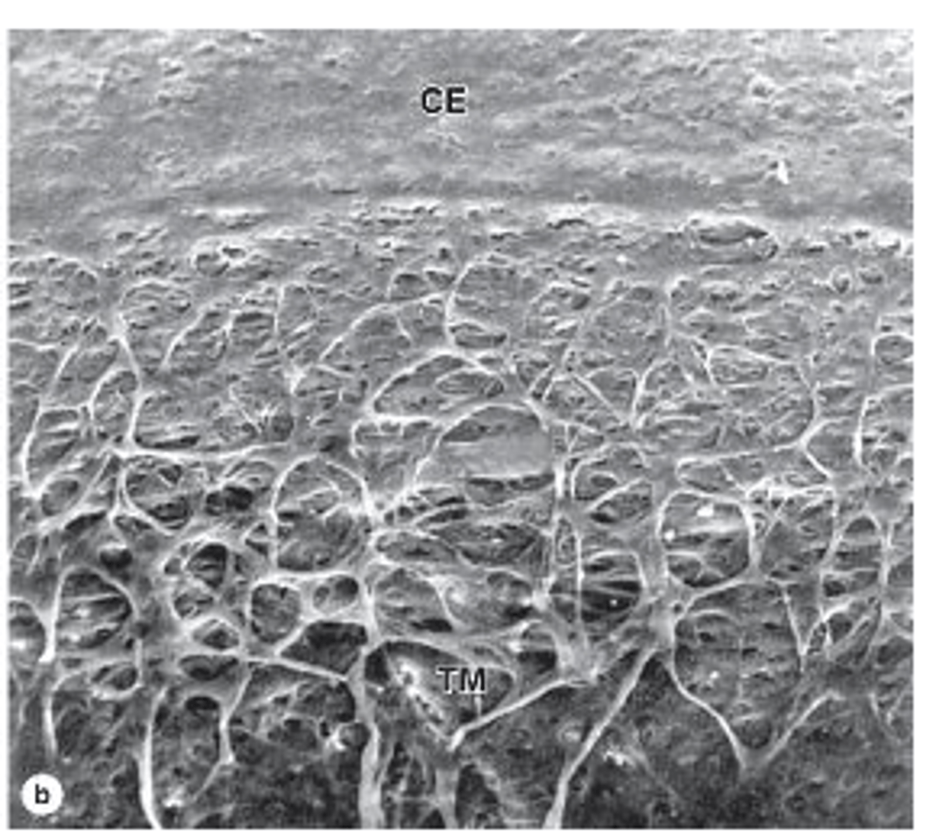
To the scleral venous sinus (Schlemm’s canal).
Where do the channels of the trabecular meshwork convey aqueous humor?

It is the most vascular layer of the eye.
What is the vascular layer (uvea) of the eye known for?
1. Choroid: posterior 2/3 of the eye
2. Ciliary body: posterior to the limbus
3. Iris: the most anterior region, covers most of the lens, has an opening (pupil)
What are the three parts of the uvea and where are they located?
2/3 of the eye; most posterior region.
What fraction of the eye does the choroid occupy and where is it located?
Loose connective tissue; well vascularized and contains abundant melanocytes.
What type of tissue composes the choroid and what are its characteristics?
1. Choroidocapillary lamina
- Innermost
- Rich in capillaries
2. Bruch membrane
- Extracellular material: collagen and elastic fibers
- Surrounds microvasculature of the choroidocapillary lamina
- Between choroid and retina
What are the two layers of the choroid?
Between the choroid and the retina.
Where is Bruch’s membrane located?
Anterior expansion of the uvea that encircles the lens; found posterior to the limbus, between the iris and the choroid.
What is the ciliary body and where is it located?
1. Ciliary muscle: three groups of smooth muscle
2. Ciliary processes: radially arranged series of about 75 ridges extending from the highly vascular region of the ciliary body
3. Ciliary zonule: system of radially oriented fibers
What are the three regions**** of the ciliary body?
The ciliary muscle.
What forms most of the stroma of the ciliary body?
Three groups:
1. Meridional (longitudinal)
2. Radial (oblique)
3. Circular (sphincteric) portions
How many groups of smooth muscle fibers are in the ciliary muscle and what are they?
Stretch the choroid and help open the iridocorneal angle to facilitate aqueous humor drainage.
What is the function of the meridional (longitudinal) fibers of the ciliary muscle?
Insert in the ciliary body; contraction flattens the lens for distant vision.
What is the function of the radial (oblique) fibers of the ciliary muscle?
Inner circular fibers; contraction reduces lens tension to accommodate for near vision.
What is the function of the circular (sphincteric) fibers of the ciliary muscle?
Radially arranged series of about 75 ridges extending from the highly vascular region of the ciliary body.
How are the ciliary processes arranged and how many are there?

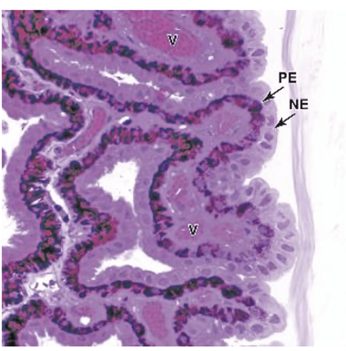
Stratified columnar epithelium (two layers of cells).
What type of epithelium covers the ciliary processes?***

Highly pigmented cells that are continuous with the pigmented epithelium of the retina.
What is the inner layer of the ciliary process epithelium like?***

Lacks melanin and is continuous with the sensory layer of the retina; cells specialize in aqueous humor secretion.
What is the superficial layer of the ciliary process epithelium like?
A system of radially oriented fibers that hold the lens in place.
What is the ciliary zonule?
Fibrillin 1 and 2, produced by non-pigmented epithelial cells.
What are the main components of the ciliary zonule?
From grooves between the ciliary processes to the surface of the lens.
Where do the fibers of the ciliary zonule extend?
The most anterior region of the uvea, covering most of the lens; the pupil is the opening of the iris.
What is the iris and where is it located?
Contacts aqueous humor; has no epithelium; dense layer of fibroblasts and melanocytes.
What is the structure of the anterior surface of the iris?
Stratified (two layers) epithelium, continuous with the ciliary body epithelium but more pigmented with melanin
What is the structure of the posterior surface of the iris?
Smooth muscle fibers arranged circularly around the pupil.
What is the sphincter pupillae muscle (SPM) and its arrangement?
Myoepithelial cells forming a partially pigmented epithelium with contractile processes extending radially
What is the dilator pupillae muscle (DPM) and its structure?
Loose connective tissue with sparse microvasculature.
What is the composition of the iris stroma?
Transparent, biconvex, highly elastic, and avascular.
What are the general characteristics of the lens?
1. Lens capsule
2. Subcapsular lens epithelium
3. Lens fibers
What are the three main components of the lens?
Thick and homogeneous, composed of proteoglycans and type IV collagen.
What is the lens capsule made of?
Only on the anterior surface; simple cuboidal epithelium.
Where is the subcapsular lens epithelium located and what type of cells does it contain?
Basally to the lens capsule and apically to the internal lens fibers.
How are the subcapsular lens epithelial cells attached?
Highly elongated, terminally differentiated cells derived from lens epithelial cells.
What are lens fibers and where do they come from?
Filled with crystallin protein granules.
What is unique about the cytoplasm of lens fibers?
They undergo autophagy.
What happens to the nucleus and organelles of lens fibers?
Packed tightly together to form a transparent tissue specialized in light refraction.
How are lens fibers arranged and what is their function?
In the vitreous chamber, behind the lens.
Where is the vitreous body located?
Transparent, gel-like connective tissue that is 99% water (vitreous humor), containing collagen fibrils and hyaluronate.
What is the composition of the vitreous body?
An external lamina called the vitreous membrane.
What surrounds the vitreous body?
A small population of mesenchymal cells called hyalocytes (synthesize hyaluronate and collagen) and a few macrophages.
What types of cells are found in the vitreous body?
1. Outer pigmented layer
2. Neural layer
What are the two layers of the Retina (innermost tunic)
Simple cuboidal epithelium.
What type of epithelium forms the pigmented (outer) layer of the retina?