Boobies Pathology
1/68
There's no tags or description
Looks like no tags are added yet.
Name | Mastery | Learn | Test | Matching | Spaced | Call with Kai |
|---|
No analytics yet
Send a link to your students to track their progress
69 Terms
(NORMAL) what age group would you normally see these characteristics?
Young age group: gland-stroma is 50/50
(NORMAL) what age group would you normally see these characteristics?
Pregnancy gland-stroma >50/50
(NORMAL) what age group would you normally see these characteristics?
Menopausal women Glan-stroma <50/50
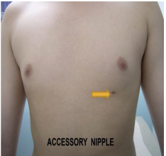
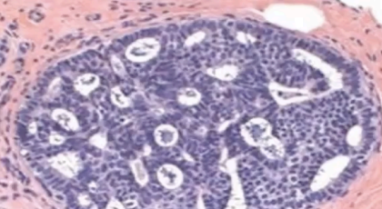
What pathology?
POLYTHELIA/SUPERNUMERARY NIPPLE
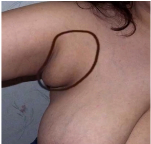
What pathology?
ACCESSORY AXILLARY BREAST TISSUE
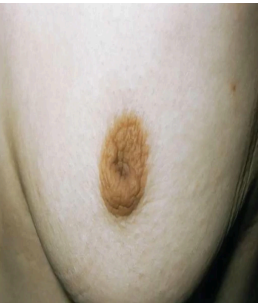
What pathology?
NIPPLE RETRACTION: Failure of the nipple to evert (can be acquired or congenital)
What pathology?
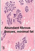
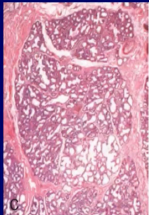
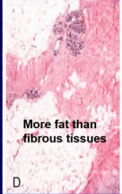
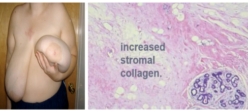
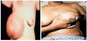
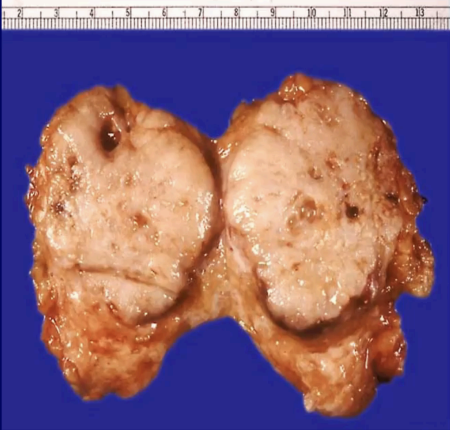
MACROMASTIA/GIGANTOMASTIA
Histology
→ Greatly increased stromal collagen
→ Broad area of moderately edem
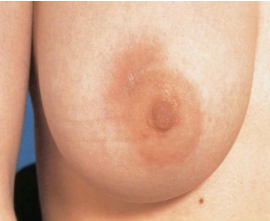
What pathology?
MASTITIS
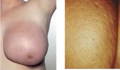
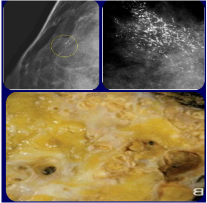
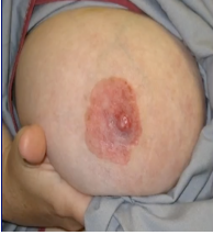
What pathology?
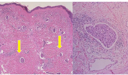
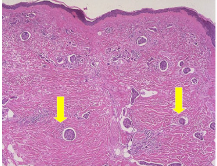
and what characteristic change is seen on the right image?
INFLAMMATORY CARCINOMA:
Peau d’orange (right image)
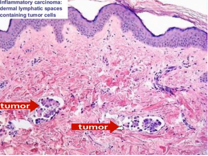
What pathology?
Inflammatory Carcinoma Histology: dermal lymphatic spaces containing tumor cells
What pathology?
ACUTE MASTITIS:
Histology
→ Infiltration of breast tissue by intraductal & periductal inflammatory cells (mostly neutrophils)
→ Localized suppurative inflammation
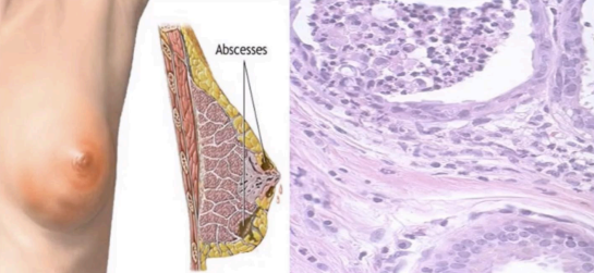
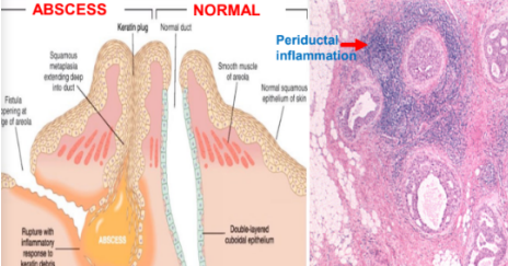
What pathology?
PERIDUCTAL INFLAMMATION:
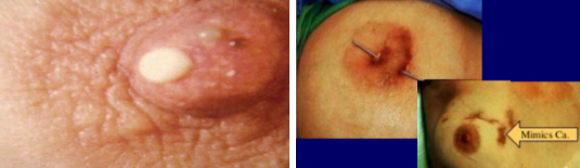
What pathology?
MAMMARY DUCT ECTASIA:
❗Poorly defined palpable periareolar mass, often with thick, white nipple secretions
❗Sometimes with skin retraction & oozing of pus in sinuses
What pathology?
Mammary Duct Ectasia[
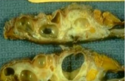
What pathology?
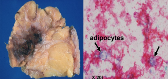
FAT NECROSIS
What pathology? What stage (Acute/ Subacute)?
FAT NECROSIS: ACUTE OR EARLY LESIONS
What pathology? What stage (Acute/ Subacute)?
Fat Necrosis: Subacute to Late Fat Necrosis
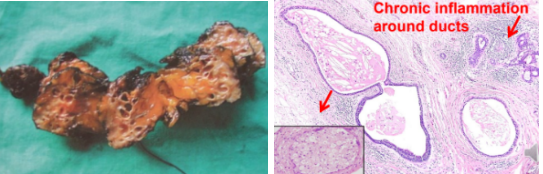
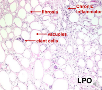
What pathology?
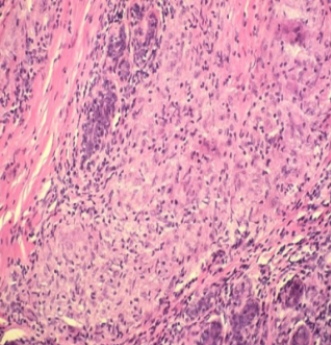
Granulomatous Mastitis
What pathology?
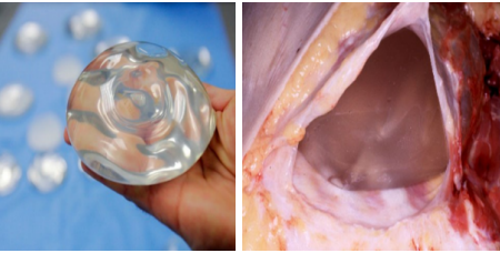
Granulomatous Mastitis from Silicone Breast Implants
What pathology?
Histologic Response in Silicone Breast Implants
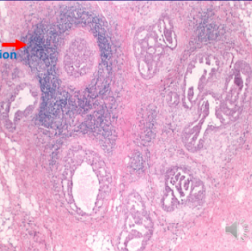
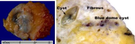
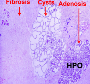
What pathology?
Non-proliferative breast changes. (L) Irregular indurations or nodules. (R) Fluid-filled cysts (colorless areas) seen in mammogram
What pathology?
Non-proliferative breast changes. Irregular areas of fibrosis with cyst
What pathology?
on-proliferative breast changes. (L) Irregular indurations or nodules. (R) Fluid-filled cysts
What pathology?
Microscopic changes of Fibrocystic changes
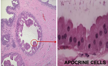
What pathology?
Apocrine Metaplasia (microscopic)
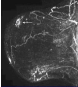
What pathology?
Mammogram of fibrocystic change
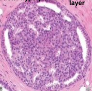
What pathology?
Ductal Hyperplasia
What pathology?
Fibrocystic change with ductal hyperplasia
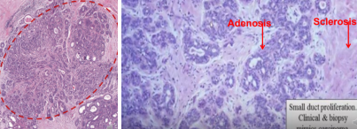
Is there atypia here?
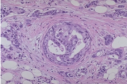
PROLIFERATIVE BREAST DISEASE WITHOUT ATYPIA: SCLEROSING ADENOSIS
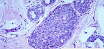
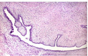
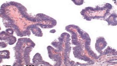
What pathology?
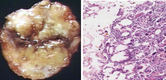
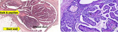
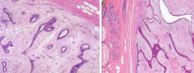
PROLIFERATIVE BREAST DISEASE WITHOUT ATYPIA: INTRADUCTAL PAPILLOMA
Tree-like microvascular stroma (L) Epithelial & myoepithelial cell lining (R)
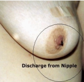
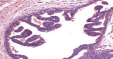
What pathology usually presents with this type of discharge?
INTRADUCTAL PAPILLOMA
Identify left vs right microscopic slide
Atypical Ductal Hyperplasia (L) and Atypical Lobular Hyperplasia (R)
both are proliferative breast diseases presenting with atypia
What pathology?
Atypical Ductal Hyperplasia (a proliferative disease that presents with atypia)
What pathology?
Atypical lobular hyperplasia (a proliferative disease that presents with atypia)

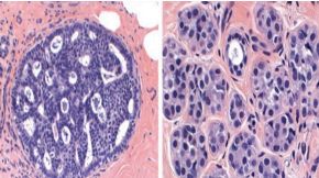
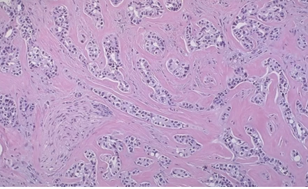
What pathology?
Fibroadenoma
Left is juvenile fibroadenoma in a 15 year old while right is a giant fibroadenma in a 23 year old
What pathology?
Fibroadenoma
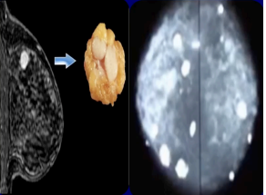
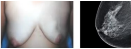
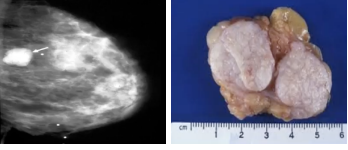
The mammography shows what lesion
Fibroadenoma Mammography
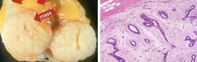
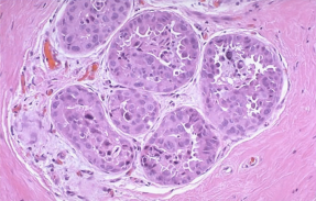
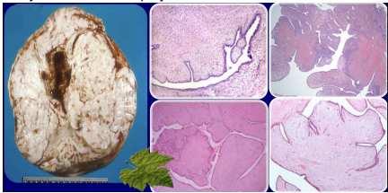
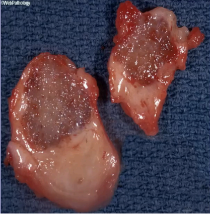
What pathology?
Gross/ Microscopic image of fibroadenoma
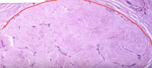
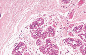
What pathology?
Fibroadenoma in older women which is more fibrous/ hyalinized with few compressed ducts
What pathology?
Phyllodes Tumor
What pathology?
Phyllodes Tumor
Are there two different pathologies? If you think yes, identify the one on the right vs the one on the left
yes besh dalawa sya Left microscopic image shows a benign fibroadenoma vs right image shows a phyllodes tumor
What pathology?
(Borderline) Phyllodes Tumor
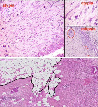
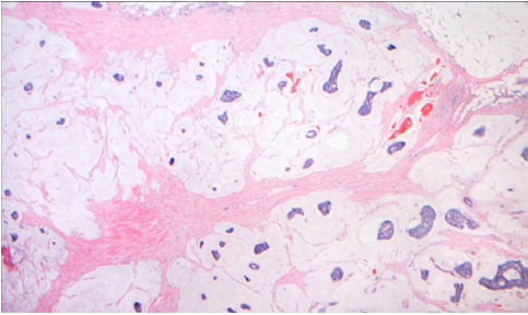
What pathology?
Malignant Phyllodes Tumor. Atypia & Mitosis (Top); Hypercellular Stroma & Infiltration of Fat (Bottom)
What pathology?
Fibroadenoma
What pathology?
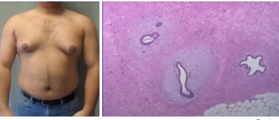
Gynecomastia
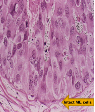
Usually seen in inflammatory carcinoma. Identify the pathology
Histology of Peau D’Orange
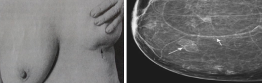
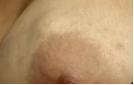
What pathology?
Nipple Retraction
What pathology?
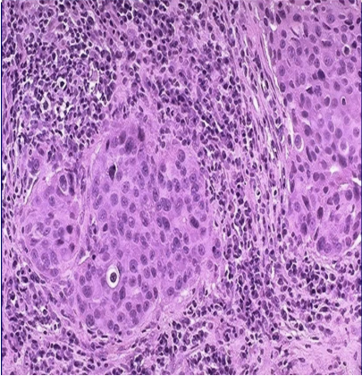
DUCTAL CARCINOMA IN-SITU (DCIS)
What pathology?
Microscopic findings of Ducctal Carcinoma in-situ
What type of DCIS?
DCIS: Comedocarcinoma (High-grade)
What type of DCIS?
DCIS (Non-comedocarcinoma): Solid DCIS
What type of DCIS?
DCIS (Non-comedocarcinoma): Cribriform DCIS
What type of DCIS?
DCIS (Non-comedocarcinoma): Papillary DCIS
What type of DCIS?
DCIS (Non-comedocarcinoma): MicroPapuillary DCIS
What type of DCIS?
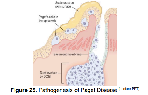
DCIS Special Type: Paget Disease of the Nipple
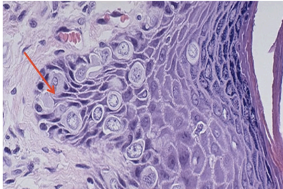
What pathology?
Microscopic Findings in Paget Disease
What pathology?
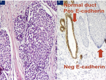
Lobular Carcinoma in-situ
What pathology?
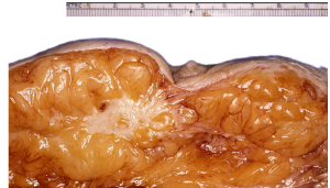
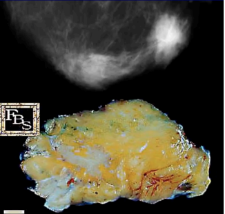
Gross morphology of intiltrating carcinoma
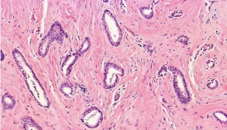
How would you grade this infiltrative ductal carcinoma?
Grade 1: well-differentiated
How would you grade this infiltrative ductal carcinoma?
Grade : well-differentiated IDC showing mostly tubules infiltrating a fibrotic or desmosplastic stroma
How would you grade this infiltrative ductal carcinoma?
Grade 2: moderately differentiated IDC
How would you grade this infiltrative ductal carcinoma?
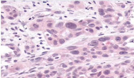
Grade 3: poorly differentiated IDC
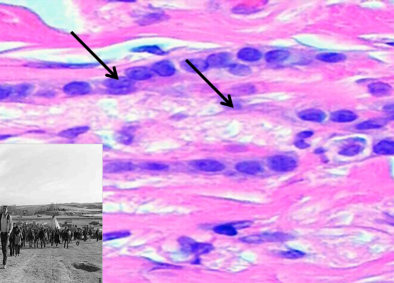
What pathology and what pattern in the pointed arrows?
Indian File Pattern of Infiltrating Lobular Carcinoma
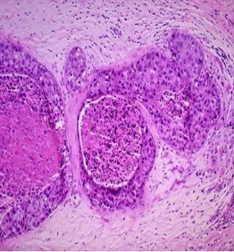
What pathology?
Medullary Carcinoma
What pathology?
Medullary Carcinoma
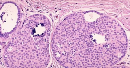
What pathology?
Mucinous (Colloid) Carcinoma
What pathology?
Mucinous Carcinoma
What pathology?
Inflammatory Carcinoma