IGCSE biology - basically everything
1/331
There's no tags or description
Looks like no tags are added yet.
Name | Mastery | Learn | Test | Matching | Spaced | Call with Kai |
|---|
No analytics yet
Send a link to your students to track their progress
332 Terms

What is the name of the part of the heart labelled as 1?
Pulmonary artery


What is the name of the part of the heart labelled as 2?
Vena cava.


What is the name of the part of the heart labelled as 3?
Right atrium.

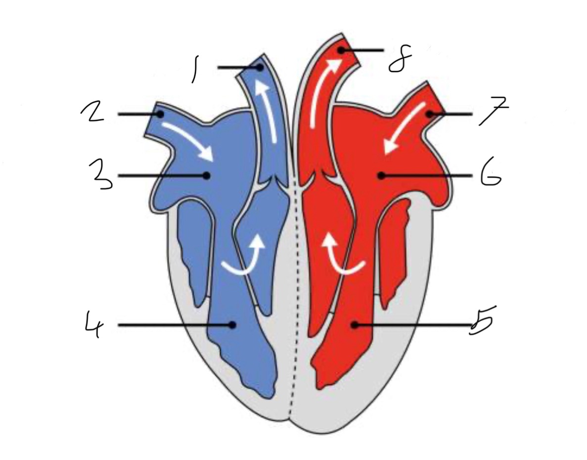
What is the name of the part of the heart labelled as 4?
Right ventricle.


What is the name of the part of the heart labelled as 5?
Left ventricle.


What is the name of the part of the heart labelled as 6?
Left atrium.


What is the name of the part of the heart labelled as 7?
Pulmonary vein


What is the name of the part of the heart labelled as 8?
Aorta.

Blood is made from:
red blood cells, white blood cells, platelets, and plasma. 55% of blood is plasma.
White blood cells contain:
Lymphocytes and phagocytes.
What is the role of platelets?
Platelets help to prevent blood loss by clumping together at the site of a wound to form a temporary plug. They then release chemicals that convert fibrinogen into fibrin, forming a mesh that creates a stable blood clot and prevents pathogens entering.
What per cent of plasma is water?
About 90%
The plasma transports the following things:
glucose
Amino acids
Antibodies
Chlosterol
CO2
Urea
Lactic acid
Hormones
The plasma also distributes heat around the body.
What is the function of red blood cells?
To transport oxygen from the lungs to body tissues and carry carbon dioxide back to the lungs.
How are red blood cells adapted for their role?
They have a biconcave shape to increase surface area for gas exchange, lack a nucleus to maximize haemoglobin content, and contain haemoglobin to bind oxygen to form oxyhaemoglobin.
The innate immune system:
first line of defence (immediate)
Non-specific
No immunological memory
It doesn’t respond differently the second time you come into contact with a pathogen
It includes physical barriers like skin and mucous membranes, as well as immune cells such as phagocytes that respond quickly to pathogens.
How does a phagocyte remove bacteria?
A phagocyte removes bacteria by engulfing them through a process called phagocytosis, where the bacteria are enclosed in a vessel and then destroyed by enzymes.
Example of a physical defence:
Ear wax = has acidic environment that kills bacteria. It traps dirt, dust and small object from damaging your ear.
Adaptive immune system
second line of defence (5-7 days)
Has immunological memory - your immmune system responds better the second time.
Just cellular defence - T and B cells (lymphocytes)
B cells produce antibodies
T cells kill virally infected cells.
Antigens
A molecule, often a protein or sugar, that is recognized by the immune system as foreign and potentially harmful, triggering an immune response to fight it.
(Fact 1: they are found on the surface of all cells
Fact 2: there are hundreds of thousands of different antigens)
Role of lymphocytes
When they come across a foreign antigen, they will produce proteins called antibodies. Memory cells are also produced in response to a foreign antigen. These remain in the body, and the next time they come into contact with the antigen, they reproduce VERY fast.
What is the role of antibodies?
Antibodies lock onto invading pathogens and mark them out for destruction by other white cells.
They stick to antigens
They can clump bacteria together and this makes them difficult to reproduce
They stick to viruses and this makes them difficult to get inside cells.
What is a vaccine?
Typically, either alive microbe, mild or weakened, or a dead microbe
Therefore, the antigens are present but the ability to cause the disease is not.
the antigens are present
Therefore, the lymphocytes multiply
Some become memory cells.
If theses memory cells meet the actual pathogens (antigens), they multiply quickly, produce more antibodies and fight the pathogens before you even feel the effects.
Active immunity
You make an immune response either following a real infection or following a vaccine
Passive immunity
You are given antibodies either naturally (in utero) or artificially.
Order of the cardiac cycle.
Atrial systole
Ventricular systole
Diastole
Atrial systole
Atria contract
bicuspid/tricuspid valves OPEN
blood flows into the ventricles
Ventricular systole
Ventricles contract
Bicuspid/tricuspid valves close
Semi-lunar valves open
Blood leaves the heart
Diastole
Heart relaxes
Semi-lunar valves close
Blood enters atria
How does the heart rate change during exercise?
More energy so there is more respiration
More carbon dioxide in your blood stream and your brain detects this
The brain sends a signal to your heart so it beats faster and more forcefully.
More blood is sent out from the heart (stroke volume) in each heart beat the blood arrives at the lungs and muscles quickeR
More oxygen arrives at the muscles and more CO2 is removed
Therefore, the muscles contract more.
How does the heart rate change under the influence of adrenaline?
adrenaline is released from the adrenal glands
It increases the heart rate (more oxygen and glucose arrive at the muscles)
The muscles can therefore contract more
Risk factors for coronary heart disease:
Smoking - nicotine + CO put strain on heart, making it work harder. Other chemicals damage the lining of your coronary arteries, causing arterial furring.
Lack of exercise - fatty deposits more likely to build up in your arteries.
High blood pressure - puts high strain on your heart
Bad diet - high amounts saturate fats in diet can cause high cholesterol
What are the three types of blood vessels?
Arteries, veins and capillaries
Arteries
carry blood away from the heart to the organs
The blood is under high pressure so the walls must be able to stretch and recoil
Generally carry oxygenated blood
Veins
carry blood from the organs back towards the heart
Under low pressure
Must allow blood to pass through easily and prevent it flowing backwards
Veins generally carry deoxygenated blood
They have ‘watch-pocket’ valves to prevent back flow
Capillaries
carry blood through organs, bringing the blood close to the cells in the organ
They are permeable so that substances are transferred between the blood and the cells
Their walls are only one cell thick

What does 1 represent?
Pulmonary artery


What does 2 represent?
Hepatic vein
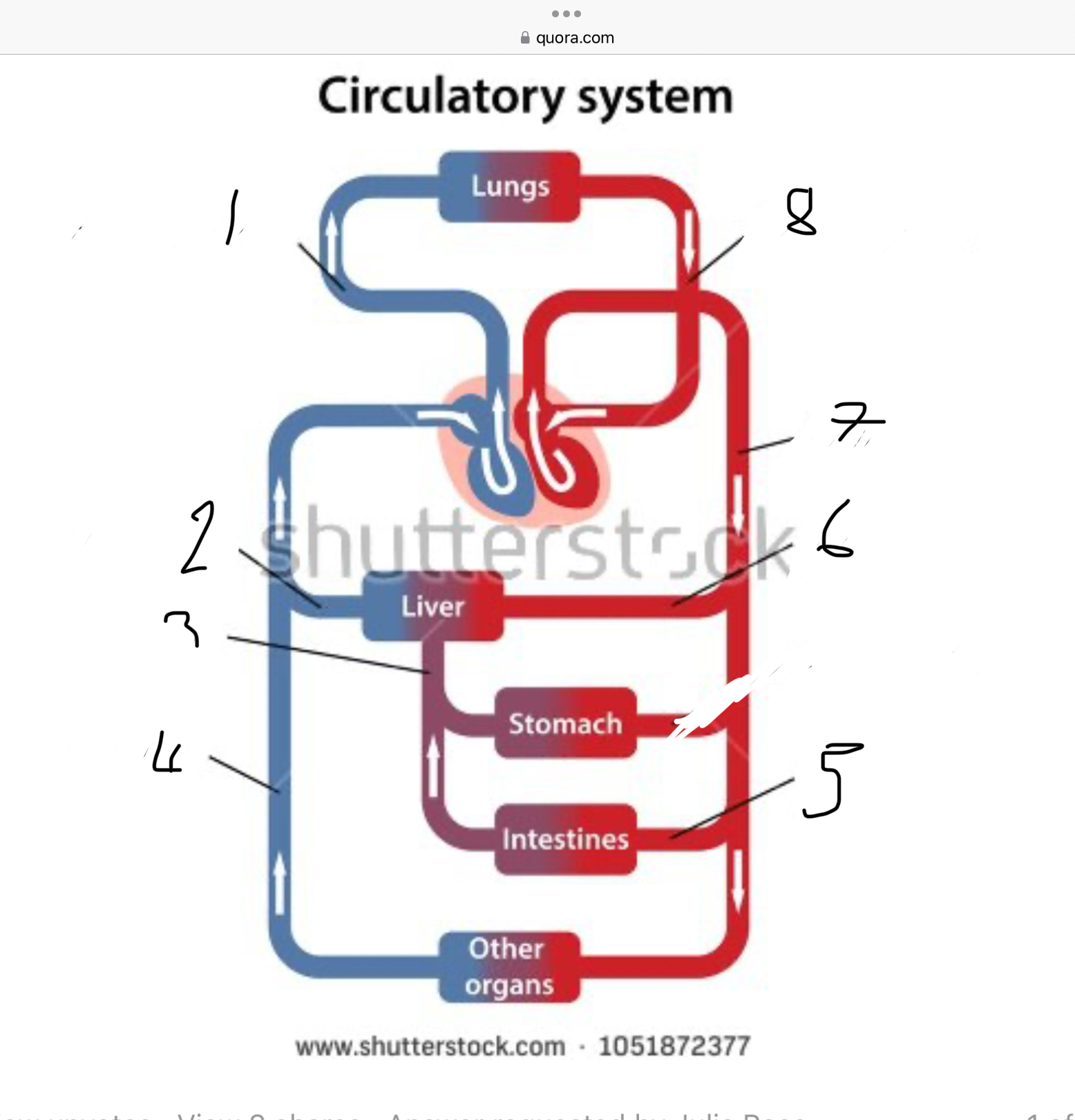

What does 3 represent?
Mesentric vein


What does 4 represent?
Inferior vena cava


What does 5 represent?
Mesentric artery


What does 6 represent?
Hepatic artery


What does 7 represent?
Aorta


What does 8 represent?
Pulmonary veins

What do the lungs excrete?
Carbon dioxide and water
What does the skin excrete?
Water and salt by sweating
What do the kidneys excrete?
Urea, water and ions from the blood. This is called urine.
How does the kidney carry out its role excretion and osmoregulation?
Excretion:
Filters blood in the glomerulus
Removes urea, excess salts, and toxins
Forms urine which is sent to the bladder
Osmoregulation:
Reabsorbs water in the collecting duct (controlled by ADH)
Adjusts how much water and salt are returned to the blood
Keeps the body’s water balance stable and blood at the right concentration
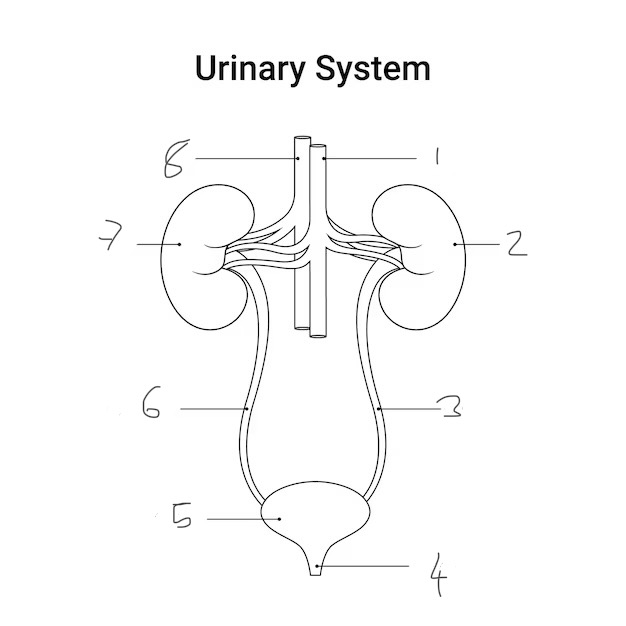
Label this diagram
Vena cava
Left kidney
Left ureter
Urethra
Bladder
Right ureter
Right kidney
Aorta
renal vein + vena cava = blue
renal artery + aorta = red

How does the kidney carry our its role of excretion?
The kidneys filter the blood to get rid of harmful substances, particularly urea, which is produced in the liver from the breakdown of amino acids

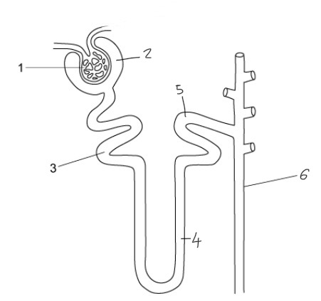
Label this diagram
Glomerulus
Bowman’s capsule
Proximal convoluted tubule
Loop of Henle
Distal convoluted tubule
Collecting duct
Ultrafiltration
The blood arriving at each glomerulus is under high pressure.
Small molecules cross over into the kidney nephron whereas large molecules stay in the blood.
The fluid moves along the nephron, it is known as the glomerular filtrate
Selective reabsorption
glucose (and other useful small molecules) mustn’t stay in the nephron and leave in the urine (it’s needed for respiration)
It is reabsorpted into the bloodstream from the proximal convoluted tubule
This happens via active transport - there are lots of mitochondria in the cells lining this part of the nephron.
osmoregulation (reabsorption of water in the loop of henle)
the wall of the nephron is permeable to water
The surrounding area has low water potential and therefore the some water will leave via osmosis.
Osmoregulation (hormonal control of water)
the body can control the water content of the blood using the kidney.
Special cells in the hypothalamus of the brain can detect the amount of water in the blood.
If you are dehydrated, the cells in the hypothalamus can cause the pituitary gland to release a hormone called the anti-diuretic hormone (ADH)
This then travels through the blood to the kidney where it make the walls of the nephron more permeable to water and therefore more water is reabsorpted into the blood.
This make the urine more concentrated (less water excreted.)
Opposite happen when there is a normal amount of water in the blood.
What does urine contain?
water, urea and ions
ADH: source, role and effect
Source: Produced in the hypothalamus and released in the pituitary gland
Role: Controls water balance in the body
Effects:
Increases permeability of kidney collecting ducts
More water reabsorbed back into the blood
Produces concentrated urine and reduces water loss
FSH: source, role and effect
Source: Pituitary gland
Role: Controls development of eggs and sperm
Effects:
In females: stimulates egg maturation in the ovary and stimulates oestrogen production
In males: stimulates sperm production in the testes
LH: source, role and effect
Source: Pituitary gland
Role: Triggers ovulation and controls sex hormone release
Effects:
In females: triggers the release of the egg and stimulates the ovary to produce progesterone
In males: stimulates testosterone production in the testes
Myelin sheath
An insulating layer, or sheath, that forms around nerves, including those in the brain and spinal cord, to speed up nerve transmissions.
Cell body
core command centre of the neuron, containing the nucleus
Axon
The long, thin extension of a neurone that carries electrical impulses away from the cell body toward other neurones, muscles, or glands.
Dendrite
The receiving or input portions of a neuron
Motor neurone
Cells in the brain and spinal cord that allow us to move, speak, swallow and breath by sending commands from the brain to the effectors (muscles) that carry out these functions.
Sensory neurone
A nerve cell that detects and transmits sensory information from the environment to the brain
Relay neurone
A cell in the CNS that acts as a messenger between sensory and motor neurones

1
Cornea


2
Aqueous humour


3
Pupil


4
Iris


5
Tear duct


6
Suspensory ligaments


7
Lens


8
Ciliary muscle


9
Sclera


10
Choroid
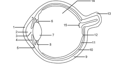

11
Retina


12
Fovea


13
Optic nerve


14
Vitreous humour


15
Blind spot

Cornea
It refracts light and protects the eye
Iris
Controls how much light enters the pupil
Lens
Focuses light onto the retina
Optic nerve
Bundle of sensory neurones that carry impulse to the brain
Retina
Layer of tissue at the back of the eye that contains light receptor cells (rods + cones)
Fovea
Area of the retina with the highest concentration of cone cells that provide sharp vision
Aqueous humour
Maintains pressure in the eye and nourishes the cornea
Vitreous humour
Maintains eye shape - ensures correct focusing of light
Supports retina - keeps photoreceptors in position
Transparent medium - allows clear passage of light
Shock absorption - protects delicate internal structures
Sclera
Strong outer layer of tissue of the eye that wraps around your eyeball.
Pupil
Hole in the center of the eye that lets light in
What happens to the eye in bright light?
circular muscle contracts
Radial muscles relax
Pupils constrict
What happens to the eye in dim light?
Circular muscles relax
Radial muscles contract
Pupil dilates (gets bigger)
What happens to the eye when it’s focusing on a distant object?
ciliary muscles relax
Suspensory ligaments pulled tight (stretched)
Lens flatten
What happens to the eye when its focusing on a nearby object?
ciliary muscles contract
Suspensory ligaments slacken
Lens more rounded (convex)
Light refracts MORE
Homeostasis
The maintenance of a constant internal environment e.g. temperature
features of nervous control system
nerve impulses
Travel fast
Short-lived effect
Localised effect
features of hormonal control systems
hormone in blood
Travel more slowly
Long-lived effect
Widespread effect
What happens to the body when we are too hot?
lots of sweat - when it evaporates it transfers energy from your skin to the environment cooling you down
blood vessels close to the surface of the skin widen - this is called vasodilation. it allows more blood to flow near the surface, so it can transfer more energy into the surroundings, which cools you down.
hairs lie flat
What happens when we are too cold?
hair - stands on end to trap an insulating layer of air, which helps you keep warm
vasoconstrictions - blood vessels near the surface of the skin contract so less blood is flowing to near the surface, so less energy is transferred to the surroundings.
shiver - increases rate of respiration, which transfers more energy to warm the body. exercise does the same.
very little sweat produced