comparative female repro
1/38
Earn XP
Description and Tags
Describe the position and form of the reproductive organs in relation to other pelvic structures. Describe the anatomy and histology of the female reproductive tract and its ligaments. Describe the structure and function of the oviduct and its secretions. Describe the structure and function of the uterus. Describe the structure and function of the cervix and its secretions. Describe the structure and function of the vagina and its secretions. Recognise sections of mammalian ovary in different reproductive states
Name | Mastery | Learn | Test | Matching | Spaced | Call with Kai |
|---|
No analytics yet
Send a link to your students to track their progress
39 Terms
theriology, veterinary gynaecology, obstetrics, adnrology
Theriogenology: Study of genesis of beasts or animals.
Veterinary Gynaecology: Physiopathology of female reproduction related to animals.
Veterinary Obstetrics: Study of normal physiology and disease condition during late pregnancy around parturition and shortly after parturition and care & management of dam and young one.
Veterinary Andrology: Study of physiopathology of male reproduction including Artificial Insemination
which organs are relatedto female repro
hypothalamus
pituitary
breast
vagina
vulva
uterus
placenta
uterinetube
ovary
what can cause differences
species
age
stage os oestrus
time of year for seasonal breeders
pregancncy
intercornual ligament
located between the 2 uterine horns
can use fingers to retract for a rectal palpation
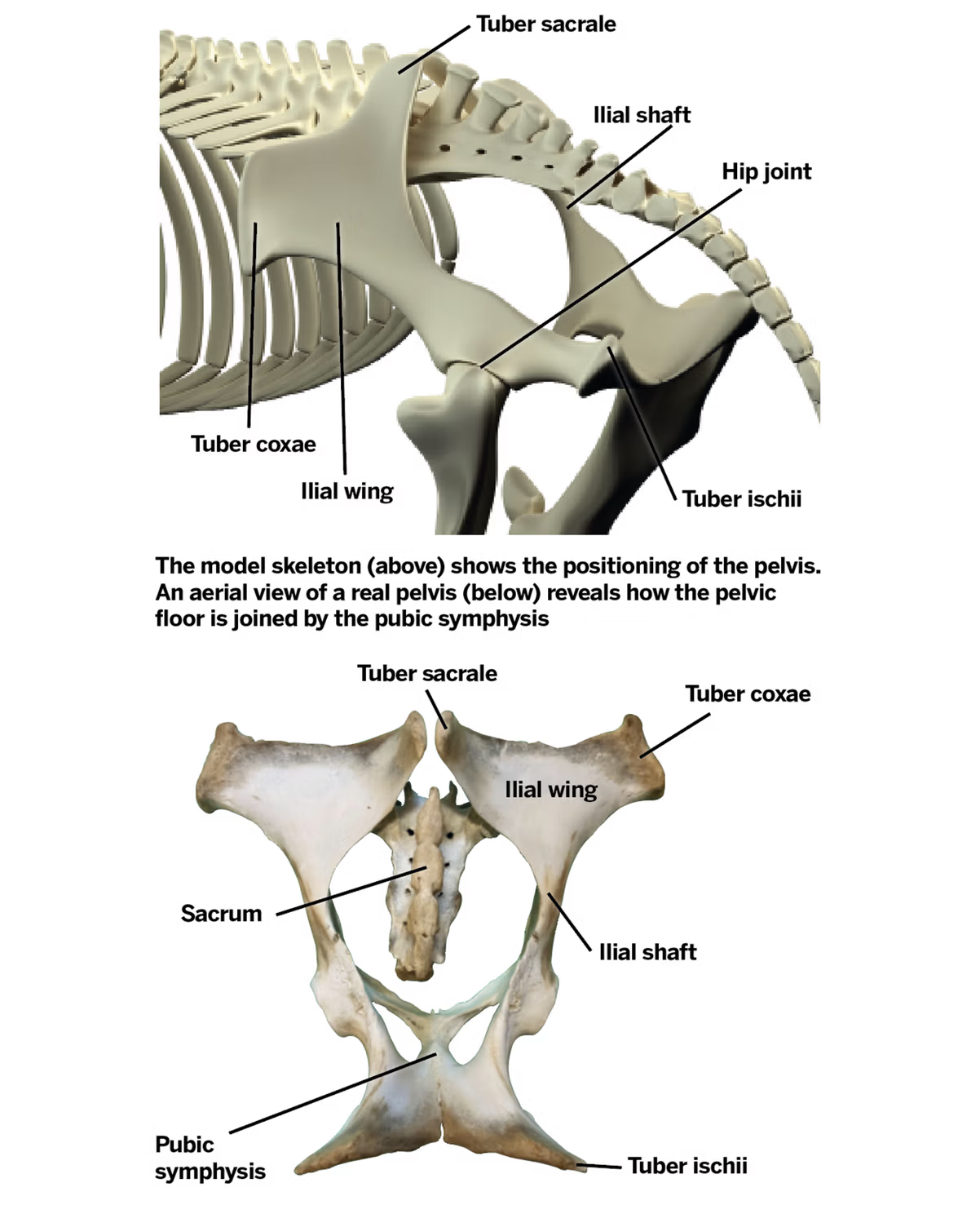
where are the ovaries
in the abdomen
relative to the kidneys
relatively to the iliac wings
which vertebra
relative to the lateral body wall
lie bilaterally in the dorsal part of the abdomen
caudoventral to the kidneys
cranioventral to the iliac wings
approx level of the 5th lumbar vertebra
adjacent to lateral body wall
where are the ovaries in a mare
which body cavities
relative to which third of the ipsilateral ileal shaft and which region
left relative to right ovary
over half of repro tract located in abdominal cavity, remainder in the pelvic cavity
often located 5-10cm directly cranial to the upper third of the ipsilateral ileal shaft in the sublumbar region
left ovary is normally situated 2-3cm futher caudal than the right
ovary in a sow and bitch
relative to kidney
relative to the last rib
caudal to the caudal pole of the associated kidney
several cm caudal to the last rib
what happens to the uterus during pregnancy in cows
uterus lies on the ventral body wall
in the cow the uterus is displaced to the right by the rumen
what are the main attachments of the reproductive tract
broad ligament (bilateral sheets anchoring organs to abdominal roof and pelvic wall)
mesometrum
mesosalphinx
mesovarian
round ligament (lateral fold)
other ligaments
inter-cornual ligament (uterine horns)
suspensory ligament- peritoneal fold attaches ovary to abdominal wall adjacent to the last rib
what is the ovarian bursa
and differences between mare and bitch
a fold of peritoneum which covers/hides ovary
mare- shallow and large opening
bitch- small and deep opening
vasculature
ovarian vein
uterine vein
vaginal vein
uterine branch of the ovarian artery
ovarian artery (off aorta)
uterine artery (off internal iliac, in mares external)
vaginal artery (internal iliac)
blood supply to the ovary
ovary
ovarian veins
uterine branch of the ovarian artery
uterine veins
ovarian artery, branch of aorta in dogs
utero ovarian veins
what is the ovarian artery in close contact with and why is this important
utero ovarian vein
transfer of PGF2 alpha from uterus to ovary for luteolysis
what is a common cause of acute death following parturition in mare
rupture of uterine artry
peripartureint arterial ruptulre
where do the lymph of the repro tract drain
aortic and sacral iliac lymph nodes
may become enlarged if tract infected but difficult to palpate
innervation
ovaries
vulva
motor and autonomic (PS and S)
ovary PS from vagus and Syp from inter mesenteric and caudal mesenteric plexus)
motor neurones supply the vulva
disruption has little apparent effect on reproduction
function of the ovary
1. Produce female gametes (ovum=singular, ova=plural; NB. Oocyte = immature ovum)
HOW: Oogenesis and Folliculogeneis
2. Hormone production (main: oestrogen and progesterone; also oxytocin, inhibin, activin)
HOW: Follicles and corpora lutea
structure of the ovary
3 layers
what do they contain
GERMINAL EPITHELIUM TUNICA ALBUGINEA- thin connective tissue capsule underlying germinal epithelium
CORTEX- surrounds the medulla and contains maturing follicles
MEDULLA- central connective tissue containing vascular supply and nervous innervation
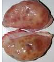
cow
almond shaped (like buffalo, ewe, doe)
sow
like bitch and queen
cluster of grapes shaped
function of ovary
oogenesis
secrete hormones like oestrogen, progesterone, oxytocin and relaxin
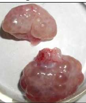
feature of mare ovaries
have a reversed structure
cortex inside and medulla outside
therefore their ovulation is internal in a specific place called ovulation fossa
mare
classification of uterus
advanced duplex- opossum
duplex- rabbit mouse
bicornuate- pig bitch
bicronute- cow ewe doe
bicornuate- mare
smplex- human
advanced duplex
opossum
2 uterine horns
2 cervice
2 vaginas
duplex
rabbit mouse
2 uterine horns
2 cervices
1 vagina
bicornuate
pig
2 long uterine horns
common uterine body
one cervx
one vagina
bicornuate
smaller uterine horns
cow ewe doe
bicornuate in mares
larger uterine body with smaller uterine horns
simplex
human
no uterine horn, all uterine body
ovuduct
cervix in cow ewe mare sow
Cow, ewe - cervix has annular rings, ewe has more obstacles
Mare- longitudinal folds
Sow- interdigitating prominences, no fornix
vagina epithelium
what type of epithelium in anterior and posterior
columnar epithelium in anterio
stratified squamous epithelium in posterior
feature of the ventral commissure of the vulva in mare
it is rounded
where does ovulation occur in the reversed cortex and medulla of a mare
the ovulation fossa
layers of the uterine wall
perimetrium (serosal layer)
myometrium (inner circular and outer longitudinal)
endometrium: produces PGF2 alpha, contains tubular glands, mucosa and submucosa, mucosa has simple columnar epithelium
draining of the ovarian vein
right
left
Ovarian Vein: The right ovarian vein drains into the caudal vena cava, while the left ovarian vein drains into the left renal vein.
draining of the uterine vein
Drains into the internal iliac veins, often via a uterine branch of the ovarian vein or uterine branches of the vaginal vein. Blood from the vagina drains into the internal iliac vein.
what is the ileal shaft
the body of the ileum