Pharyngeal apparatus and their derivatives
1/53
There's no tags or description
Looks like no tags are added yet.
Name | Mastery | Learn | Test | Matching | Spaced | Call with Kai |
|---|
No analytics yet
Send a link to your students to track their progress
54 Terms
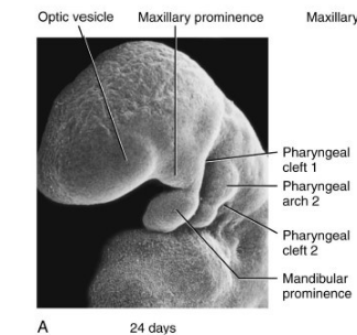
When do pharyngeal arches develop
week 4
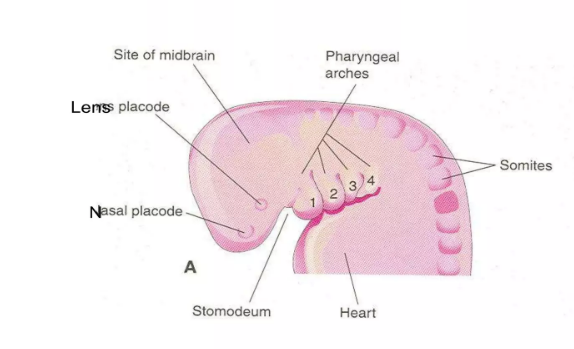
What do pharyngeal arches develop from
growth of mesenchymal tissue in the cranial region of the embryo
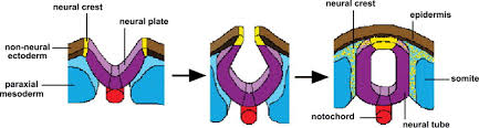
What is mesenchymal tissue
loose embryonic connective tissue that gives rise to almost all the connective tissue in the body
What is mesenchyme derived from
-most is derived from the mesoderm (specifically the paraxial mesoderm)
-some derived from the neural crest cells which are made up of ectoderm
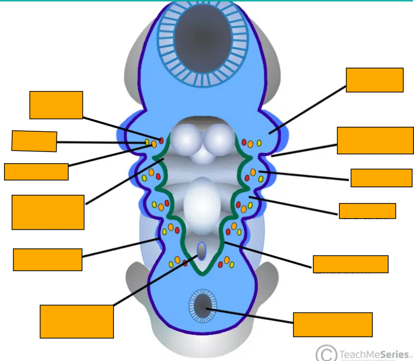
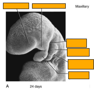
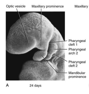
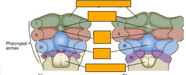
Label this image
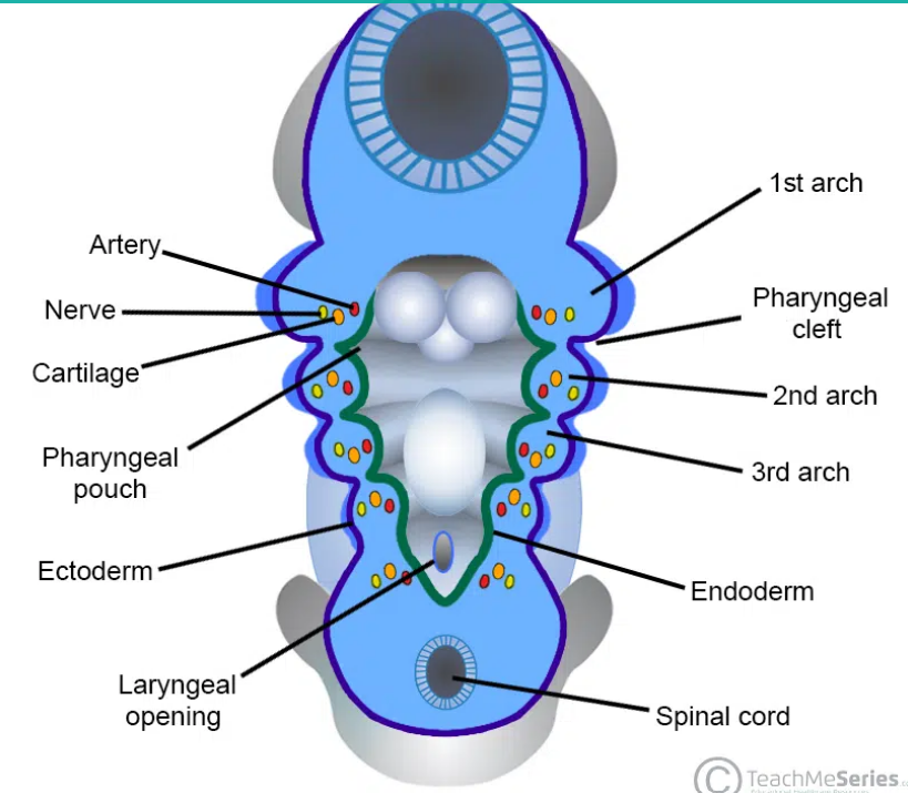
Describe the structure of the pharyngeal arches
-outer covering of ectoderm (pharyngeal cleft)
-mesenchyme core
-lined internally by endoderm (pharyngeal pouch)
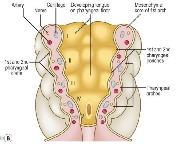
What happnes to the pharyngeal arches in week 5
they become distinct and numbered (1-6 however the 5th pharyngeal arch either doesn’t form or regresses)
What are the different components of each pharyngeal arch
-the mesenchyme differentiates into cartilage, the associated muscle and an an aortic arch artery
-each pharyngeal arch also contains a cranial nerve that enters from the developing brain stem
Label this image
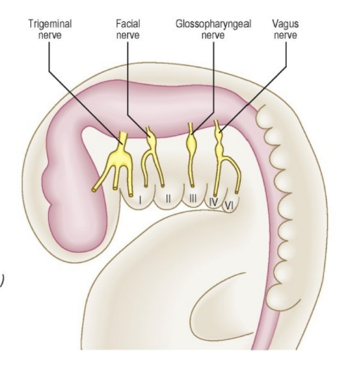
Which cranial nerves innervate each pharyngeal arch
1st arch- trigeminal nerve
2nd arch- facial nerve
3rd arch- glossopharyngeal nerve
4th arch- vagus nerve (superior laryngeal branch)
6th arch- vagus nerve (recurrent laryngeal branch)
Function of the cranial nerves innervating the pharyngeal arches
-carry motor fibres to supply the muscles derived from the pharyngeal arches
-carry sensory fibres from the developing skin covering these muscles and the mucosal tissue lining them
What are the 2 main parts of the 1st pharyngeal arch
maxillary prominence- associated with the maxillary cartilage and gives rise to the maxilla, zygomatic and the squamous part of temporal bone
mandibular prominence- associated with Meckel’s cartilage and gives rise to the mandible
Which muscles are derived from the 2nd pharyngeal arch
muscles of facial expression
stylohyoid, posterior belly of digastric (helps with swallowing)
stapedius (anchors stapes)
Which cartilagenous structures are derived from the 2nd arch
bones: stapes (ossicle), styloid process, lesser horn and upper body of hyoid,
ligaments: stylohyoid ligament
Which muscles are derived from the 1st pharyngeal arch
muscles of mastication (temporalis, masseter and pterygoid msucles)
mylohyoid, anterior belly of digastric, tensor veli palatini (helps with swallowing)
tensor tympani (blocks chewing noises)
Which cartilagenous structures are derived from the 1st pharyngeal arch
bones: malleus and incus (ossicles), mandible, maxilla, zygomatic and squamous part of temporal bone
sphenomandibular ligament
Which muscles are derived form the 3rd pharyngeal arch
stylopharyngeus muscle
Which cartilagenous structures are derived from the 3rd arch
lower body and greater horn of the hyoid
Which muscles are derived from the 4th and 6th pharyngeal arches
muscles of the pharynx and larynx
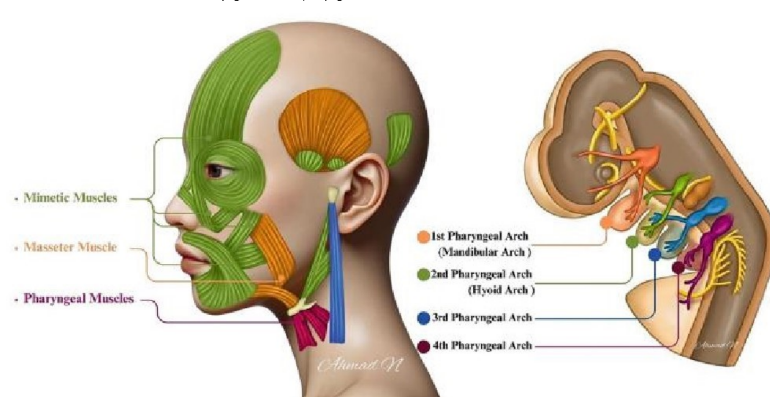
Which cartilagenous structures are derived from the 4th and 6th pharyngeal arches
pharyngeal and laryngeal cartilages
no bones
What happens to the 2nd pharyngeal arch in week 6
enlarges and grows rapidly formind a flap over the 3 remaining clefts
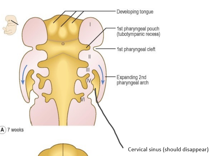
What is the cervical sinus
formed from remnants of lower clefts lined with ectoderm that remian beneath the flap
What happens to the cervical sinus
normally obliterated in week 8
Which structures develop from the 1st pharyngeal pouch
eustaichan tube (auditory tube), middle ear cavity
What forms from the 1st pharyngeal cleft
external auditory meatus
What forms from the 2nd pharyngeal pouch
palatine tonsils
What forms from the 2nd cleft
cervical sinus which disappears

What forms from the 3rd pouch
splits into a dorsal (posterior) and ventral (anterior) region
dorsal- inferior parathyroids
ventral- thymus
What is the thymus
small gland in the lymphatic system
where T-lymphocytes mature
What forms from the 3rd cleft
cervical sinus (disappears)
What forms from the 4th pouch
dorsal- superior parathyroid glands
ventral- parafollicular cells of the thyroid (produce calcitonin which lowers Ca2+ levels)
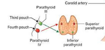
What forms from the 4th cleft
cervica sinus (disappears)
What are branchial cysts
-sealed off sac under the skin
Why do branchial cysts form
occur if the 3 pharyngeal clefts are not obliterated by the 2nd pharyngeal arch due to the formation of the cervical sinus
Where are branchial cysts usually found
lateral aspect of the neck
along the anterior border of the sternocleidomastoid muscle
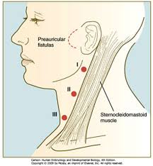
Where are branchial cysts often not manifested until after puberty
as they expand due to increased amounts of secretions by the epithelium lining the inner surface of the cyst
What are branchial sinuses and fistulas
formed for the same reasons as the branchial cysts
sinuses- tract connecting the skin of the neck and pharynx that is only open on one side
fistulas- a complete tunnel connecting the skin of the neck and the pharynx
What is DiGeorge syndrome
birth defect that affects the immune system and can lead to the absence or underdevelopment of the thymus and parathyroid glands due to defective development of the 3rd and 4th pharyngeal pouches
results in immune deficiency from defective T-cell function
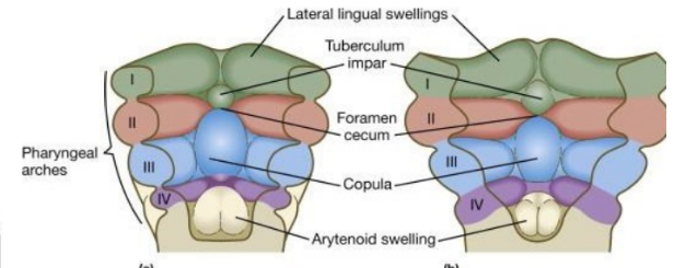
When does the tongue begin to form
end of week 4
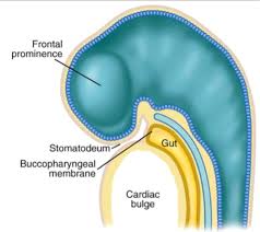
What is the stomatodeum
future oral cavity
space betweent he cardiac bulge and the developing brain of the embryo
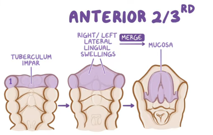
Whta does the anterior 2/3 of the tongue develop from
1st pharyngeal arch
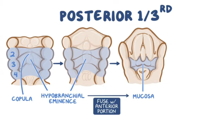
What does the posterior 1/3 of the tongue develop from?
3rd and 4th pharyngeal arches
How does the anterior 2/3 develop
-mesoderm of first pharyngeal arch proliferates forming a midline swelling (tuberculum impar)
-mesoderm also gives rise to right and left lateral lingual swellings which enlarge, overlap the tuberculum impar and merge together forming the mucosa over the anterior 2/3 of the tongue
-the line where they fuse is the median sulcus of the tongue
How does the posterior 1/3 develop
copula forms (midline swelling of the 2nd arch)
hypobranchial eminence develops (midline swelling from 3rd arch)
hypobranchial eminence grows upwards, over the copula and forms the mucosa for the posterior 1/3 of the tongue and fuses with the anterior portion
the terminal sulcus separates the anterior and posterior tongue
at the tip of the teminal sulcus is the foramen cecum which is a pit that represents the place of origin of the thyroid gland
the most posterior part of the tongue develops from a 3rd median swelling arising from the 4th pharyngeal arch
Label this image
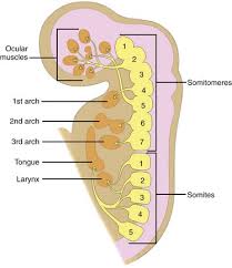
What are the muscles of the tongue derived from
-derived from occipital somites that migrate anteriorly and differentiate into myoblasts which give rise to the muscles of the tongue
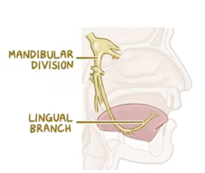
What innervates the anterior 2/3 of the tongue
lingual branch of CNV3
What innervates the posterior 1/3 of the tongue
glossopharyngeal nerve
What innervates the most posterior part of the tongue formed from the 3rd median swelling
vagus nerve (via the internal laryngeal nerve)
Which nerve provides motor innervation to the muscles of the tongue
hypoglossal nerve
except palatoglossus muscle (vagus nerve)
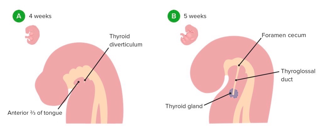
When does development of the thyroid gland begin
week 4
How does the thyroid gland develop
-midline endodermal thickening at the foramen cecum in the floor of the primitive pharynx
-a small outpouching called the thyroid diverticulum forms and it descends from the floor of the pharynx
-the thyroid descends anteriorly to the pharynx and its migration is guided by the thyroglossal duct
-thyroid reaches its destinatino int he neck by week 7, formed of 2 lobes connected by a central isthmus
the thyroglossal duct that usually regresses by week 7-10
What are thyroglossal duct cysts
cysts that form along the former thyrogossal duct if it doesn’t regress
usually asympotmatic and may contain some thyroid tissue
where are thyroglossal duct systs commonly located
in the tongue or anterior neck inferior to the hyoid bone