Lecture 10.1 - Sensory III; Photoreceptors, Opsins, Rhabdomeric and Ciliary
 Call Kai
 Learn
Learn Practice Test
 Spaced Repetition
 Match
Match Flashcards
 Knowt Play
1/11

There's no tags or description
Looks like no tags are added yet.
Name | Mastery | Learn | Test | Matching | Spaced | Call with Kai | Chat |
|---|
No analytics yet
Send a link to your students to track their progress
12 Terms
Photoreceptors
Convert photons into graded potentials
Change light energy → chemical → electrical energy
Specie detection of wavelengths
Humans: 650 nm - 350 nm
Humans and animals only detect form electromagnetic spectrum
Exact wavelengths that can be detected by an animal depend on the type of photoreceptors
Rhabdomeric Photoreceptor
Specialized accessory photoreceptor neuron
Similar to bipolar neuron in terms of structure
Off of the cell body - microvilli, have photoreceptor particles on them, which receive light messages, send messages via graded potential through cell body and to terminal, release NT via exocytosis
Specialized proteins inside microvilli that detect light - rhodopsin
NT - usually glutamate*
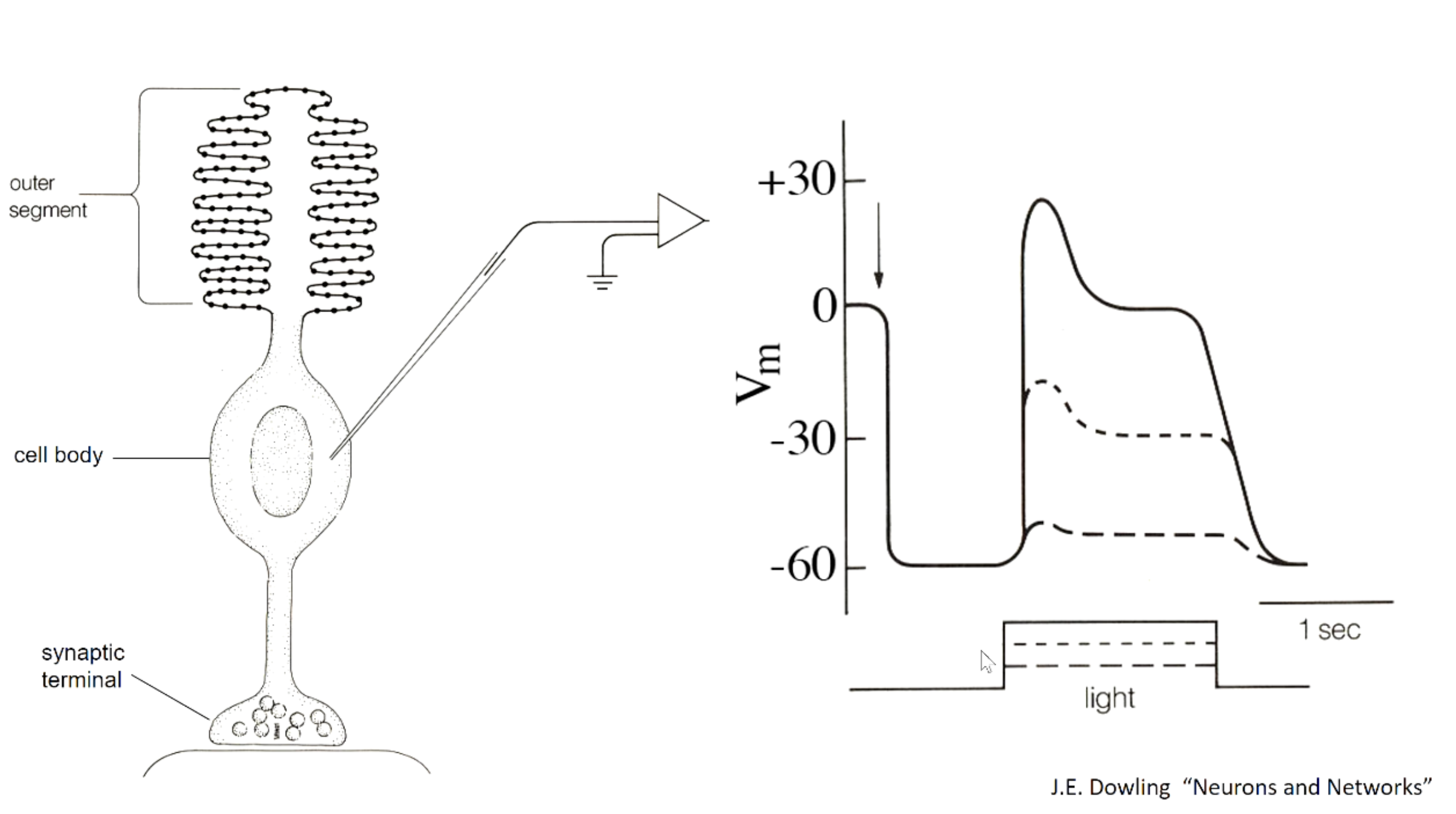
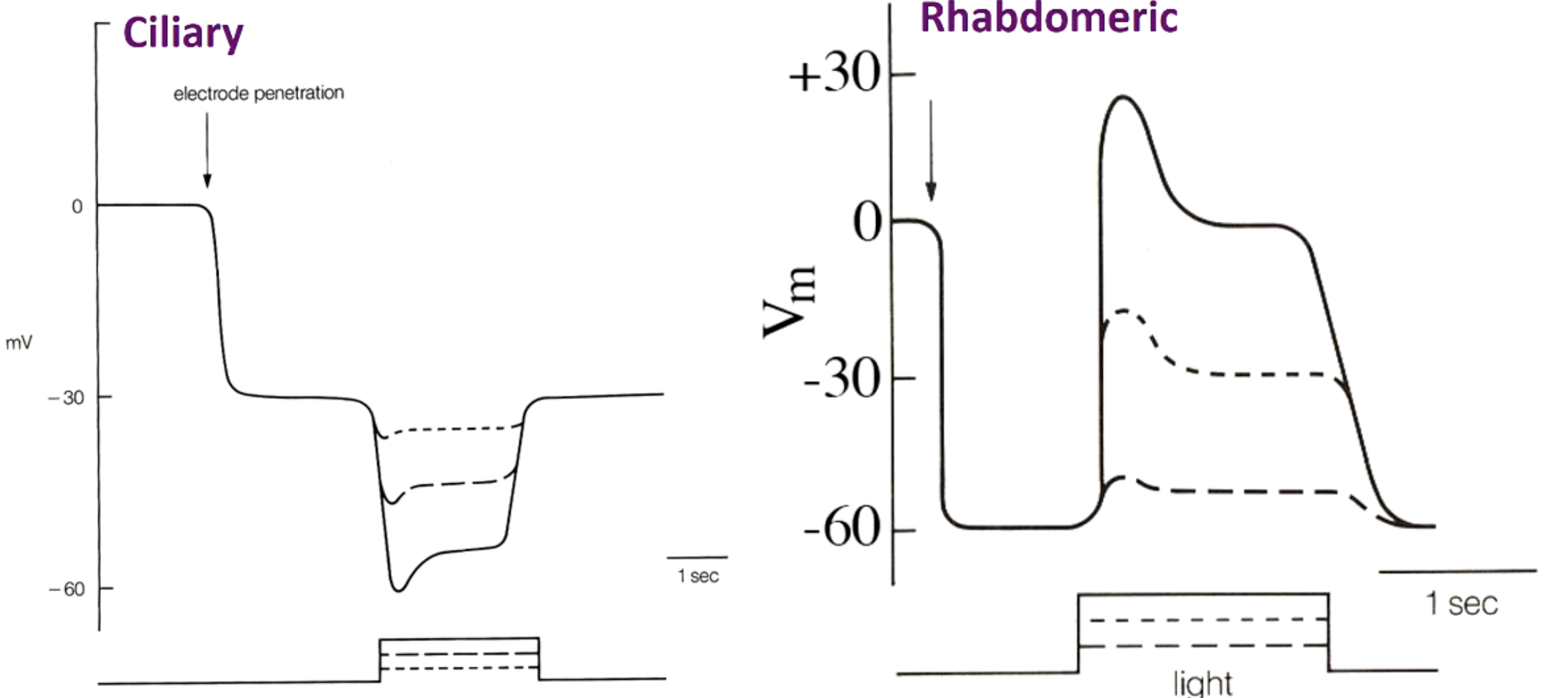
Arrow = electrode probe
RMP = -60 mV
Light stimulation, increases mV
Higher light, higher graded potential
Ciliary Photoreceptors
Ciliated photoreceptors
Cilia membrane folded many times to increase SA
1 outer segment
1 Cilia with Ciliary
Many microvilli in Rhabdomeric
Hyperpolarizes with stronger stimulus
2 types:
Rod
Cone
Rods and Cones (Ciliary)
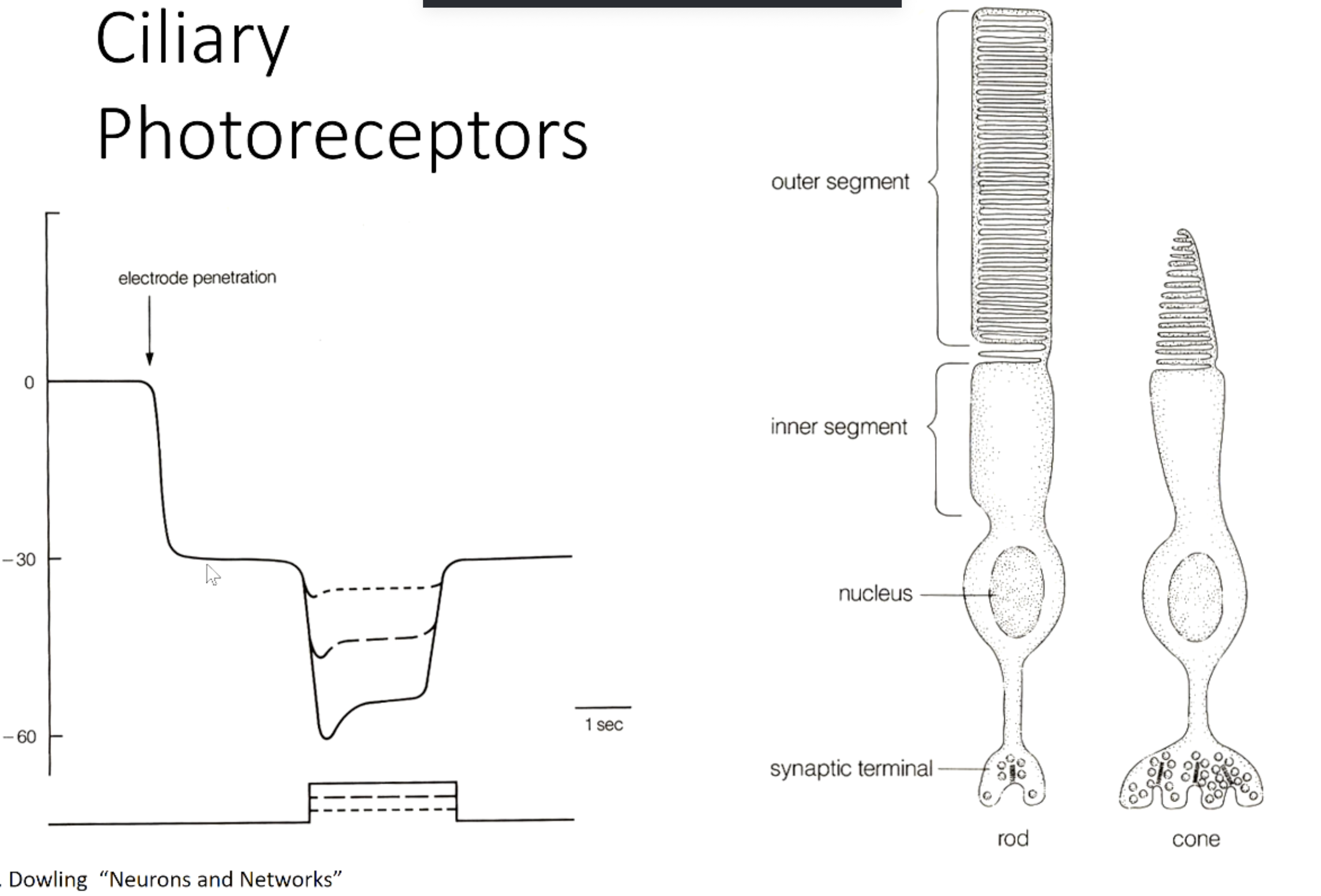
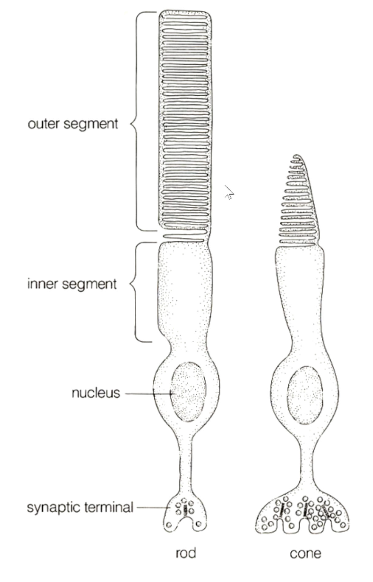
Shape determined by cytoskeleton inside cilia
Discs inside the membranes contain photo pigments = “antennae” for rods and cones
RMP = -30 mV
Light intensity increases, membrane potential becomes more hyperpolarized
Rhabdomeric vs Ciliary
(1) 2 types of Ciliary vs 1 Rhabdomeric
Ciliary can be rods or cones
(2) Extensions
Ciliary = 1 cilia folded
Rhabdomeric = many microvilli
(3) RMP
Ciliary = -30 mV
Rhabdomeric = -60 mV
(3) Stimulus effect
Ciliary = mV becomes more negative
Rhabdomeric = mV becomes more positive
Photoreceptors in Animal kingdom
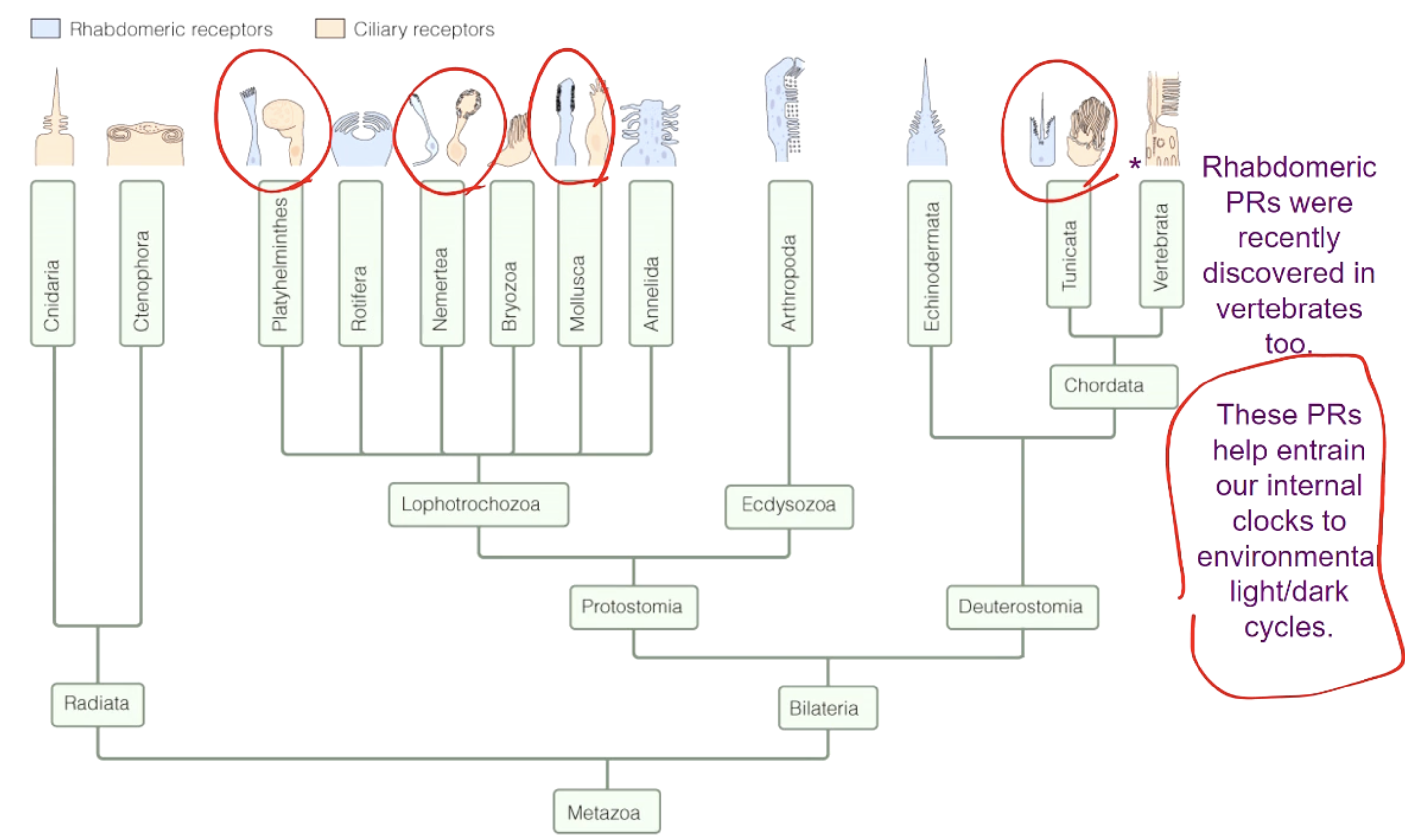
Present in all types of species
Some have 1 or the other, others have both
Can’t tell which animals have which photoreceptors
G-protein receptors - Opsin
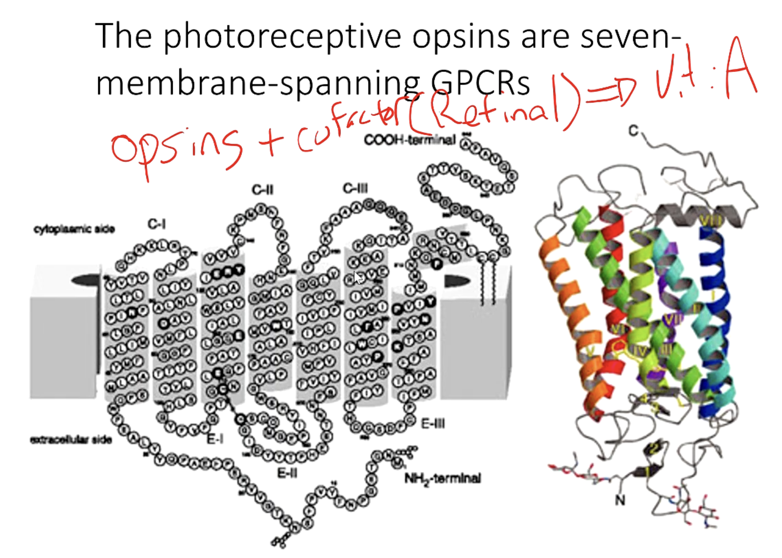
Photoreceptors are coupled by G-protein receptors (GPCRs)
7 transmembrane protein in G-coupled receptors usually
Folded:
Inside = NP amino acids
Outside = P amino acids, causes the folding
Carboxylic terminal - associated with G-protein
Opsin - are receptors / GPCRs
Always associated with a cofactor, usually retinal, which is derived from Vitamin A
Opsins
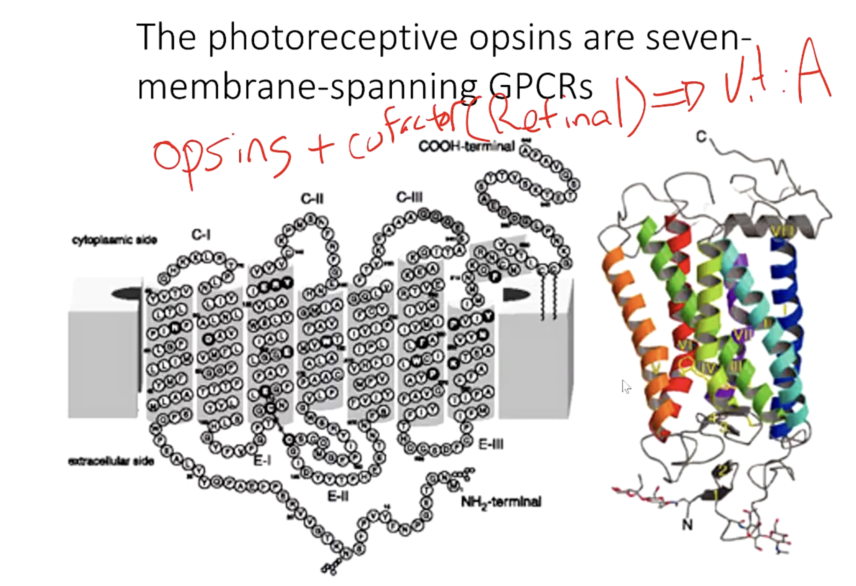
A type of GPCR (7-transmembrane)
Have a cofactor/chromophore/retinal
Opsin is covalently bound to a chromophore (pigments)
Opsins - Chromophore stimulation
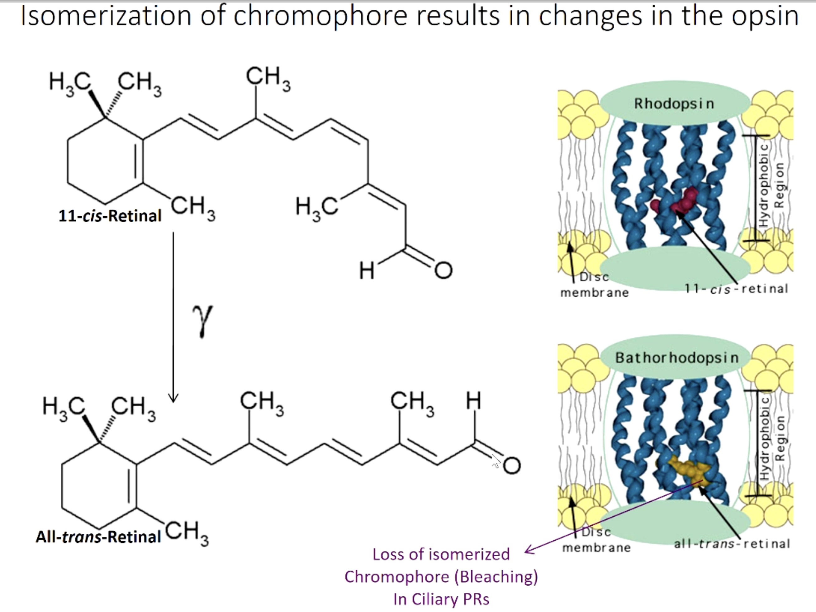
Ex: 11 - Cis - retinal configuration (of the chromophores) = standard for when not stimulated
Cis bond formation is typical for non stimulated / non active
Rhodopsin = 1 Opsin + 1 retinal (chromophore)
Cis retinal bound inside covalently in the Opsin
When light strikes the retinal:
(1) Rhodopsin senses it, isomerize into 11 - All - Trans - retinal
Cis bonds convert into trans bond
(2) Depends on the type of Rhodopsin
(2A) Chromophore becomes trans, leaves opsin, conformational change
(2B) Chromophore becomes trans, conformational change of opsin occurs while still inside Rhodopsin
*Disassociation of chromophore causes the conformation change
When chromophore is no longer with opsin → Bathorhodopsion
Process of retinal leaving opsin = BLEACHING
Means opsin had been stimulated, and chromophore had left it
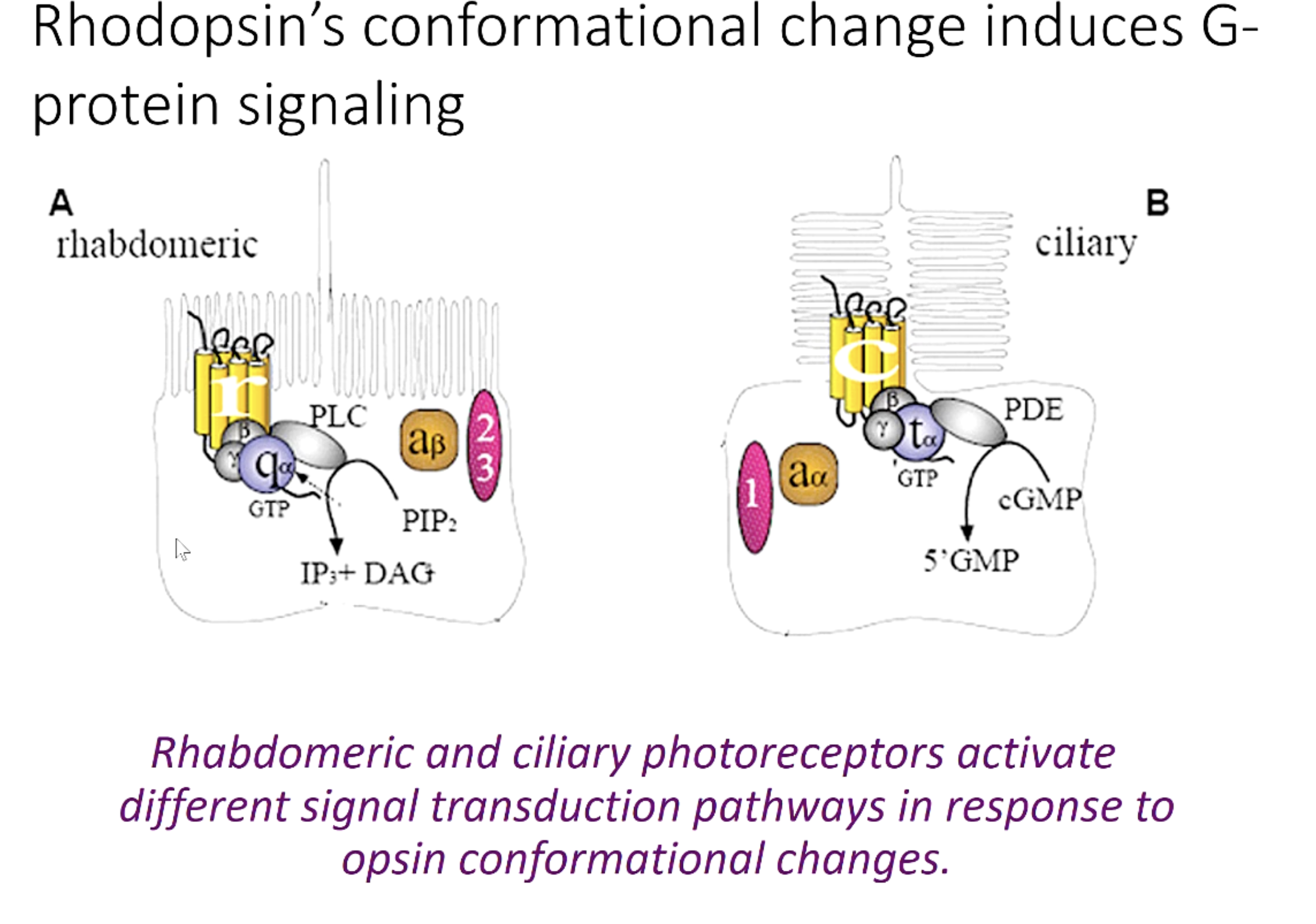
Rhodopsin conformational change → protein signaling
Change in conformation induces G-protein activation
Rhabdomeric and Ciliary have different photon receptors
Rhabdomeric Transduction Pathway
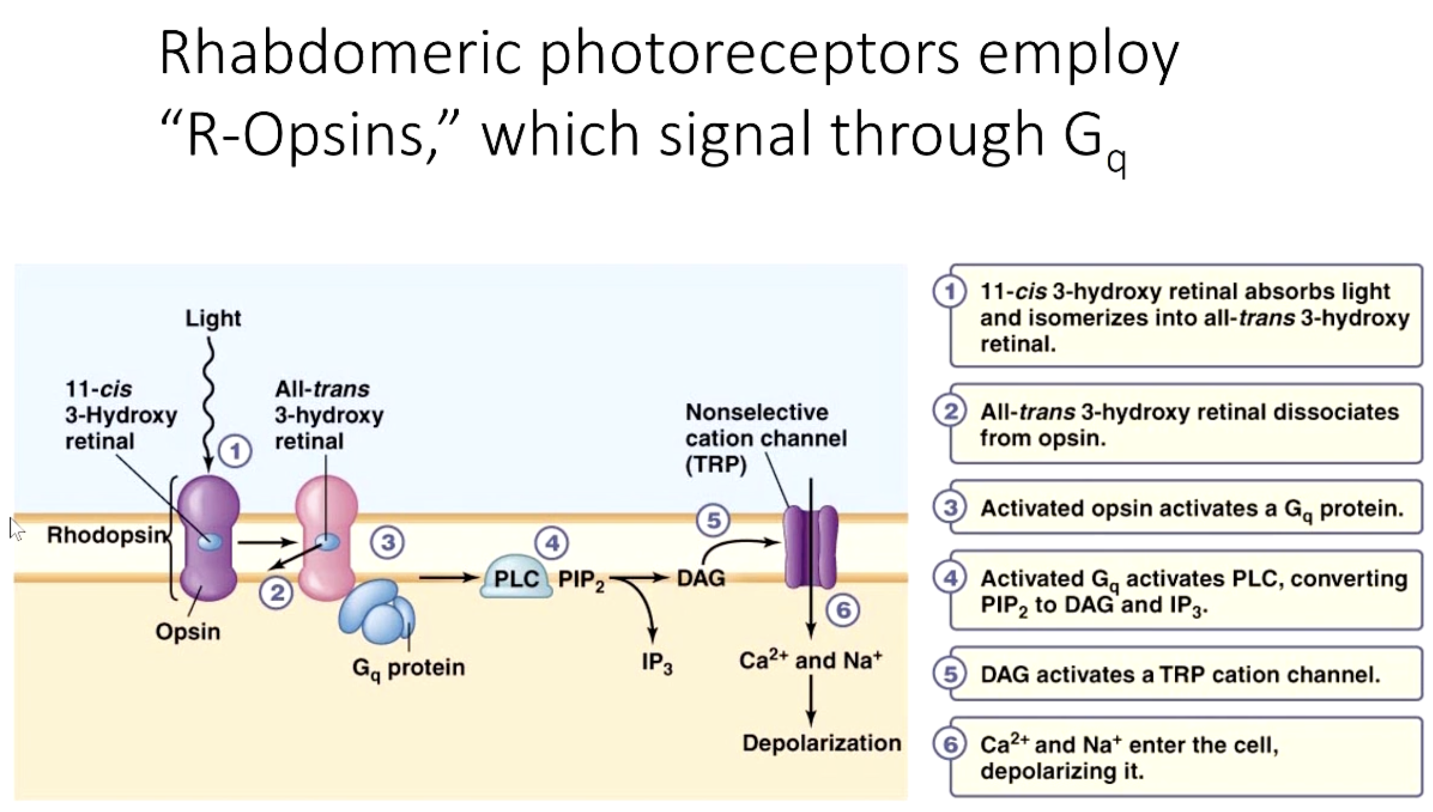
R-Opsins through Gq protein
Steps:
Light sensed and absorbed by cis chromophore, which becomes trans
Trans chromophore dissociates from opsin
**Conformation change of opsin after cis → trans
Rhodopsin → Bathorhodopsin, opsin + trans
Activated Opsin (activated rhodopsin) activates Gq protein
Gq protein activates Photolipase C
PLC converts PIP2 into DAG and IP3
*Similar to NE B1/Gq and Bitter Taste mechanism
DAG activates Nonspecific TRP Cation channel
Ca2+ and Na+ enter cell, depolarizing it
All occurs in Rhabdomeric photoreceptors’ microvilli
Ciliated Receptors Transduction Pathway
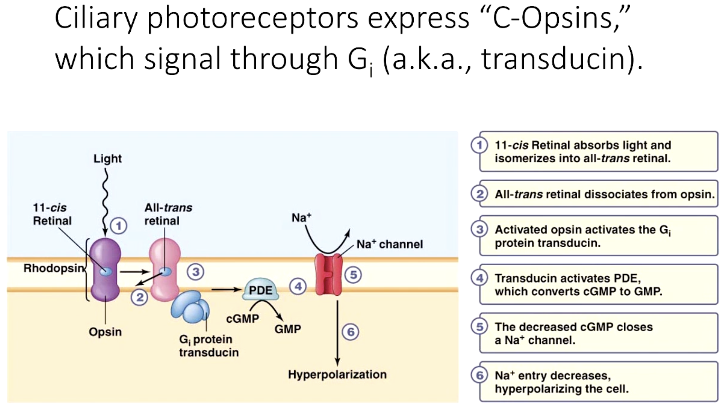
C-Opsin
Signal through Gi protein (transducin)
Steps:
Light sensed and absorbed by cis chromophore, which becomes trans
Trans disassociates from Rhodopsin - bleached
Activated Rhodopsin activates Gi protein / Transducin
Transducin (Gi) activates phosphodiesterase (PDE)
PDE converts cGMP to GMP
Less cGMP closes Na+ channels
Na+ entry decreases, hyperpolarizing the cell
Occurs in cone/rod cilia