Chapter 42 - Animal Circulation and Gas Exchange
1/29
There's no tags or description
Looks like no tags are added yet.
Name | Mastery | Learn | Test | Matching | Spaced | Call with Kai |
|---|
No analytics yet
Send a link to your students to track their progress
30 Terms
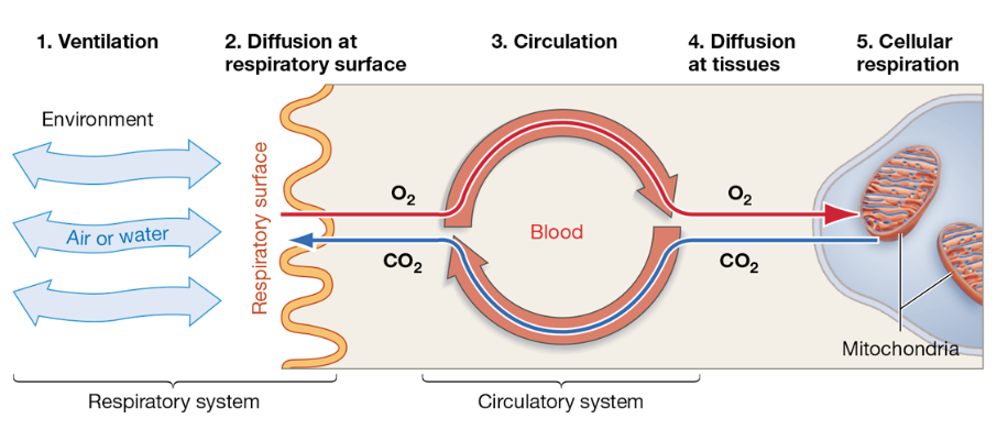
Five Steps of Gas Exchange from Environment → Cells
Ventilation: movement of air or water over a respiratory surface (i.e. lungs or gills)
Diffusion at respiratory surface: O2 moves into blood and CO2 moves out (both down their partial pressure gradients)
Circulation: transports O2 and CO2 throughout the body via the bloodstream
Diffusion at the tissues: O2 leaves the blood and enters cells, CO2 enters the blood from cells.
Cellular respiration: uses O2 to produce ATP (generates CO2 as a waste product)
Functional Link Between Respiratory and Circulatory Systems
Respiratory system: responsible for gas exchange b/w organism and external environment.
Circulatory system: transports oxygen from respiratory surfaces to tissues, returns carbon dioxide back to those surfaces
Continuous blood flow maintains steep partial pressure gradients by removing oxygen and delivering carbon dioxide.
Partial Pressure: Driver of Gas Exchange
The effective availability of gas molecules for diffusion
Gases diffuse based on differences in partial pressure, not overall concentration.
Oxygen and carbon dioxide move from regions of higher partial pressure to lower partial pressure.
At high altitudes, reduced atmospheric pressure lowers oxygen partial pressure, decreasing diffusion into the lungs.
Gas Exchange in Air vs. Water
Air: higher concentration of oxygen and is less dense and viscous than water
Water: less dissolved oxygen, significantly denser → making ventilation cost more energy
Aquatic organisms must move large volumes of water to obtain sufficient oxygen; require highly efficient gas-exchange structures such as gills.
Increasing temp and salinity reduce O2 availability in water, but mixing helps improve
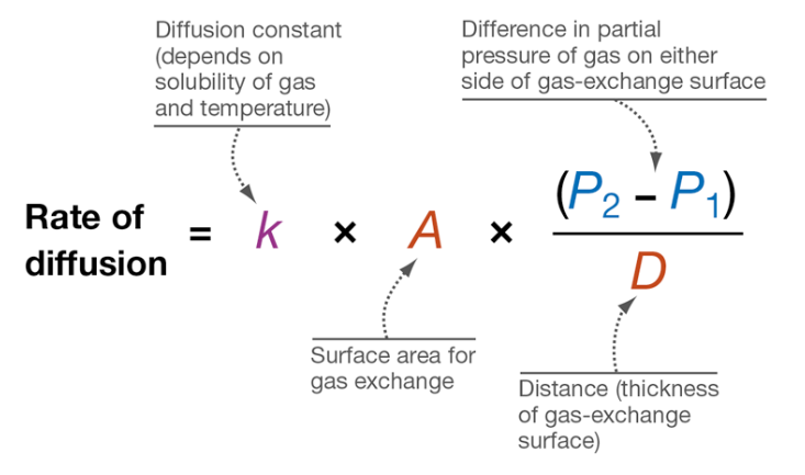
Fick’s Law
Diffusion rate = k × A × (P₂ − P₁) divided by D.
The constant k represents the effects of temperature and gas solubility.
Surface area (A) determines how much area is available for gas exchange. •
The partial pressure gradient (P₂ − P₁) drives diffusion across the surface.
Thickness (D) reduces diffusion rate as it increases, meaning thinner surfaces enhance diffusion.
Fick’s Law Example of High Diffusion Rate
P1 = 5
P2 = 15
A = 2
D = 2
Large partial pressure difference drives diffusion
Although surface area is relatively low, the stronger partial pressure gradient drives diffusion more efficiently
Respiratory Structures: Optimizing Fick’s Law
Gills maximize surface area and maintain gradients through countercurrent exchange
Lungs increase surface area through extensive branching and thin alveolar membranes
Tracheal systems minimize diffusion distance by delivering air directly to cells
Importance of Respiratory Surfaces Being Moist/Thin
Gases must dissolve in water before diffusing (moisture)
Thinness helps minimize diffusion distance
Often increase risk of water loss; many terrestrial animals have internal respiratory structures to reduce dehydration
Gills: Maximizing Oxygen Uptake
Contain numerous filaments and lamellae that greatly increase surface area
Gill epithelium is extremely thin → minimizes diffusion distance
Continuous water flow maintains a strong oxygen gradient across the surface
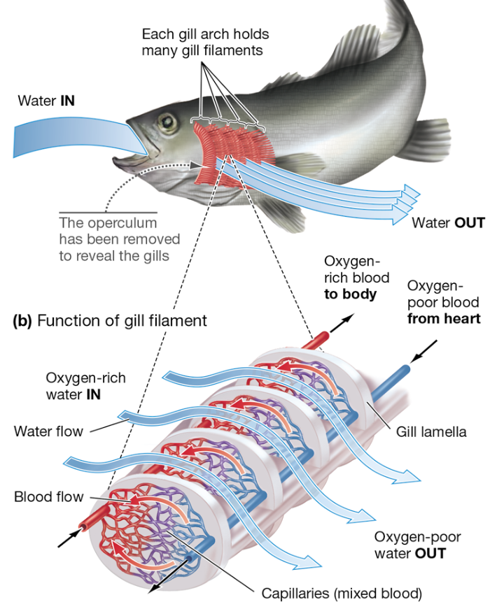
Countercurrent Exchange
Occurs when blood flows in the opposite direction of water across gill surfaces
Helps maintains a consistent partial pressure gradient along the entire exchange surface
O2 diffuses into blood across the entire length of the gill
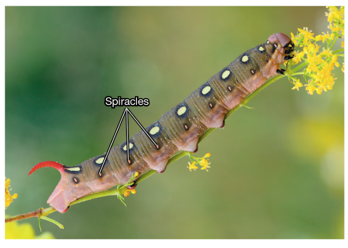
Insect Tracheal System
Tracheae: network of air-filled tubes that open to the exterior through spiracles
Branch extensively into tracheoles that reach individual cells
Oxygen diffuses directly from tracheoles into cells without the need for blood transport
Bypasses the circulatory system
Insects: How Do They Ventilate?
Via rhythmic contractions and relaxations of muscles
Spiracles relax → tracheal volume increase → internal pressure drops → air flows in
Spiracles contract/close → tracheal volume decreases → internal pressure increases → air is forced out
Boyle’s Law (inverse relationship of pressure and volume)
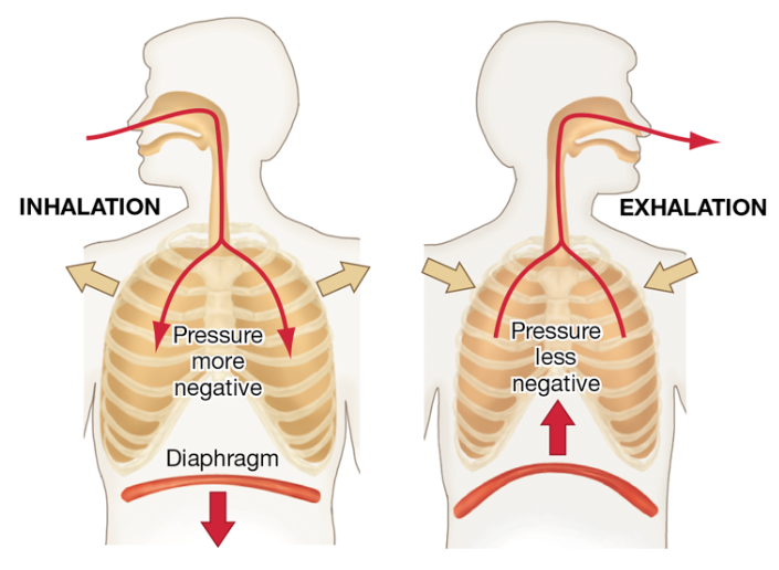
Positive vs. Negative Pressure Ventilation
Positive: forces air into the lungs by increasing pressure in the oral cavity
Seen in amphibians such as frogs
Negative: draws air into the lungs by expanding the chest cavity and lowering internal pressure
Seen in mammals
More efficient and supports higher metabolic demands
Regulation of Breathing Rate
Controlled by the medullary respiratory center in the brain
Primary stimulus for breathing: elevated CO2 levels in blood.
CO2 reacts w/ H2O to make H+, lowering pH
A detectable change that is sensed → increases breathing rate + depth to restore balance
Open vs. Closed Circulatory Systems
Open: allow hemolymph to leave vessels and directly contact tissues
Operate at low pressure
Have limited ability to direct flow
Closed: keep blood confined within vessels
Allows higher pressure and faster flow
Enable precise regulation of blood distribution to tissues.
Insects: Exception to the Open System
Insects have an open circulatory system but do not rely on it for oxygen transport.
Their tracheal system delivers oxygen directly to tissues
This bypasses the limitations of low-pressure hemolymph flow.
Arteries, Capillaries, and Veins
Arteries have thick, elastic walls that allow them to withstand and maintain high pressure as blood leaves the heart.
Capillaries have extremely thin walls that facilitate exchange of gases, nutrients, and wastes.
Veins have thinner walls and contain valves that prevent backflow as blood returns to the heart.
Pathway of Blood to the Heart
Deoxygenated blood enters the right atrium from the body
Blood moves to the right ventricle and is pumped to the lungs.
Oxygenated blood returns to the left atrium.
Blood moves to the left ventricle and is pumped to the body.
Pulmonary vs. Systemic Circuits
The pulmonary circuit carries blood between the heart and the lungs for gas exchange.
The systemic circuit carries blood between the heart and body
This separation allows efficient oxygenation and delivery
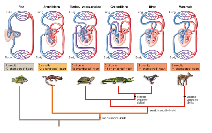
Evolution of Heart Chambers
Fish: two chambers and a single circuit, limiting pressure and flow
Amphibians and reptiles: three chambers and two circuits, allowing some mixing of blood.
Birds, crocodilians, and mammals: four chambers and two circuits, fully separating oxygenated and deoxygenated blood.
Increased chamber number improves oxygen delivery and supports higher metabolism.
Systole vs Diastole
Systole: cardiac cycle phase where muscle contracts → pumps blood out of the chambers.
Diastole: cardiac cycle phase where muscle relaxes and the chambers fill with blood.
Blood Pressure
Determined by cardiac output (HR x stroke volume) and resistance within blood vessels.
Resistance: influenced by vessel diameter (smaller = more resistance)
BP decreases as blood moves through capillary beds b/c of increased cross-sectional area.
Regulated by baroreceptors located in major arteries/heart
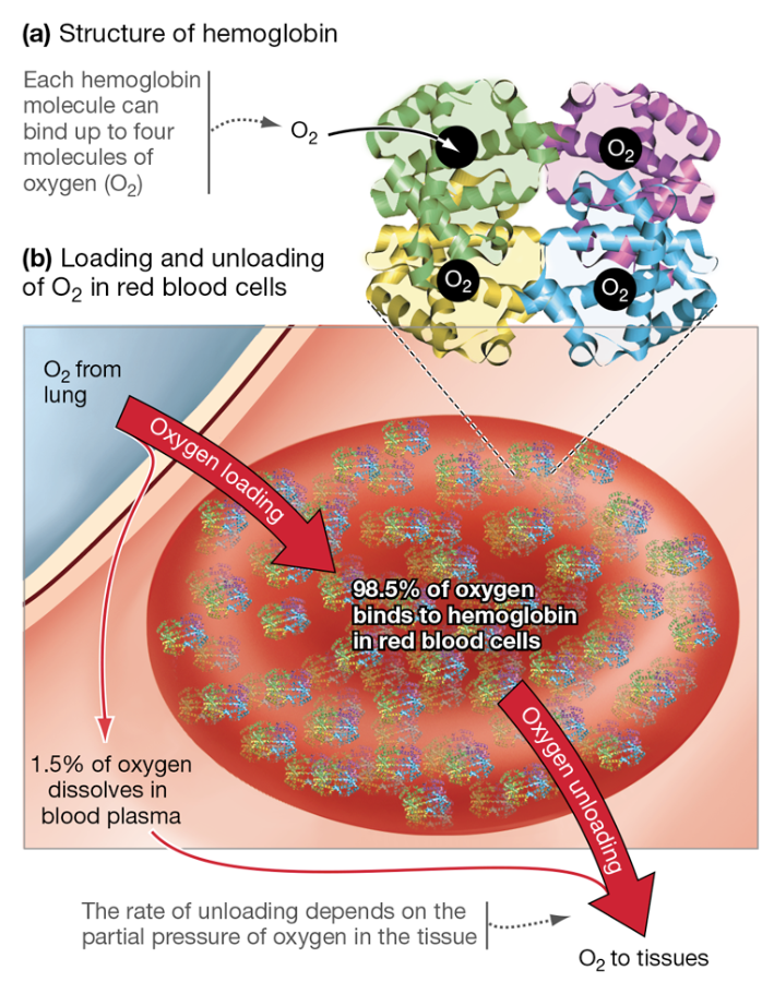
Structure + Function of Hemoglobin
Protein found in RBCs that consists of four polypeptide chains
Each chain contains a heme group with an iron ion that can bind one oxygen molecule
Each hemoglobin can carry 4 O2 molecules
Significantly increases the oxygen-carrying capacity of the blood
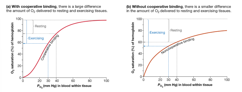
Cooperative Binding
Binding of one oxygen molecule increases the affinity of hemoglobin for additional oxygen molecules
Causes conformational changes in the hemoglobin protein after each oxygen binds
So, O2 binding becomes progressively easier
Produces a sigmoidal oxygen dissociation curve
Cooperative Binding Importance
Allows hemoglobin to load oxygen efficiently in the lungs where oxygen partial pressure is high
It also allows hemoglobin to release oxygen efficiently in tissues where partial pressure is lower.
Small changes in oxygen partial pressure result in large changes in hemoglobin saturation.
This makes oxygen delivery highly responsive to tissue demand.
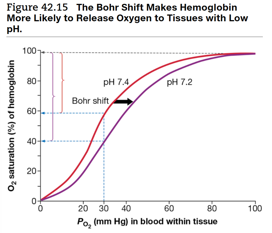
Bohr Shift
Occurs when hemoglobin’s affinity for oxygen decreases which promotes oxygen release.
Caused by increased CO2 levels, decreased pH, and increased temperature.
The shift ensures that more oxygen is delivered to tissues that need it most.
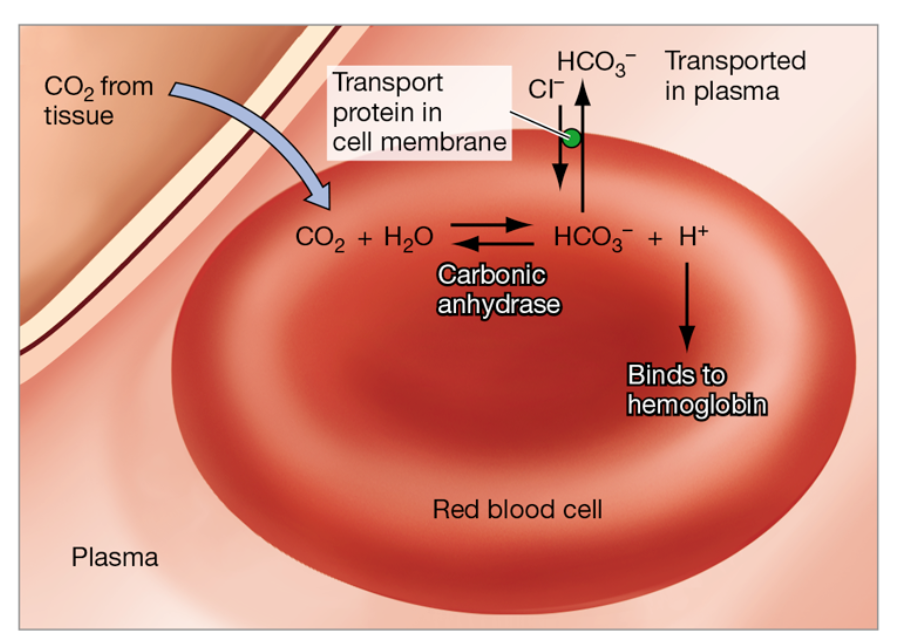
What Happens to CO2 in the Blood?
Most CO2 diffuses into RBCs and is converted into bicarbonate ions (HCO3-) by carbonic anhydrase.
This reaction also produces hydrogen ions which bind to hemoglobin
Converting carbon dioxide into bicarbonate helps maintain a diffusion gradient for continued CO₂ uptake
CO2 in the Lungs
Bicarbonate ions and hydrogen ions recombine to form carbon dioxide and water
Carbon dioxide then diffuses from blood → alveoli → expelled during exhalation
As CO2 leaves blood → pH rises → hemoglobin affinity for oxygen increases
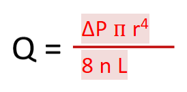
Hagen-Poiseuille
Blood flow increases with a greater pressure difference between the ends of a vessel.
Flow increases dramatically with increases in vessel radius because radius is raised to the fourth power.
*Most influential factor
Small changes in radius can have large effects on flow rate
Blood flow decreases with increased viscosity and increased vessel length.
Endotherms: Requiring More Efficient Systems
Endotherms maintain body temperature through metabolic heat production, which requires high energy expenditure.
This increased metabolic rate creates a greater demand for oxygen and removal of carbon dioxide.
As a result, endotherms require highly efficient respiratory and circulatory systems.
These systems typically include large surface areas for gas exchange and high pressure circulation.