Mycology and Parasitology Morphology
 Call Kai
 Learn
Learn Practice Test
 Spaced Repetition
 Match
Match Flashcards
 Knowt Play
1/46

There's no tags or description
Looks like no tags are added yet.
Name | Mastery | Learn | Test | Matching | Spaced | Call with Kai |
|---|
No analytics yet
Send a link to your students to track their progress
47 Terms
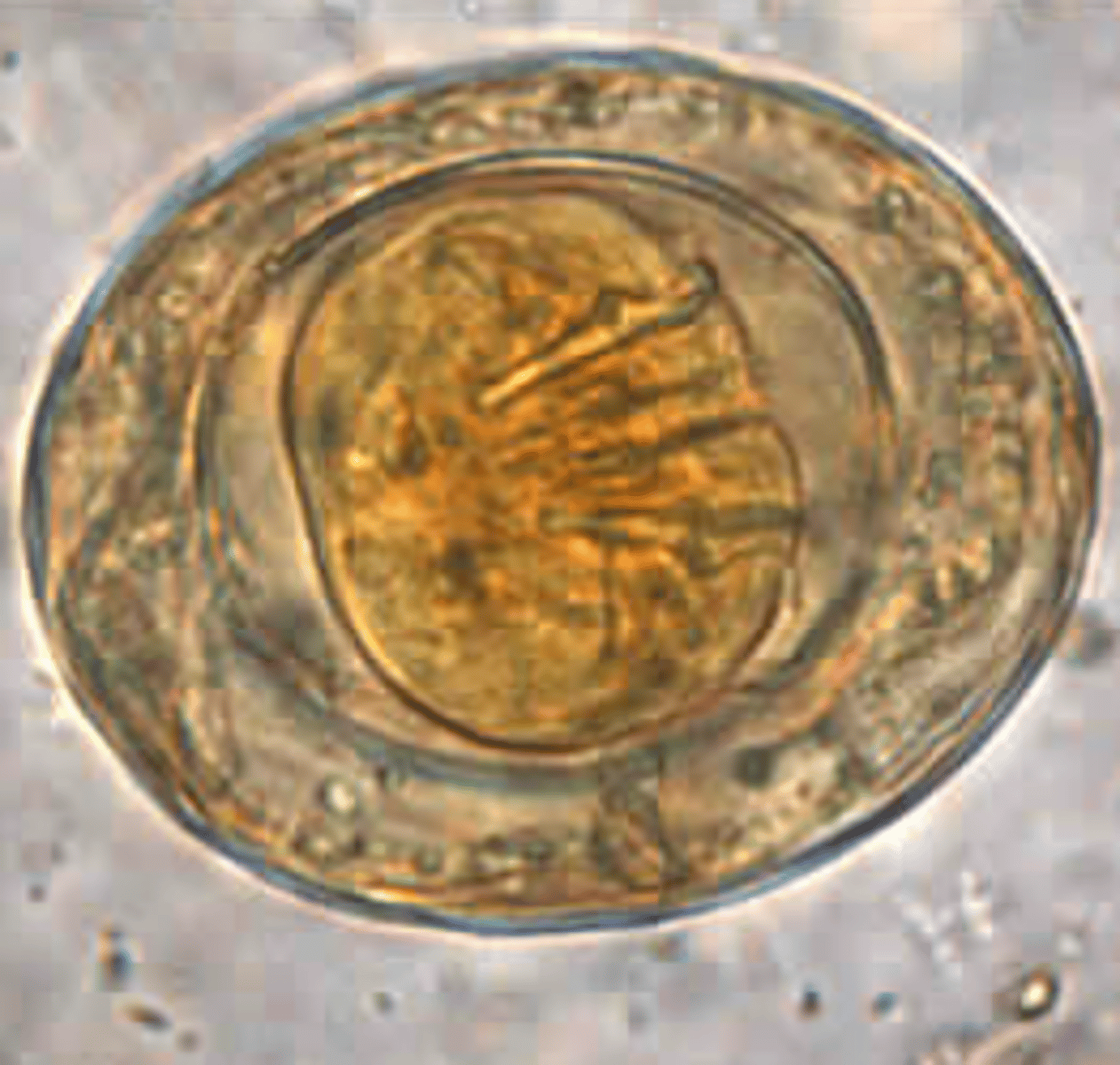
Malassezia furfur; Yeast
Causes tinea versicolor
• Requires lipid source
(sterile olive oil)
• Yeast with "spaghetti and
meatballs" morphology
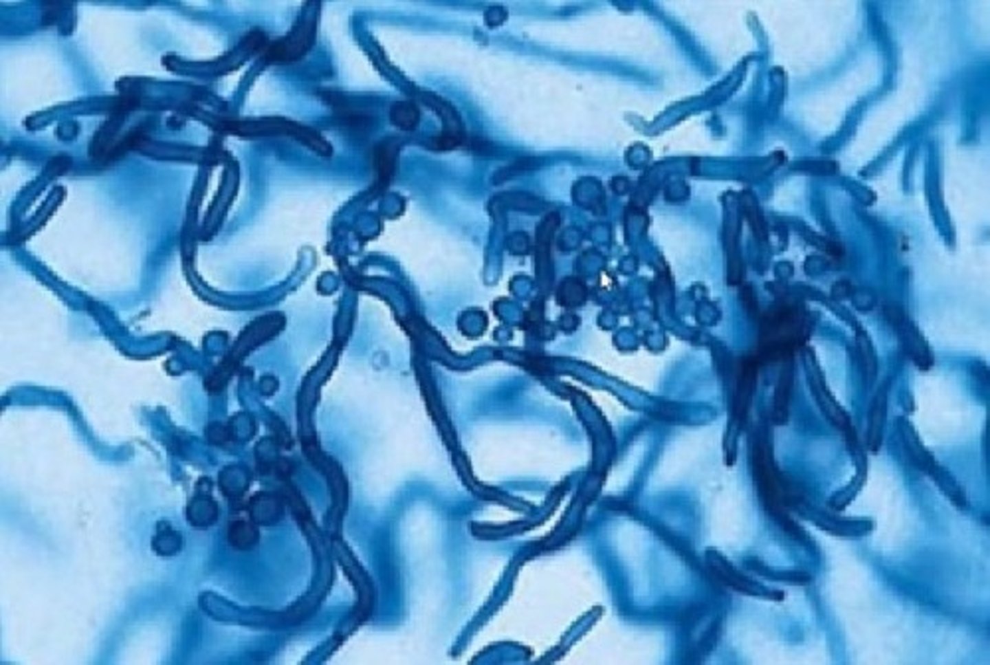
Trichophyton rubrum (dermatophyte)
Skin and nail infections
(most common worldwide)
• Colony surface is
white/buff
• Colony bottom is red
(rubrum)
• Tear shaped microconidia
"birds on a wire"
• Negative for hair shaft
penetration
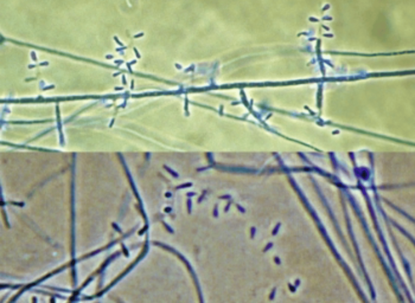
Trichophyton mentagrophytes (dermatophyte)
Looks very similar to T.
rubrum
• Positive for hair perforation
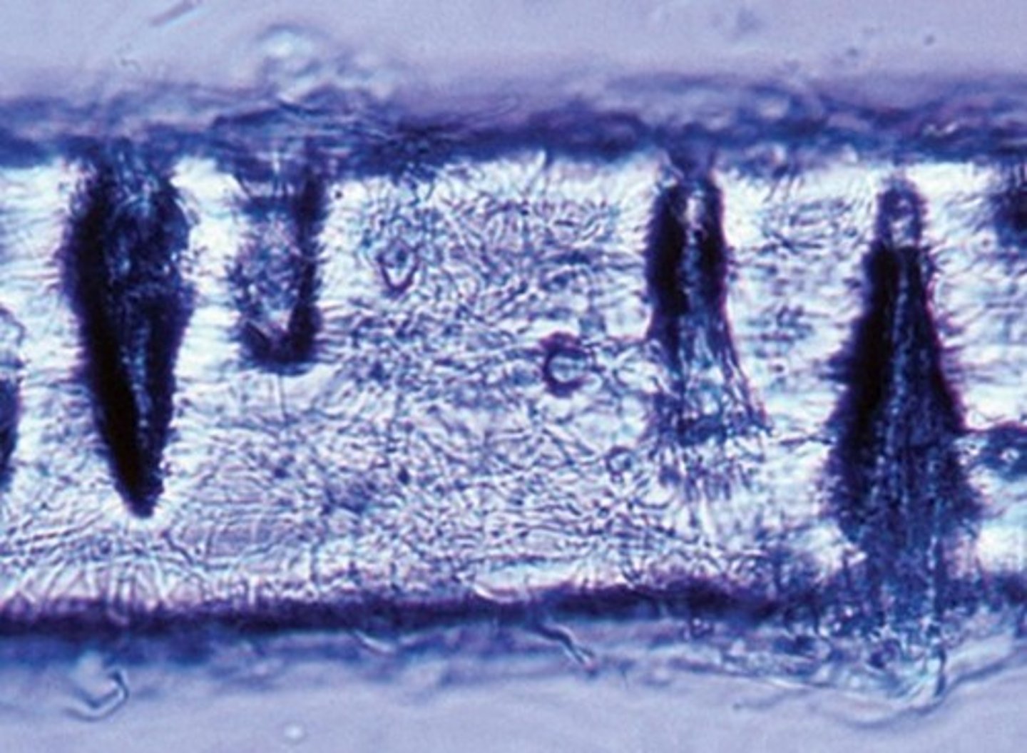
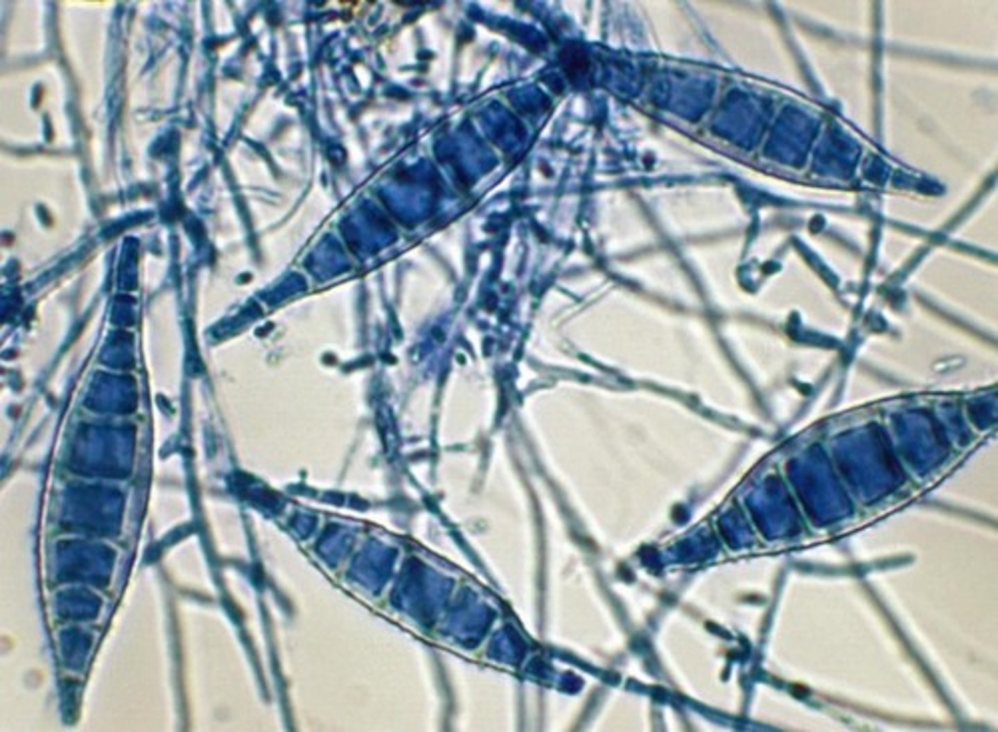
Microsporum canis (dermatophyte)
• Skin and scalp infections
usually from pets
• Colony surface is white
with yellow edges
• Spindly, rough
macroconidia with >6
cells
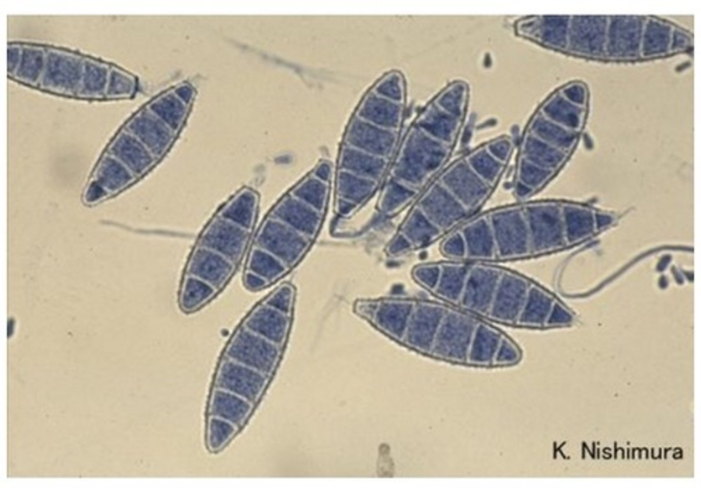
Microsporum gypseum (dermatophyte)
• Scalp and skin infections,
common in animals
• Oval, rough walled
macroconidia with <6 cells
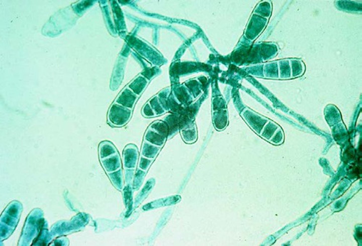
Epidermophyton floccosum (dermatophyte)
• Skin and nail infections
• Colony is yellow, olive-gray,
or khaki
• Reverse of colony is orange-
brown
• Smooth club shaped
macroconidia with rounded
ends, often found in
clusters
o 2-6 cells
• No microconidia
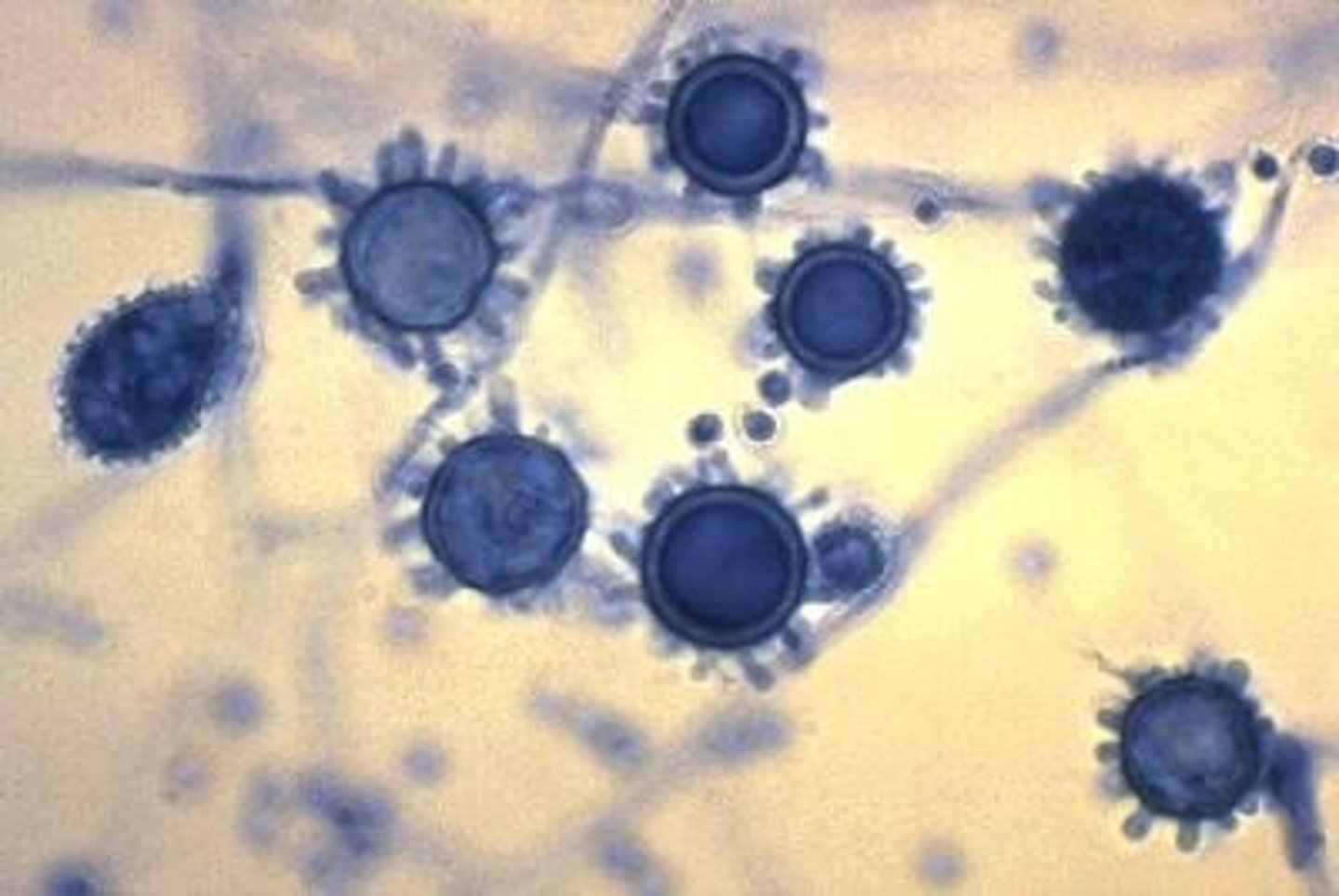
Histoplasma capsulatum
• Small, narrow yeast in tissues
(easy to confuse with other
pathogens)
• Septate, hyaline hyphae with
spiny macroconidia
Blastomyces dermatitidis
• Broad based budding yeast (BBB)
often with double contours
• Mold form is not very distinct
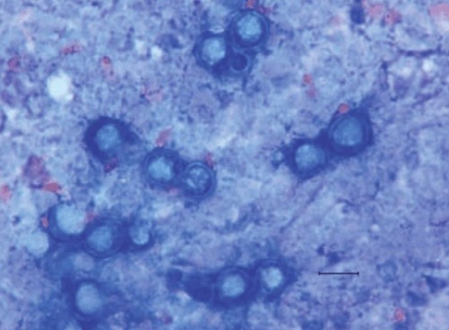
Coccidioides immitis
Spherule form at 37 C
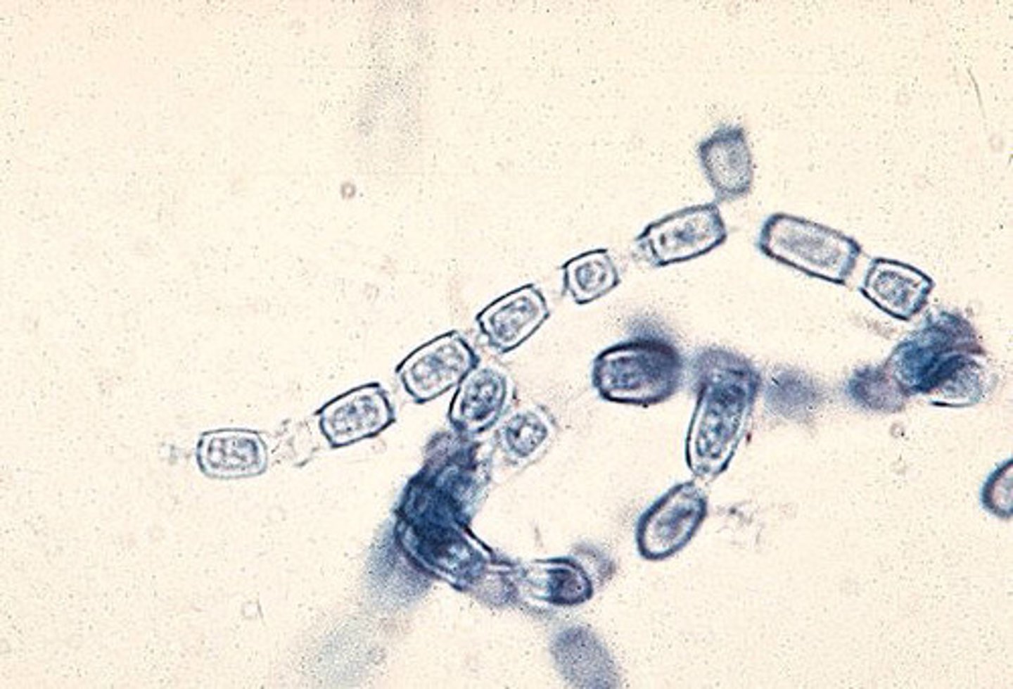
• Mold form
o Hyaline, septate with "Train
track" arthroconidia
Paracoccidioides brasiliensis
Yeast: large, round "mariner's
wheel" with circumferential
budding
o Don't mix this up with H.
capsulatum's mold form
macroconidia
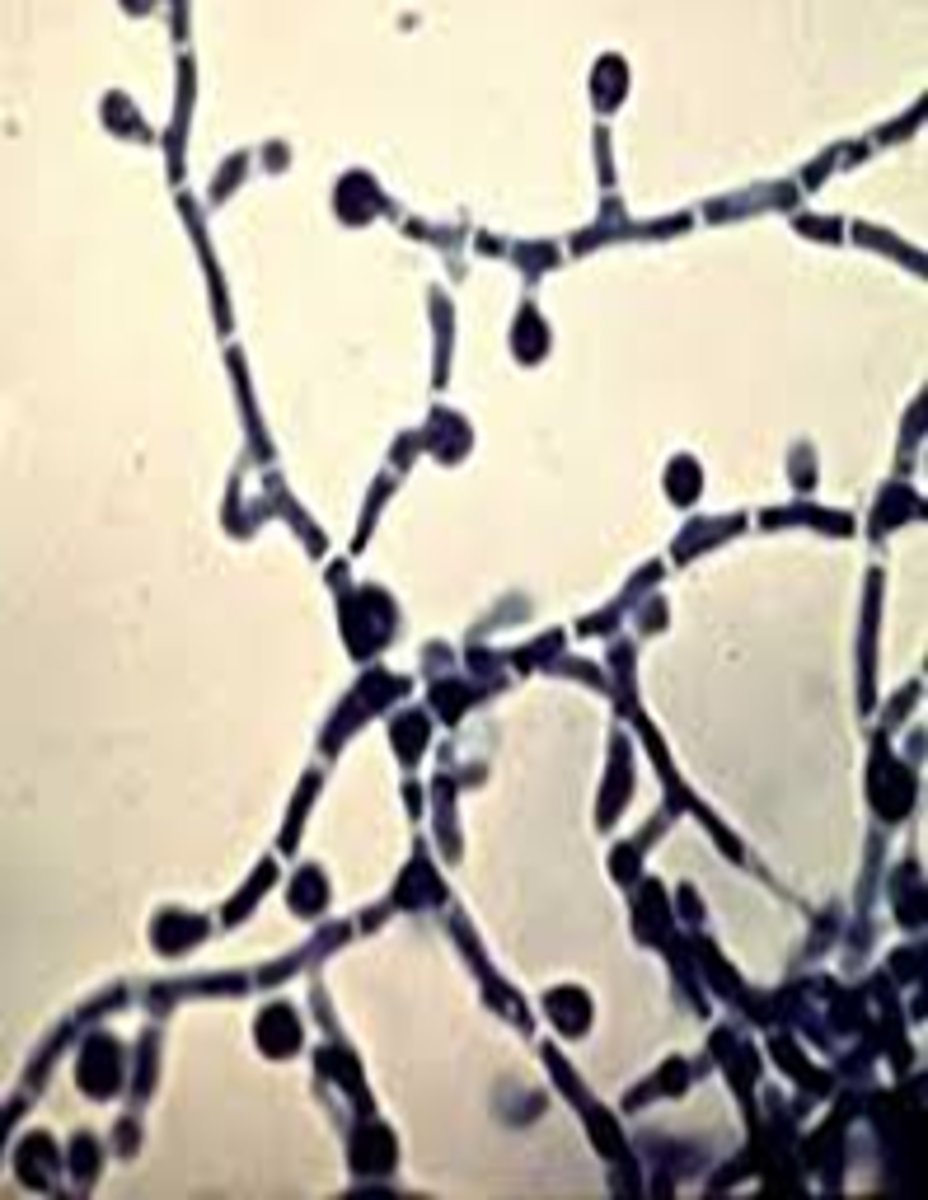
• Mold: septate, hyaline,
terminal and intercalary
chlamydospores
Sporothrix schenckii
• Causes "rose gardener's disease"
• Yeast: cigar shaped, narrow based
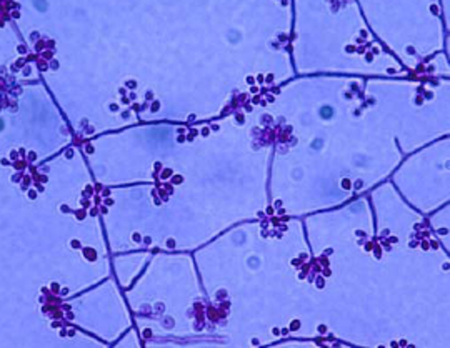
• Mold: hyaline, septate hyphae with
"rosettes" of microconidia
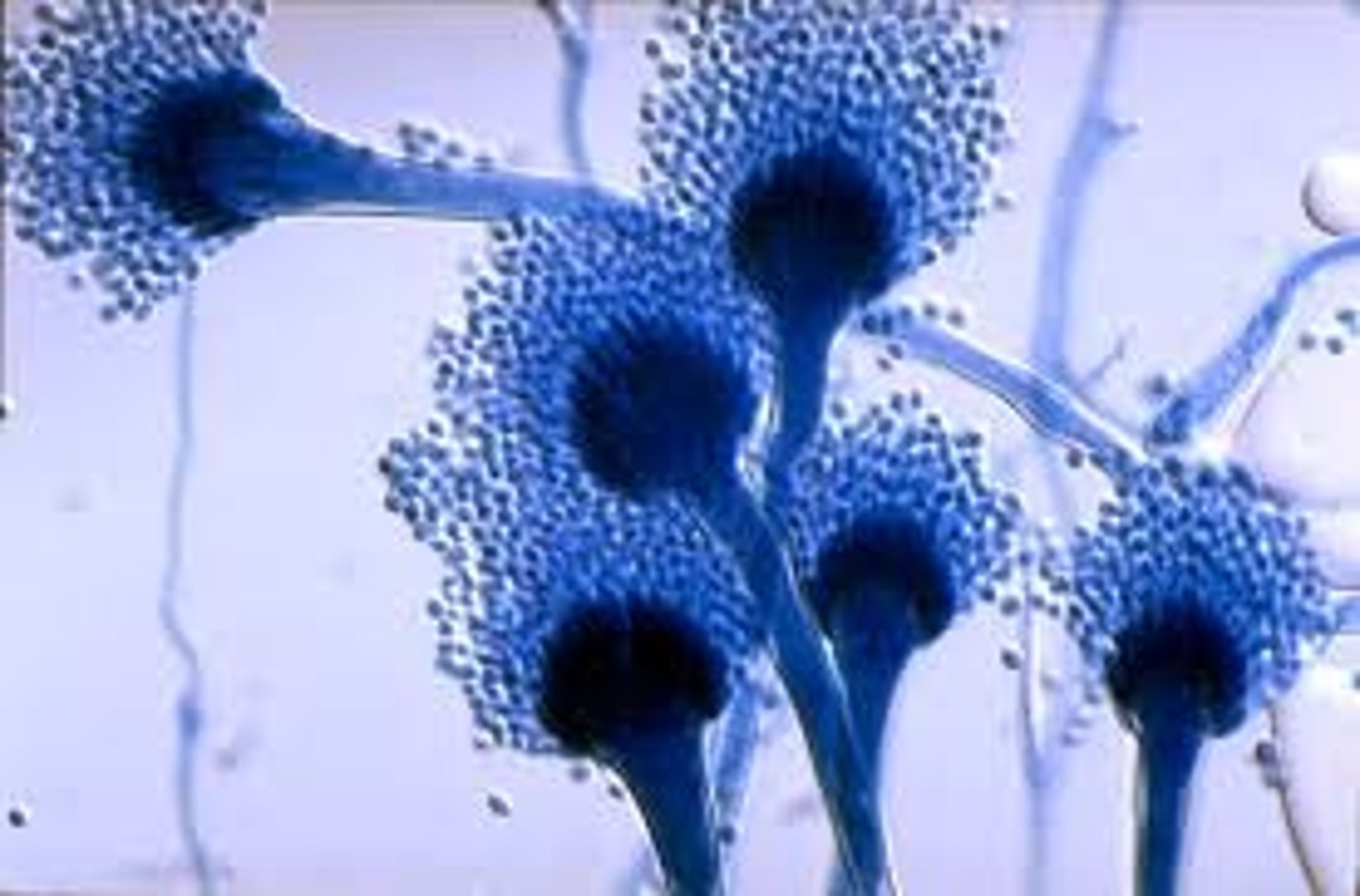
Aspergillus fumigatus
• Blue-green colonies
o White apron and reverse
• Single row of phialides
covering top 2/3 of vesicle
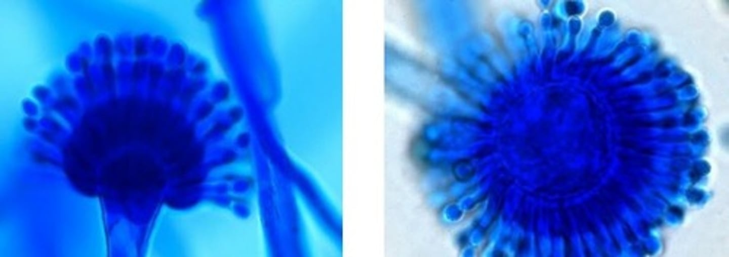
Aspergillus flavus
• Yellow-green or olive colonies
o Light reverse side
• Circumferential phialides
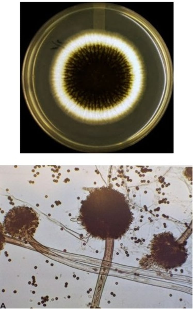
Aspergillus niger
• Dark brown/black colonies
o Light reverse side
• Vesicle has two rows of
phialides covering the whole
surface
• Black conidia
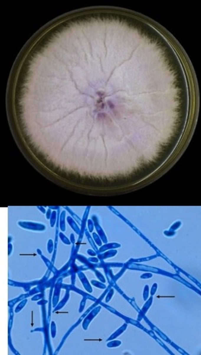
Fusarium spp.
• Violet colonies
• "Canoe" or "banana"
microconidia
Acremonium spp.
• Gray, white, or pink
colonies
• Long, narrow,
unbranched phialides
with clusters of
microconidia
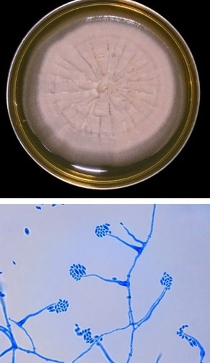
Penicillium spp.
• Usually a
contaminant, but can
cause infections
• Blue-green powdery
colonies with white
border
• "Skeleton hand"
phialide and conidia
arrangements
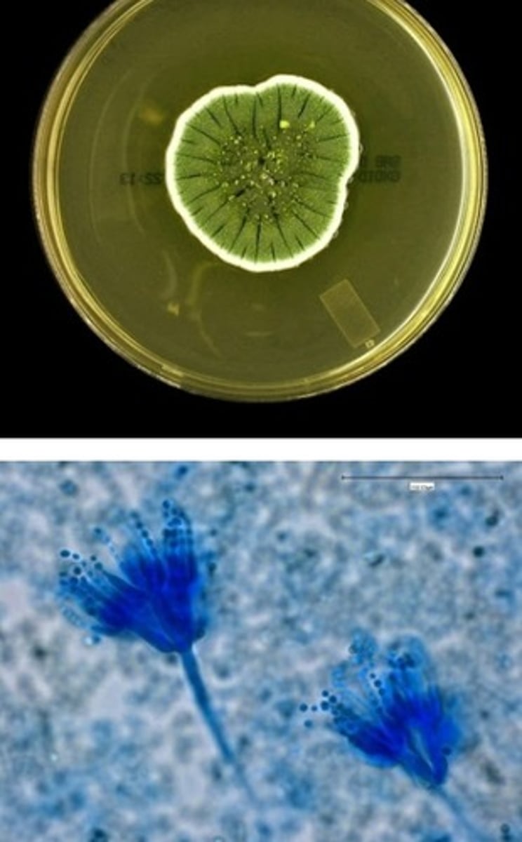
Scorpulariopsis spp.
• Contaminants but can cause
infection
• Powdery brown/tan colonies
• Rough "lemon drop" conidia
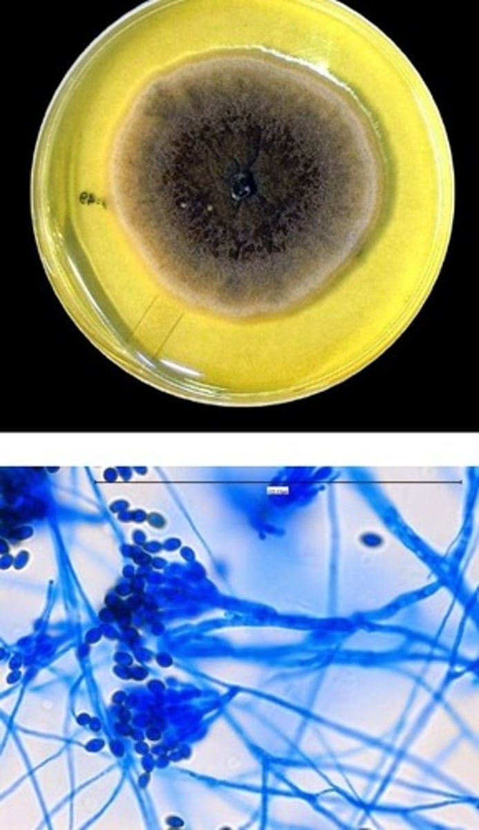
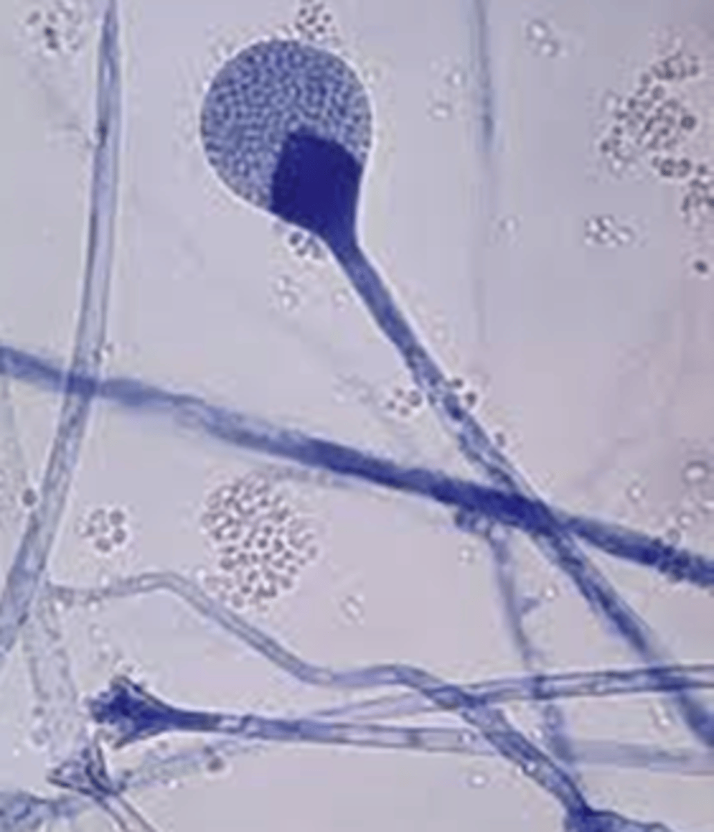
Rhizopus spp.
• Rhizoids and
unbranched
sporangiophores
• Sporangiophore = sac of
spores
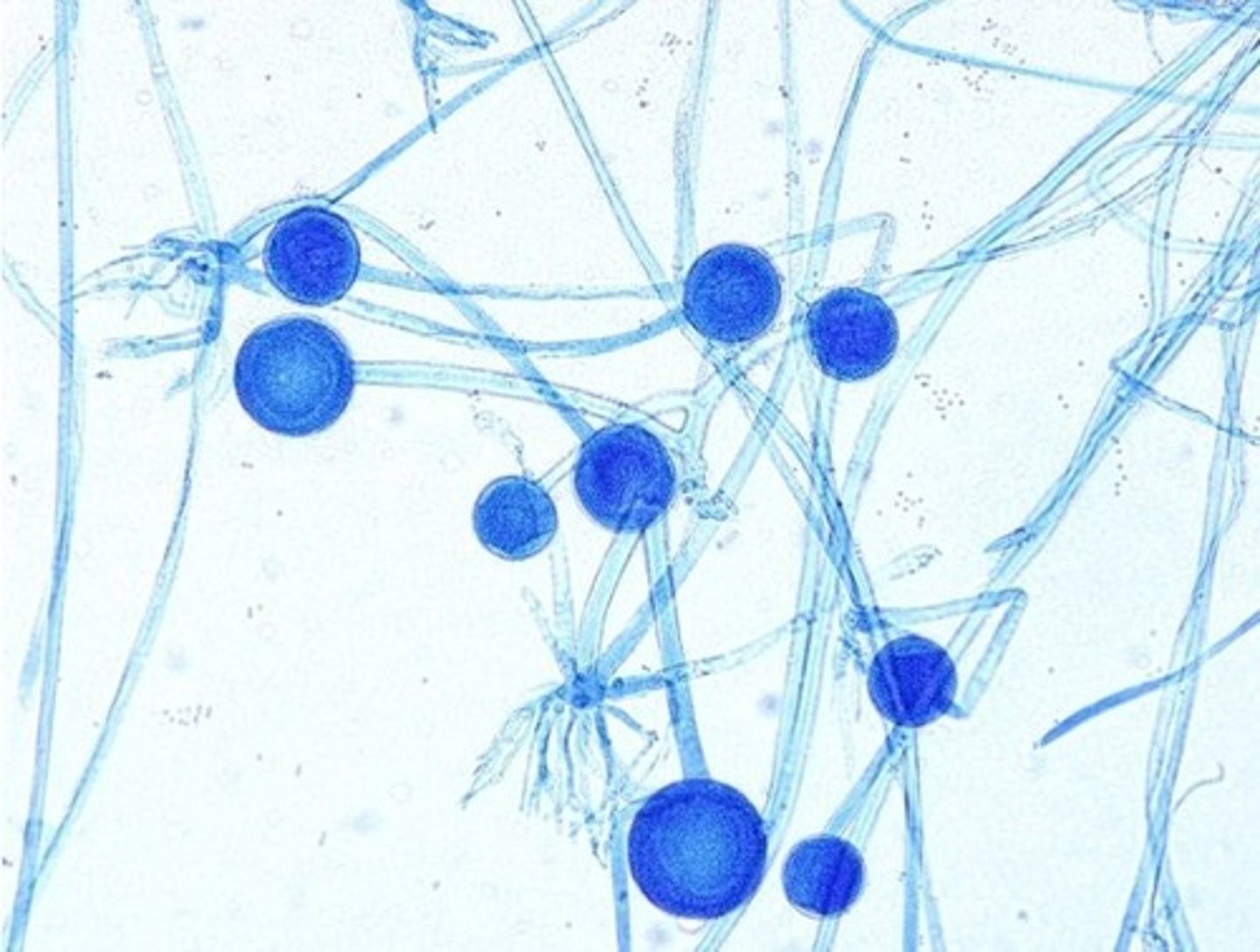
Mucor spp.
No rhizoids
• Sporangia fall apart
easily
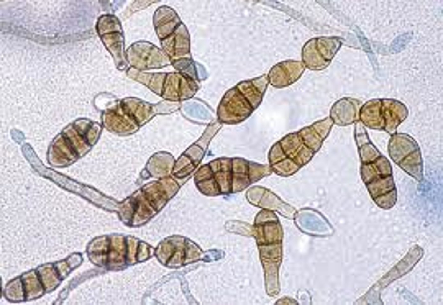
Alternaria (dematiaceous mold)
• Brownish black colonies
• Chains of club-shped conidia
with transverse and longitudinal
separations
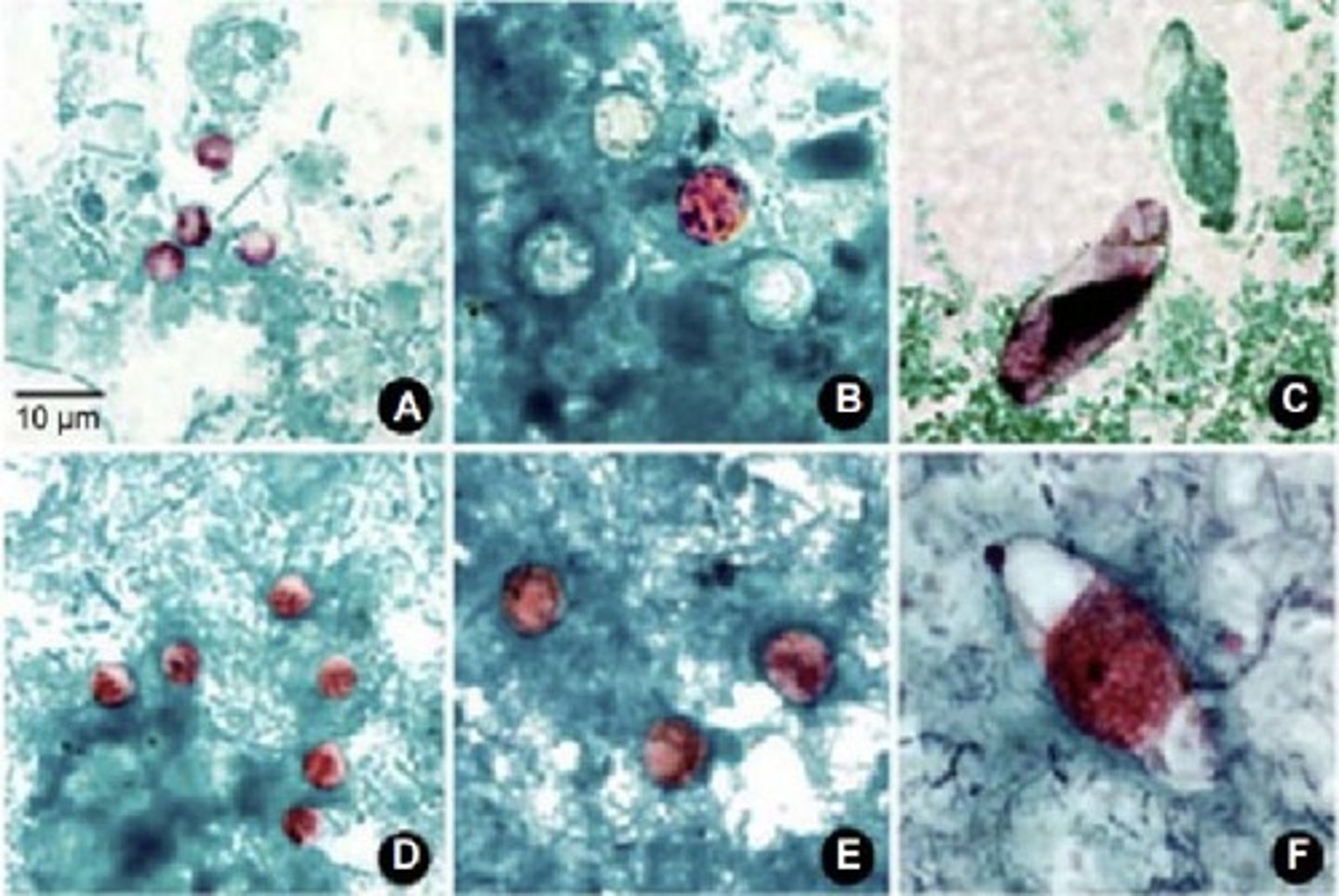
Coccidia
• Stool samples
• Modified acid fast
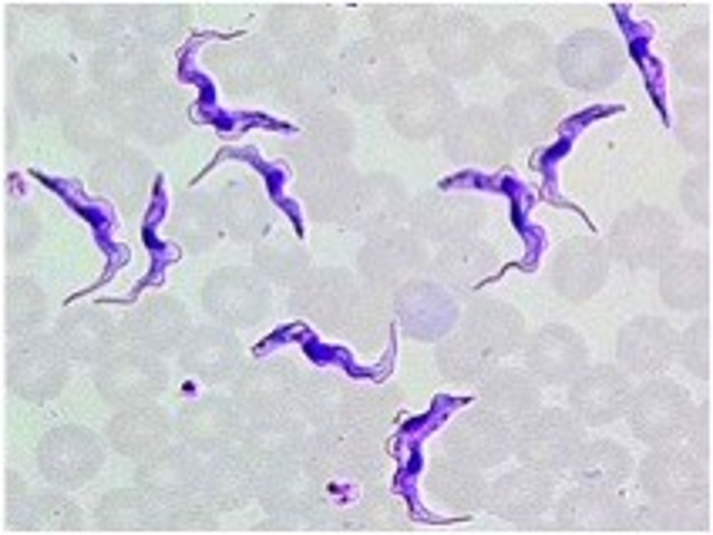
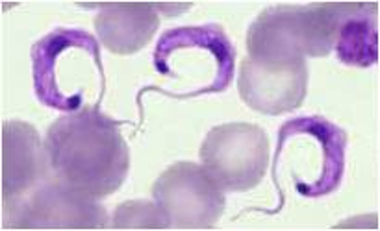
Trypanosoma brucei
• Larger organism
• S- or C-shaped
• Small posterior kinetoplast
Trypanosoma cruzi
• Smaller
• C-shaped
• Large posterior
kinetoplast
Ted cruz
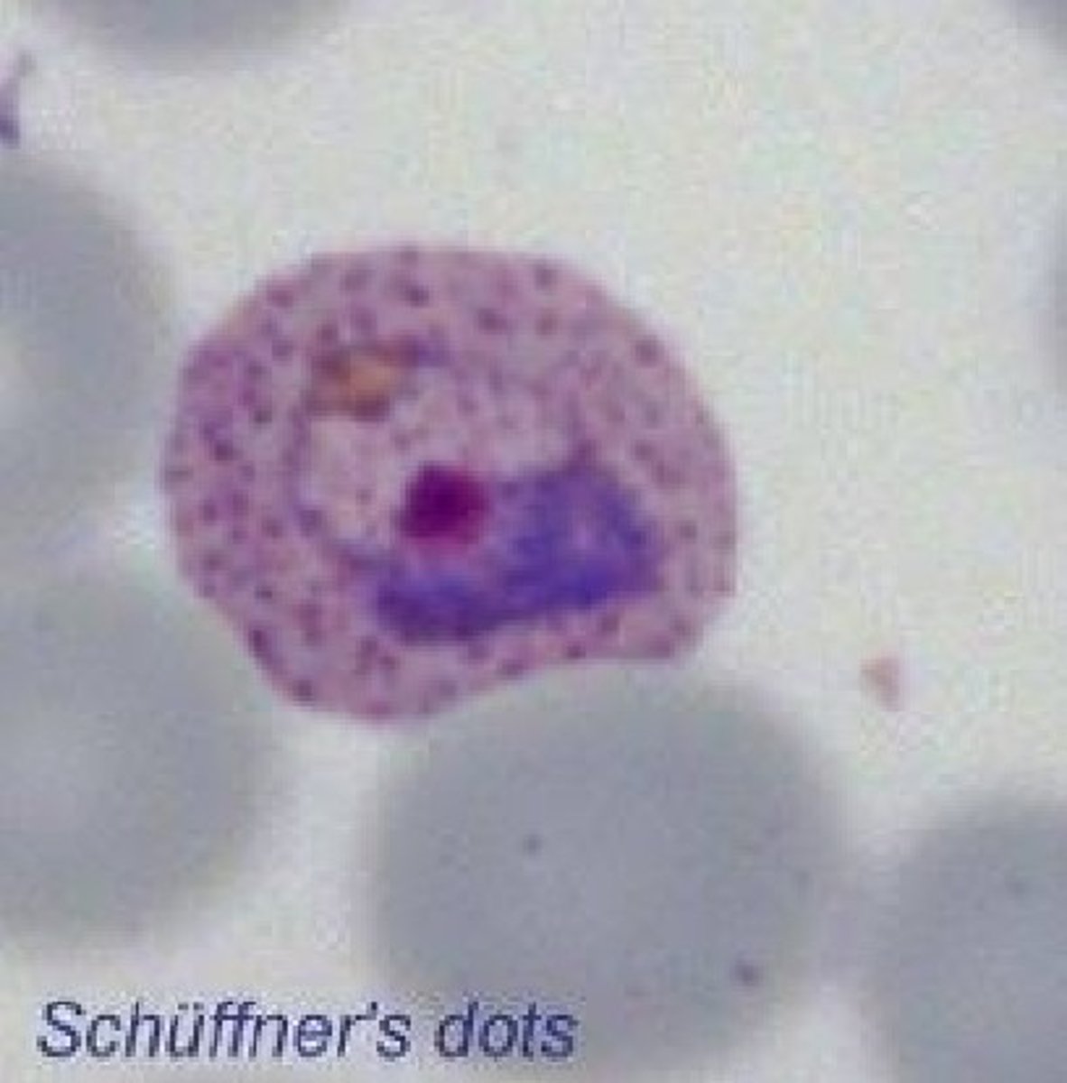
Plasmodium vivax
• Retics infected (large RBCs)
• Ring forms >1/3 size of RBC
• Mature troph is ameboid, fills RBC
• Schizont has 12-24 merozoites
• Gametocytes are large and oval
• Schuffner's dots
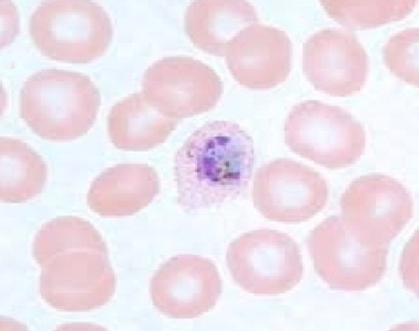
Plasmodium ovale
• Retics infected (large RBCs)
• Ring forms >1/3 size of RBC
• Mature troph more compact than P. vivax "
o "Comet forms"
• Schizont has 6-14 merozoites
• Gametocytes are large and oval
• Schuffner's dots
Plasmodium malariae
• Infects senescent RBCs (small to
normal)
• Ring form 1/3 size of RBC, thick
o Bird's eye
• Mature trophs = band and basket
forms
o Do not fill RBC
o Prominent pigment
• Schizont has 6-12 merozoites
o Rosette forms
• Gametocyte can fill RBC
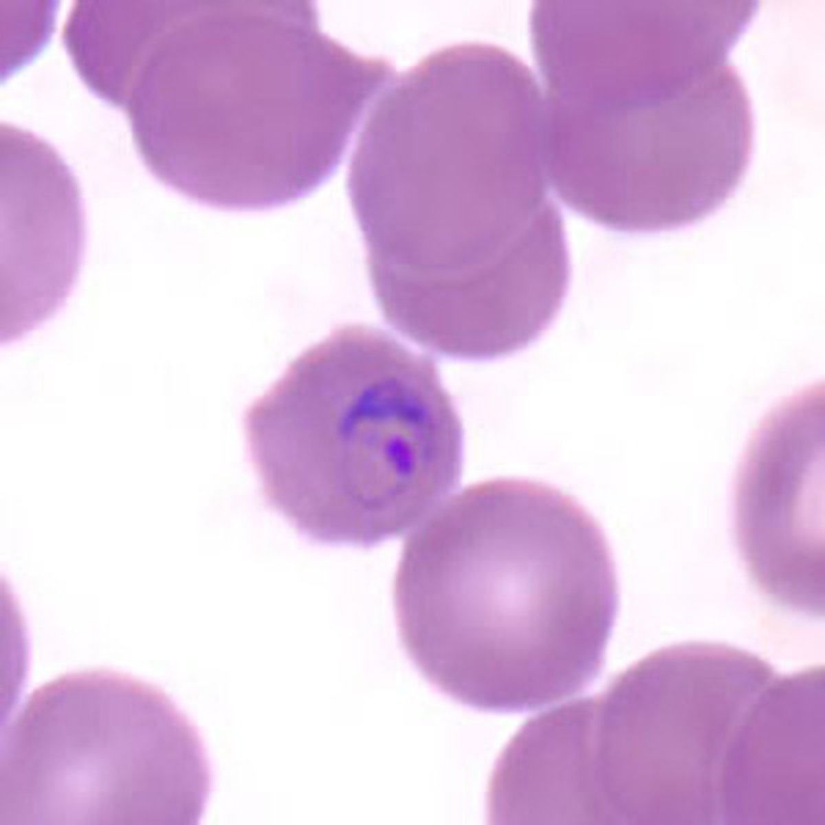
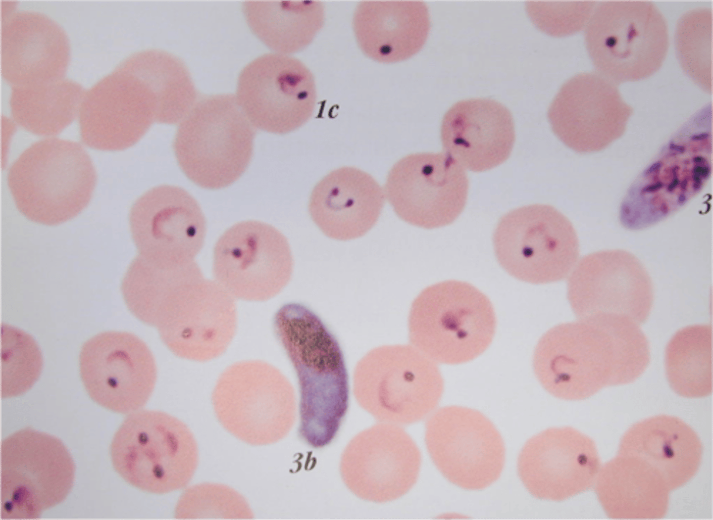
Plasmodium falciparum
Only ring forms and gametocytes seen
in blood
• Infects all sizes of RBC
• Small, delicate ring forms
o Applique forms
o Multiple infections
o Multiple chromatin dots
• Gametocyte = banana shaped
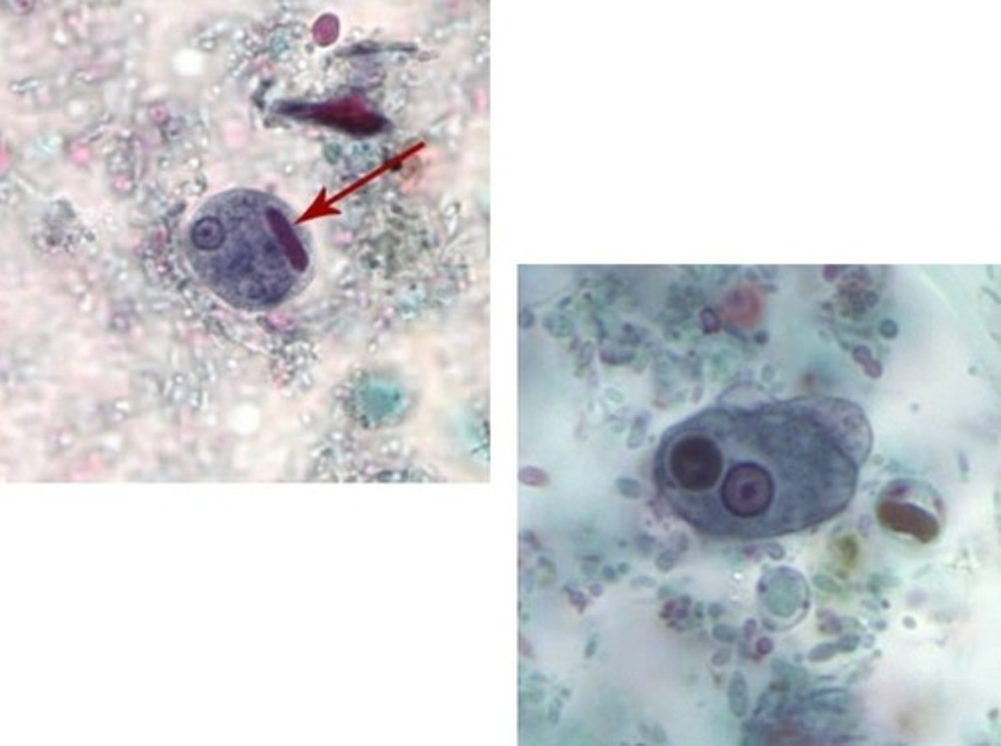
Entamoeba histolytica
• Cyst (left)
o Up to 4 nuclei
o Chromatoidal body with
smooth ends
• Trophozoite (right)
o 15-20 microns
o Small, central karyosome
and fine peripheral
chromatin
o May see
erythrophagocytosis
(diagnostic)
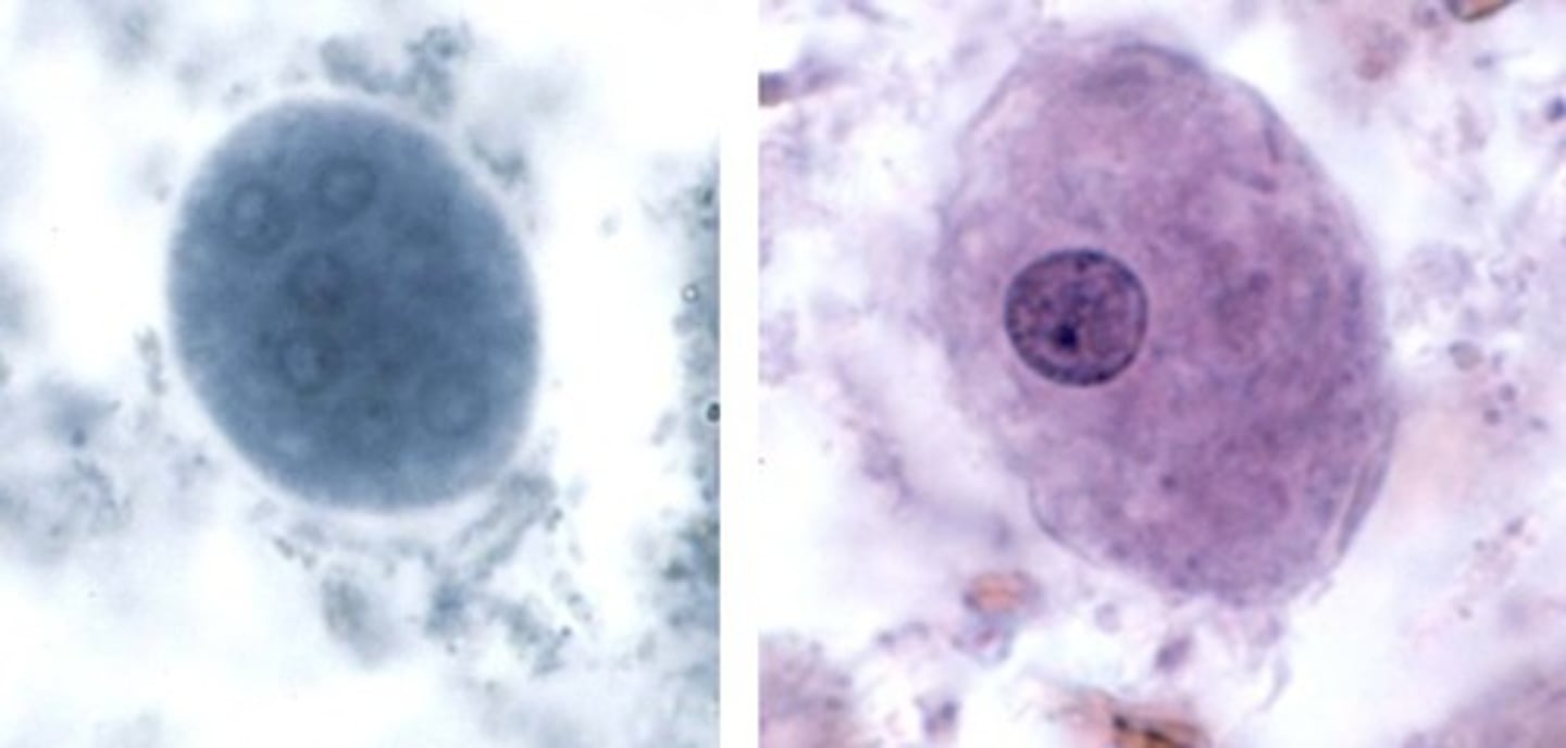
Entamoeba coli
• Cyst (left)
o Up to 8 nuclei
• Trophozoite (right)
o 20-25 microns
o Eccentric karyosome
o Clumpy peripheral
chromatin
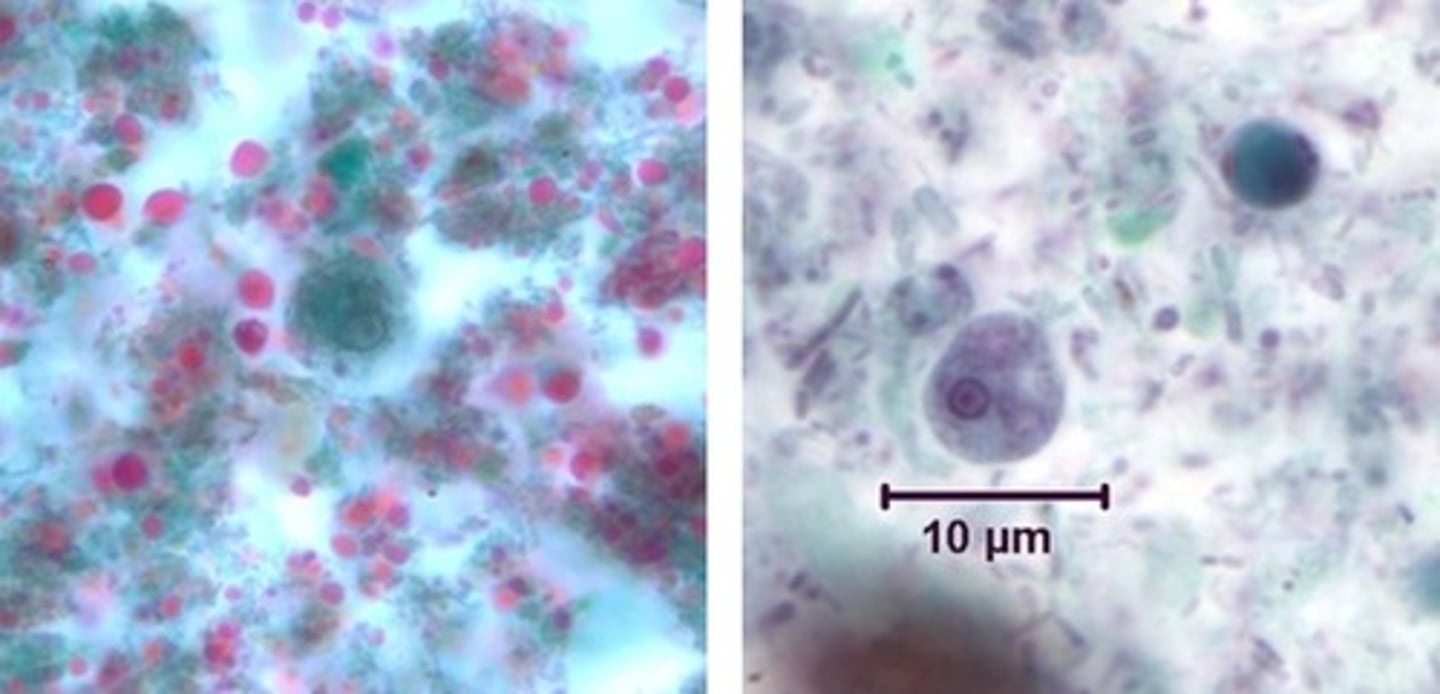
Entamoeba hartmanni
• Cyst (left)
o Looks like
aminiature E.hist
olytica
o Up to 4 nuclei
• Trophozoite (right)
o 5-10 microns
o Looks like a
miniature E.
histolytica
o No ingested
RBCs
Endolimax nana
• Cyst (left)
o No peripheral chromatin
o 4 nuclei with karyosomes
• Trophozoite (right)
o 6-10 microns
o Hard to distinguish from
Iodamoeba buetschlii
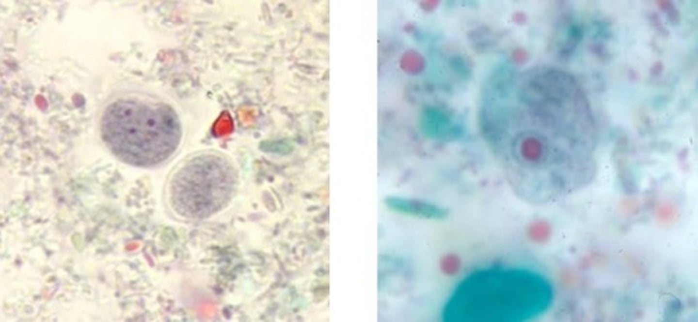
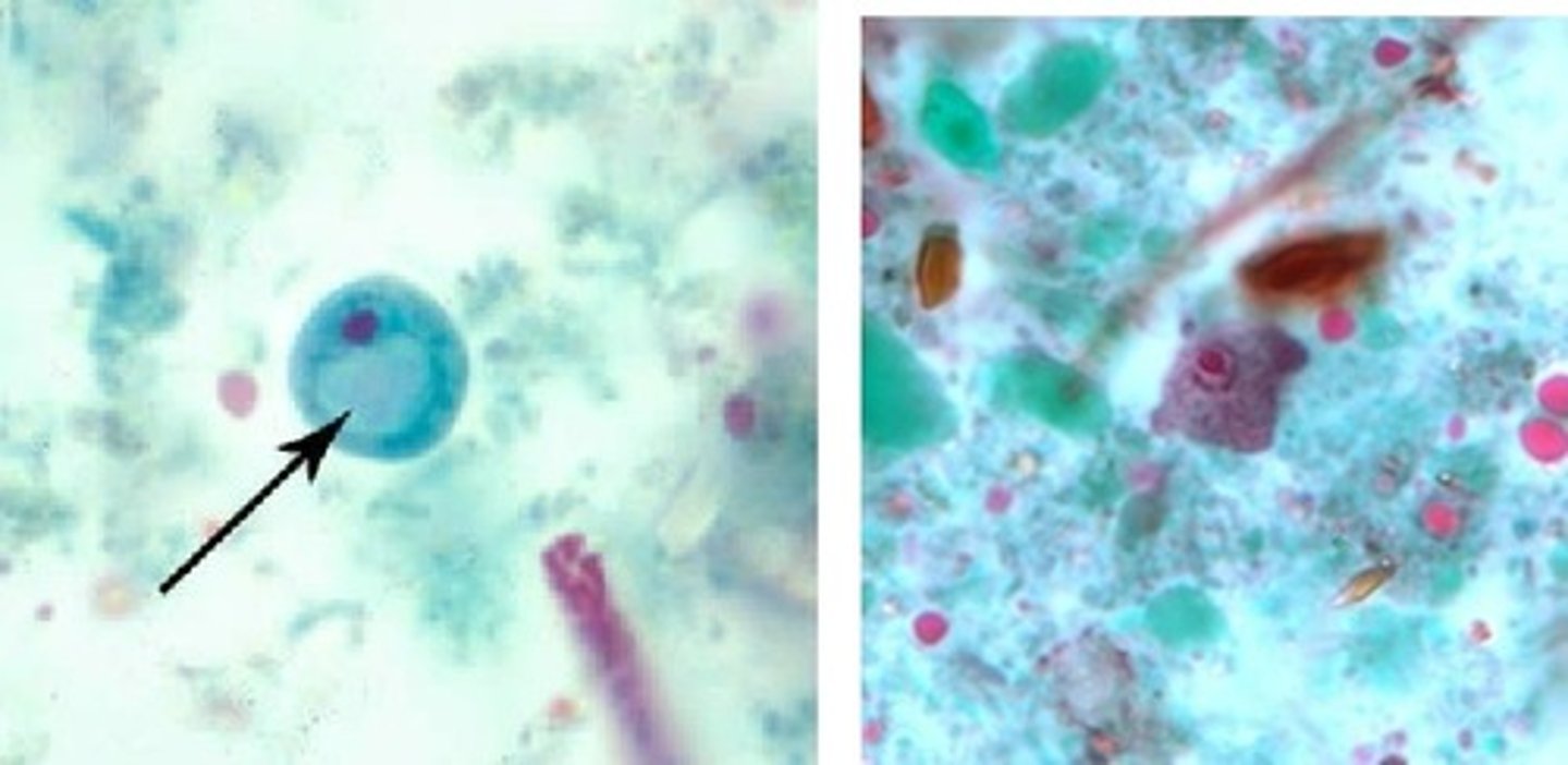
Iodamoeba buetschlii
• Cyst (left)
o No peripheral chromatin
o Prominent vacuole
• Trophozoite (right)
o 8-20 microns
o Vacuolated and granular
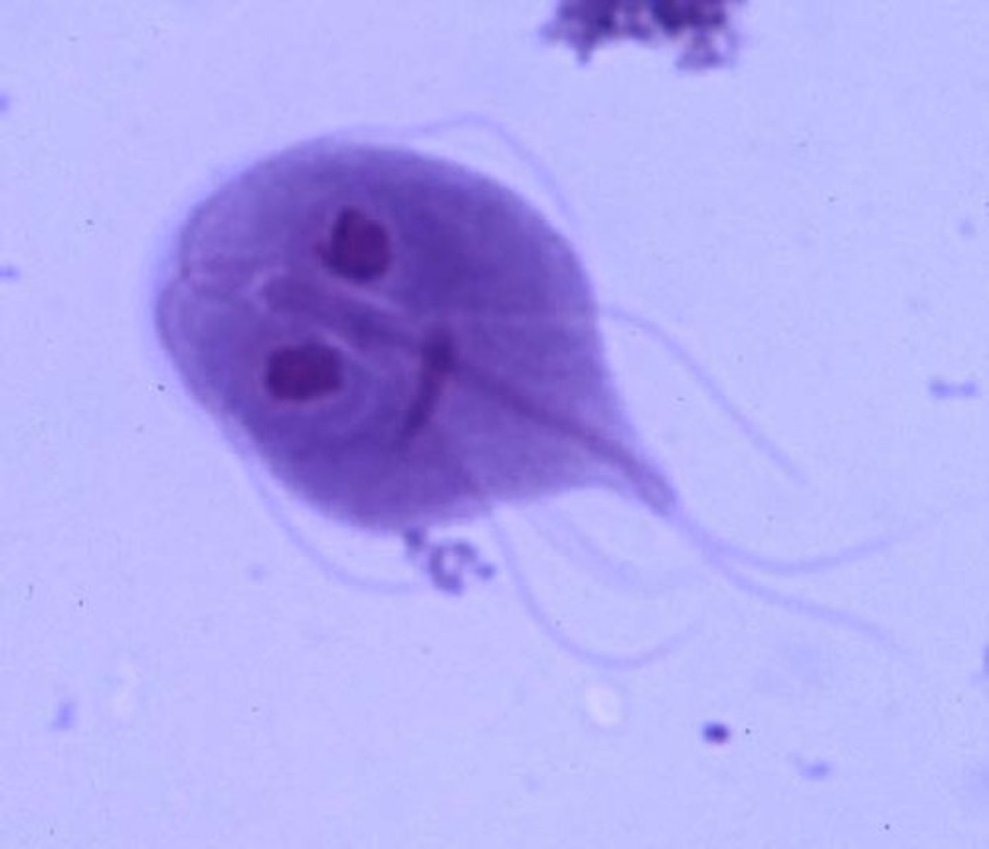
Giardia intestinalis (flagellate)
• Cyst (left)
o 4 nuclei
• Trophozoite (right)
o Pear shaped
o Central axoneme
o 2 nuclei
o 2 sucking disks
o 2 parabasa
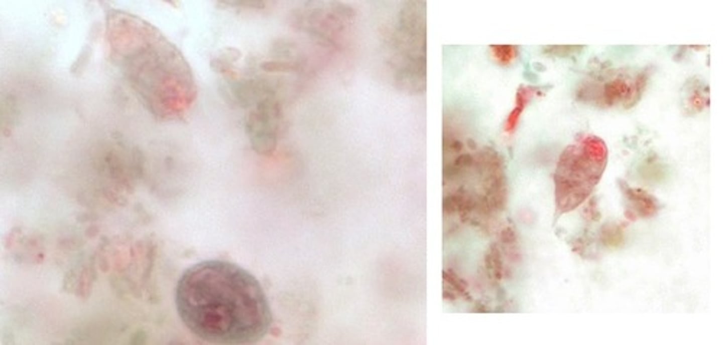
Chilomastix mesnili (flagellate)
• Cyst (lower)
o "Shepherd's crook"
cytostome
• Trophozoite (upper
and right)
o Pear shaped
o Anterior end is
rounded, nucleus
o Posterior end is
tapered
o "Shepherd's crook"
cytostome
Dientamoeba fragilis
• No cyst
• Trophozoite
o Round
o Binucleate
o Internal flagellum
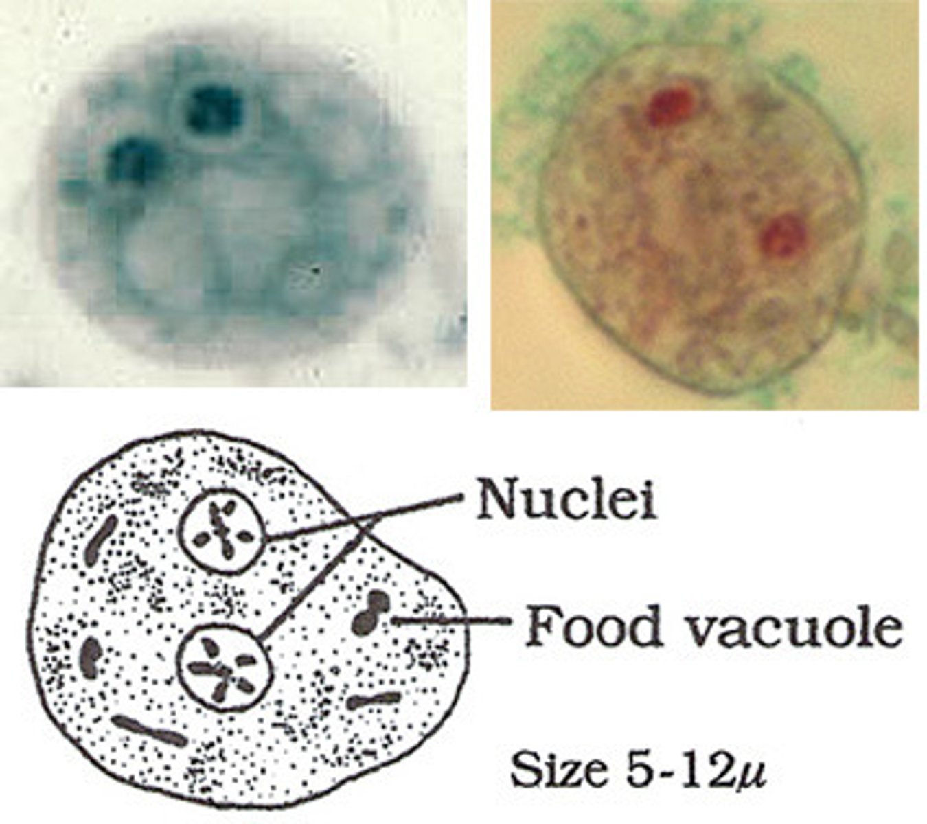
Balantidium coli (cilliate)
• Cyst
o Infrequently seen
• Trophozoite
o Large, up to 200 microns
o Cytostome and cilia
Kidney bean
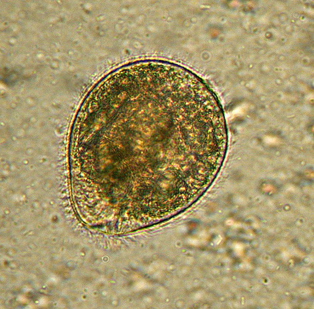
Enterobius vermicularis
• Egg
o Thin walled oval with
flat side
o Found in scotch tape
prep rather than stool
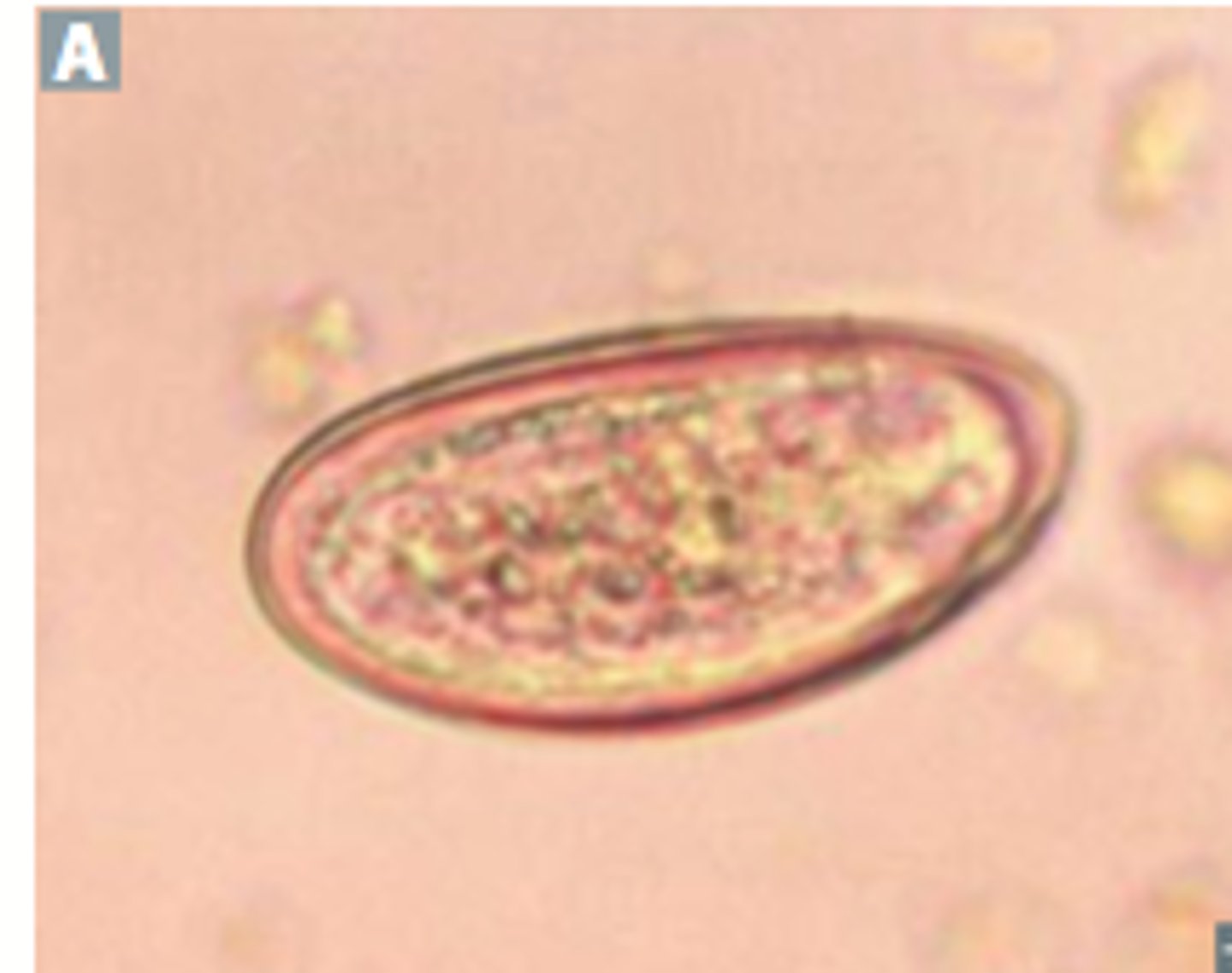
Trichuris trichiura
• Egg
o Thick walls
o Bilateral polar plugs
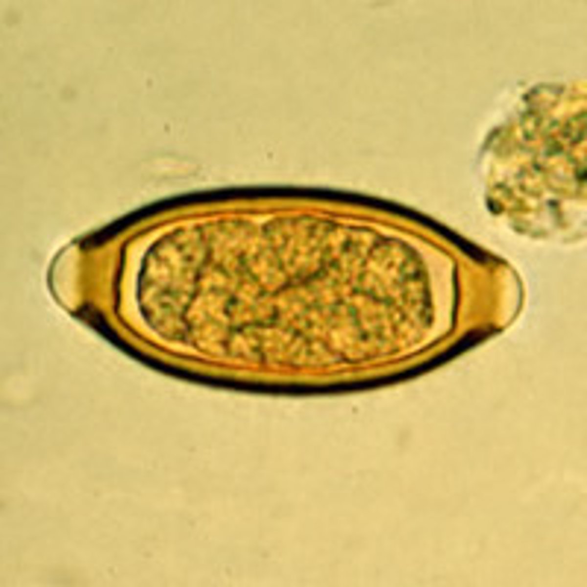
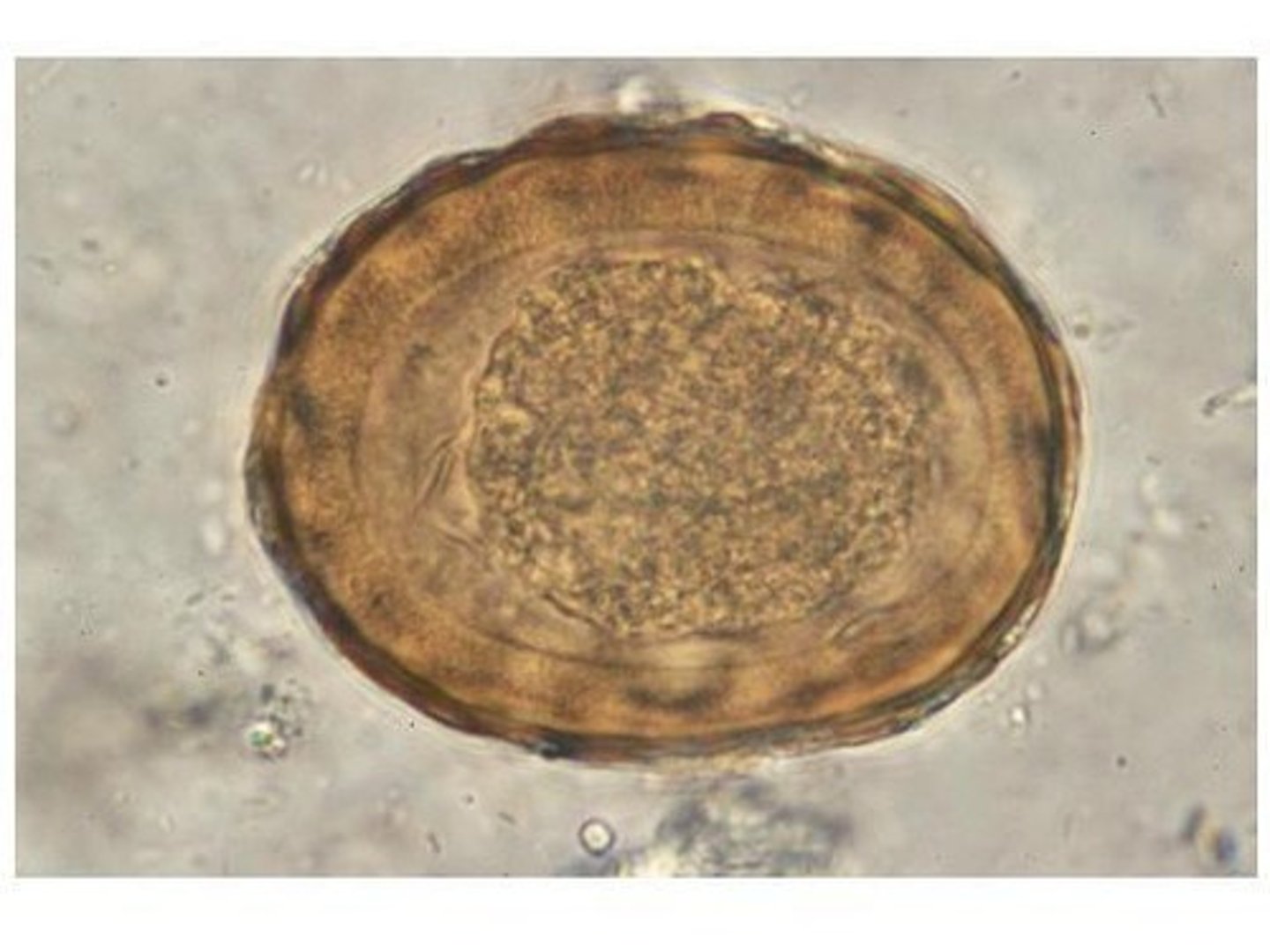
Ascaris lumbricoides
• Egg
o Rough, bumpy surface
o Thick shell, stained with
bile
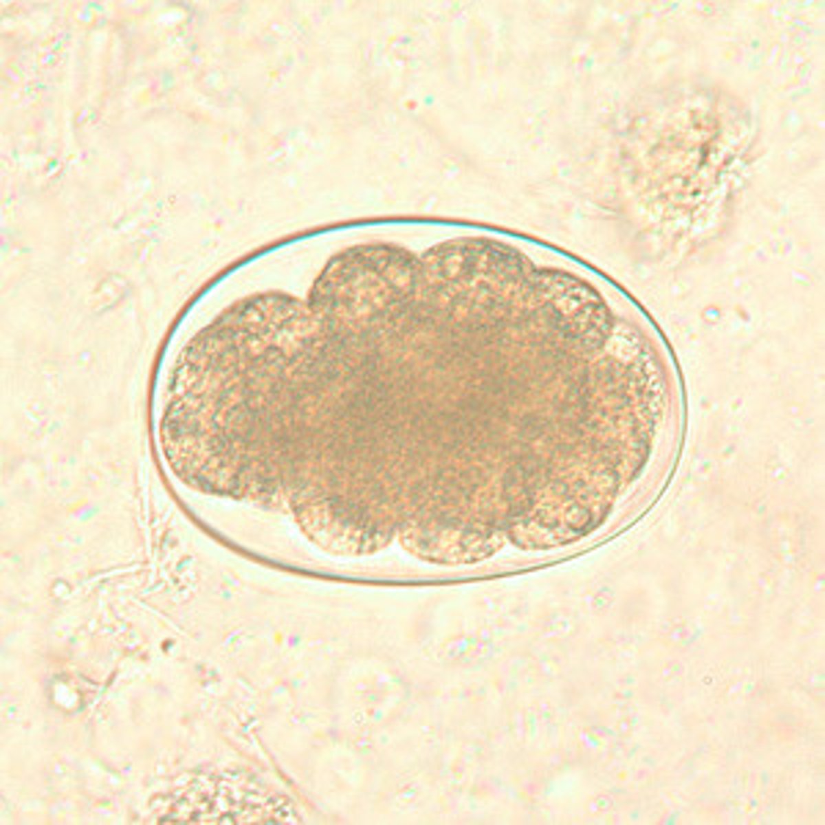
Hookworm
• Egg
o Necatur and Ancylostoma
eggs are indistinguishable
o "Steak on a plate"
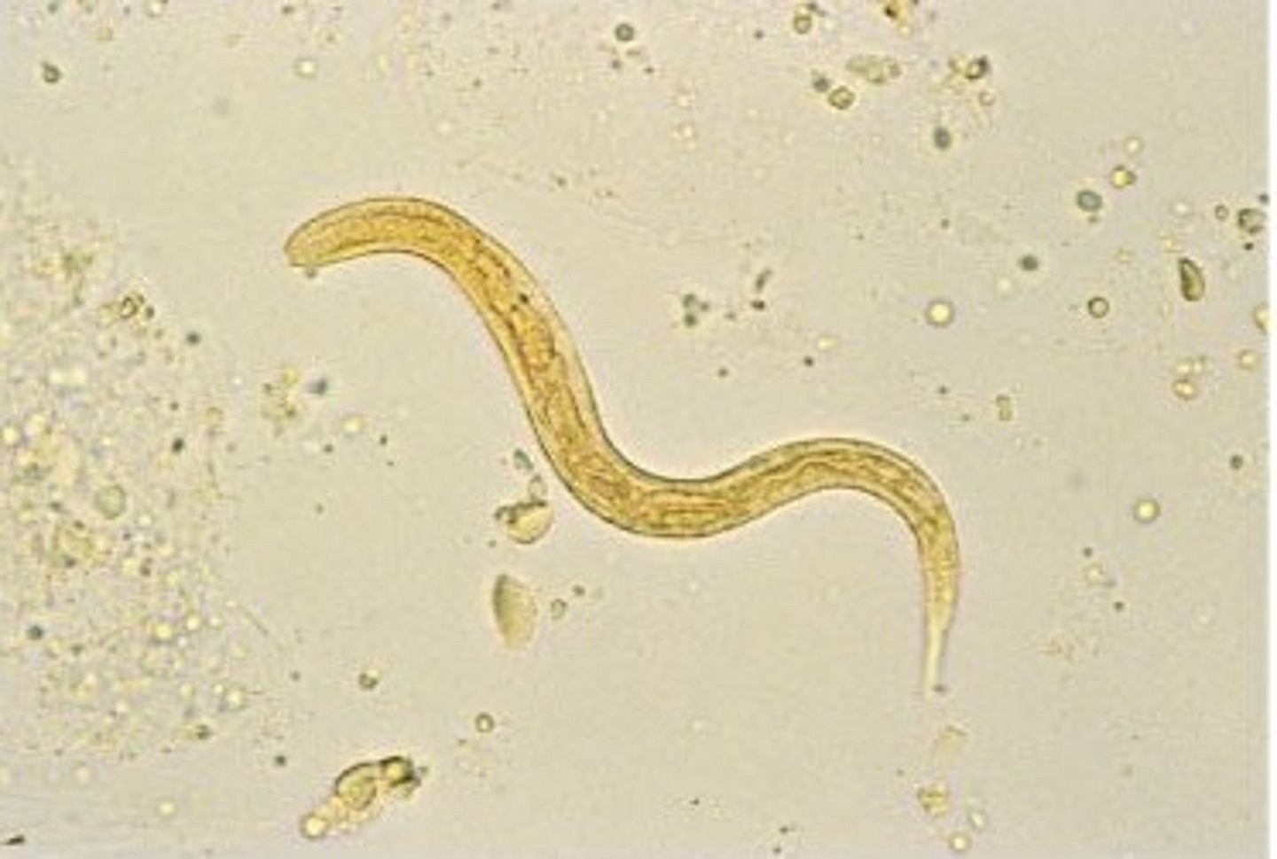
Strongyloides stercoralis
• Worm (rhabditiform larva)
o Short buccal canal
o Bulbar portion of
esophagus
o Genital primordium
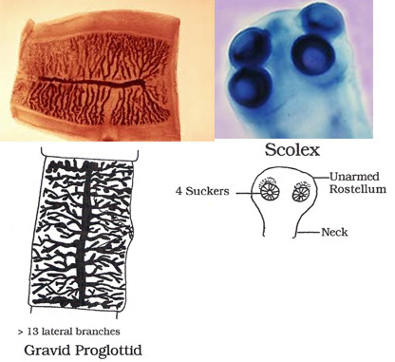
Taenia saginata
• Eggs are identical to T.
solium
o Around 30-40 microns
• Worm
o Smooth scolex with
four suckers
o Proglottids
Longer than they are
wide
>13 uterine branches
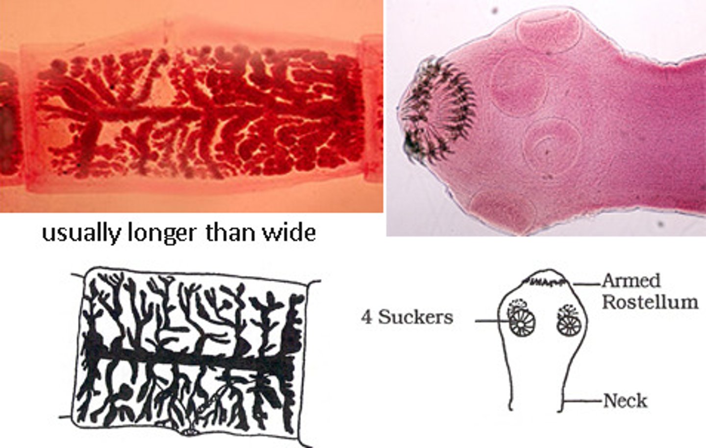
Taenia solium
• Eggs are identical to T. saginata
• Worm
o Scolex with four suckers with armed
rostellum
o Proglottids
Longer than they are wide
<13 uterine branches
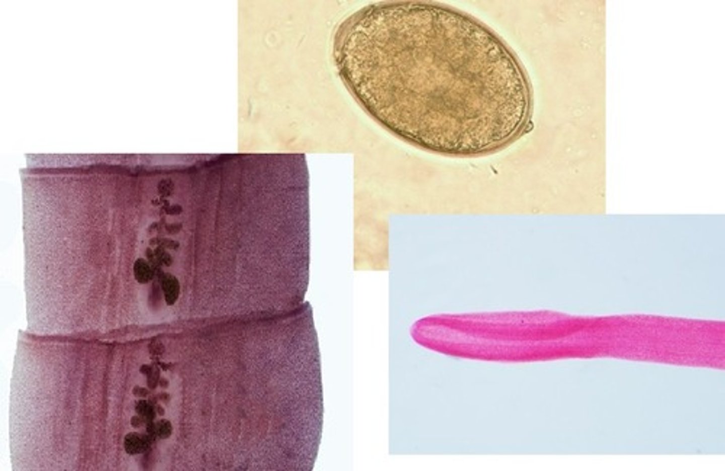
Diphylobothrium latum
• Eggs smooth with
shoulderless
operculum and
small knob
o Around 60 microns
• Worm
o Scolex is almond-
shaped with 2
sucking grooves
o Proglottids wider
than long
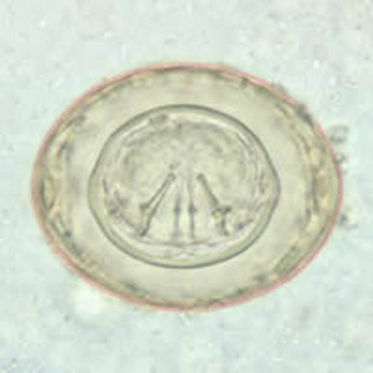
Hymenolepis diminuta
• Eggs have thin inner and
outer shells
o About 80 microns
o Polar filaments between
shells
o Embryo with hooklets
inside inner shell
Hymenolepis nana
• Eggs
o About 40 microns
o Two shells with polar filaments between
o Oncosphere has six hooklets