biology 2401 exam 2
1/175
There's no tags or description
Looks like no tags are added yet.
Name | Mastery | Learn | Test | Matching | Spaced | Call with Kai |
|---|
No analytics yet
Send a link to your students to track their progress
176 Terms
tissue
a group of similar cells working together to perform a particular function
4 major groups of tissue
epithelial, connective, muscle, and nervous
histology
the study of tissues
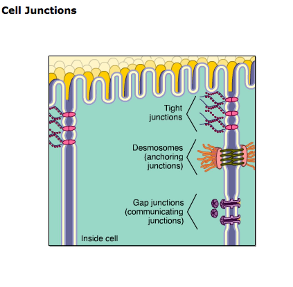
2 most significant ways cells are stuck together
1. via the "sticky" glycocalyx (sugar coats) on the surfaces of the cells
2. via specialized junctions (include desmosomes, tight junctions and gab junctions)
desmosomes
- filamentous connections that not only hold adjacent cells together but also give internal structural support to the cells
- do not prevent materials from passing in between the connected cells
plaque
- each desmosome contains one located on the cytoplasmic surface
- consists of glycoproteins and resembles a tiny button
cadherins
filamentous proteins that radiate out from the plaque and intertwine with cadherin filaments from the adjacent cell
where are desmosomes found?
in tissues that experience stretching, such as skin
tight junctions
- continuous bands of protein that form an almost impenetrable barrier to prevent substances from passing through the intercellular space between the connected cells
- consist of occludins
occludins
extend out from the plasma membrane of adjoining cells and fuse together
where are tight junctions found?
in cells that line the stomach and intestines, and in cells that line blood vessels in the brain
gap junctions
tiny channels that allow materials to pass from the cytoplasm of one cell into the cytoplasm of an adjacent cell
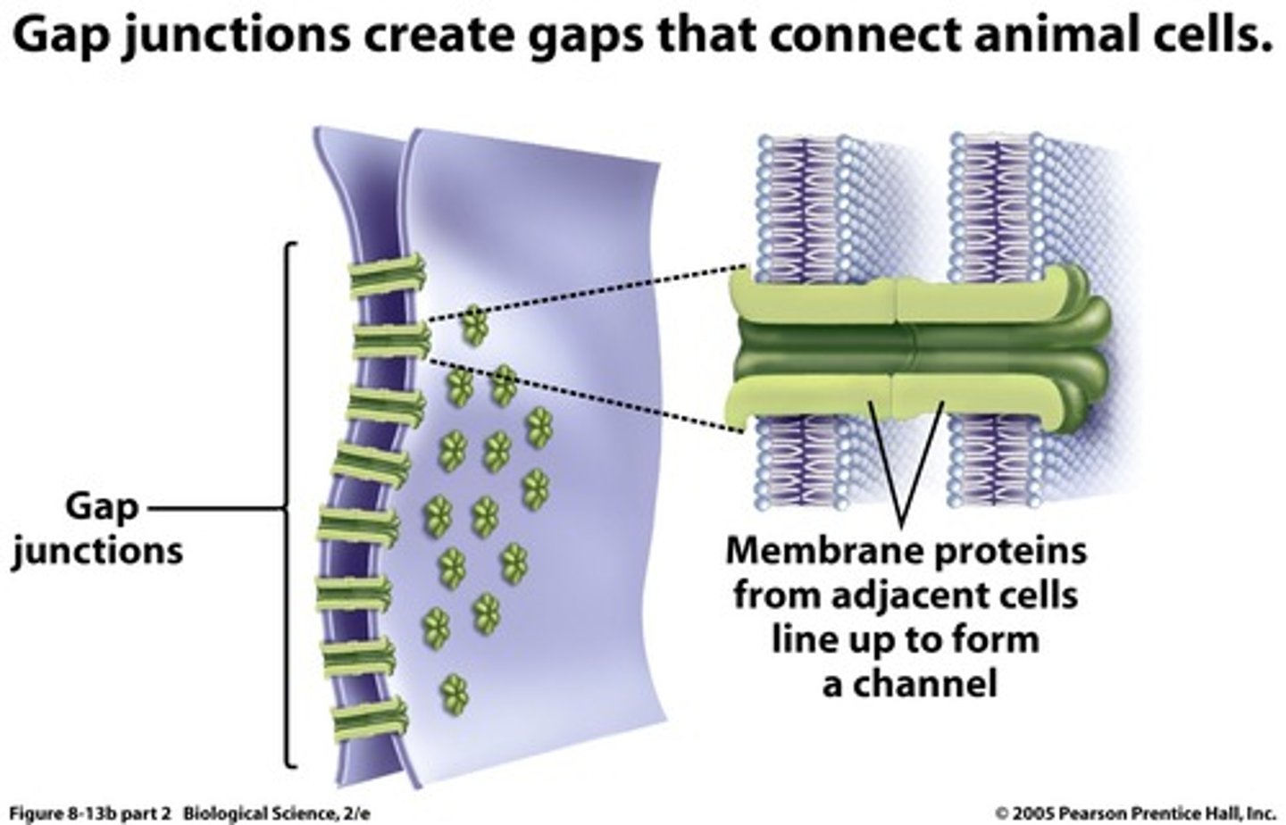
when do gap junctions form?
when transmembrane proteins called connexons in adjacent cells bind to one another
where are gap junctions found?
in cardiac and smooth muscle cells
epithelial tissue (epithelium)
forms thin membranous coverings around and inside various organs and it forms glands
how is epithelial tissue classified?
as either membranous or glandular
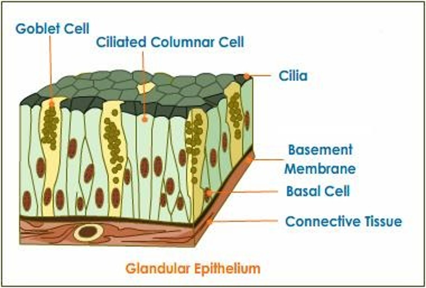
glandular epithelium
forms specialized structures called glands that release substances beneficial to the body
secretion
is a noun when it refers to the substance released from the gland, and it is a verb when referring to the process of releasing the substance
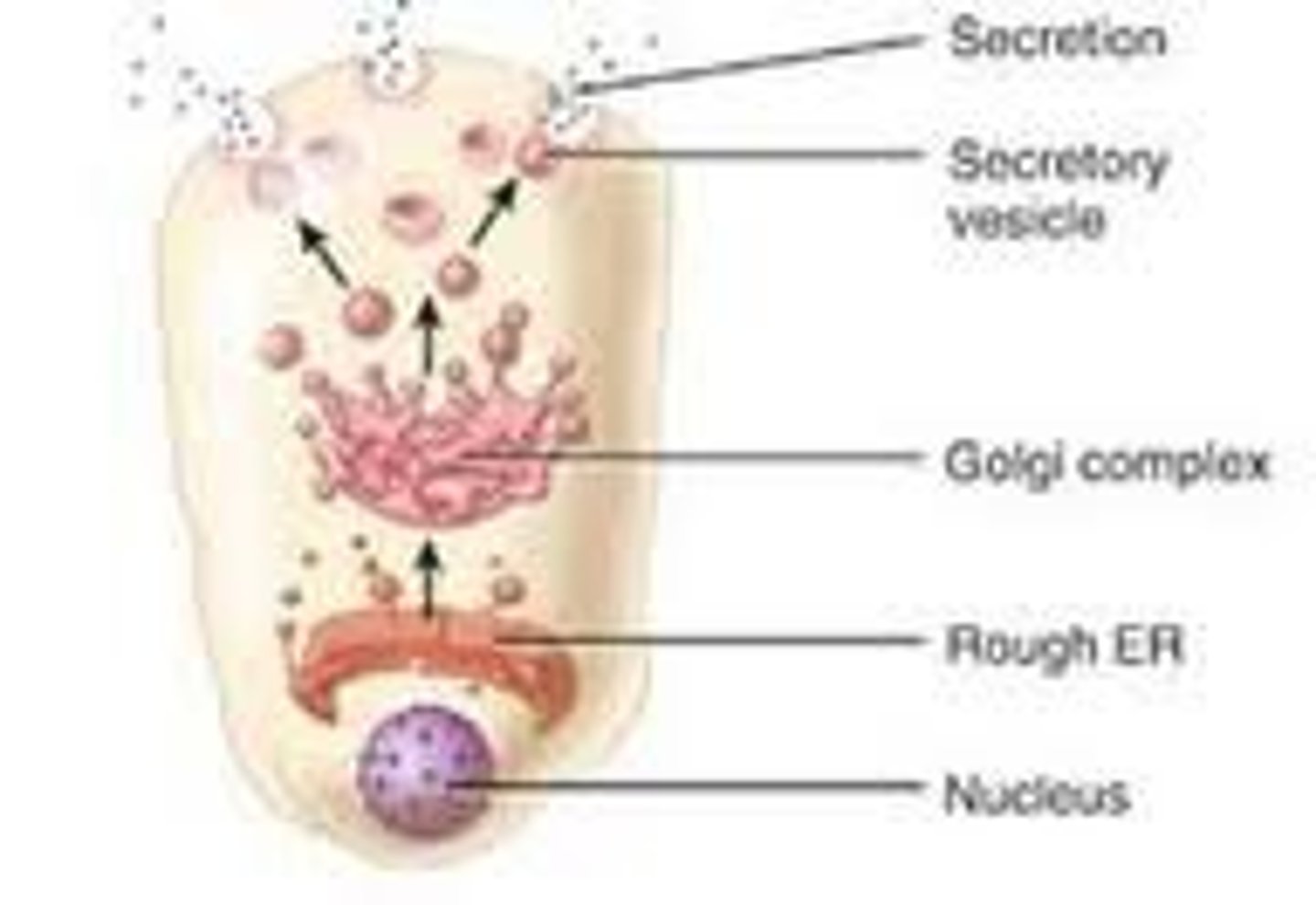
endocrine glands
secrete chemicals called hormones into tissue fluid or the bloodstream (secretions of these glands remain inside the body)
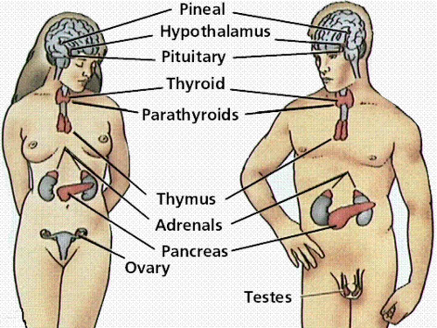
hormones
function as chemical messengers sent through the blood from endocrine glands to other organs in the body
exocrine glands
expel secretions into a cavity or tube that has connections with the outside of the body
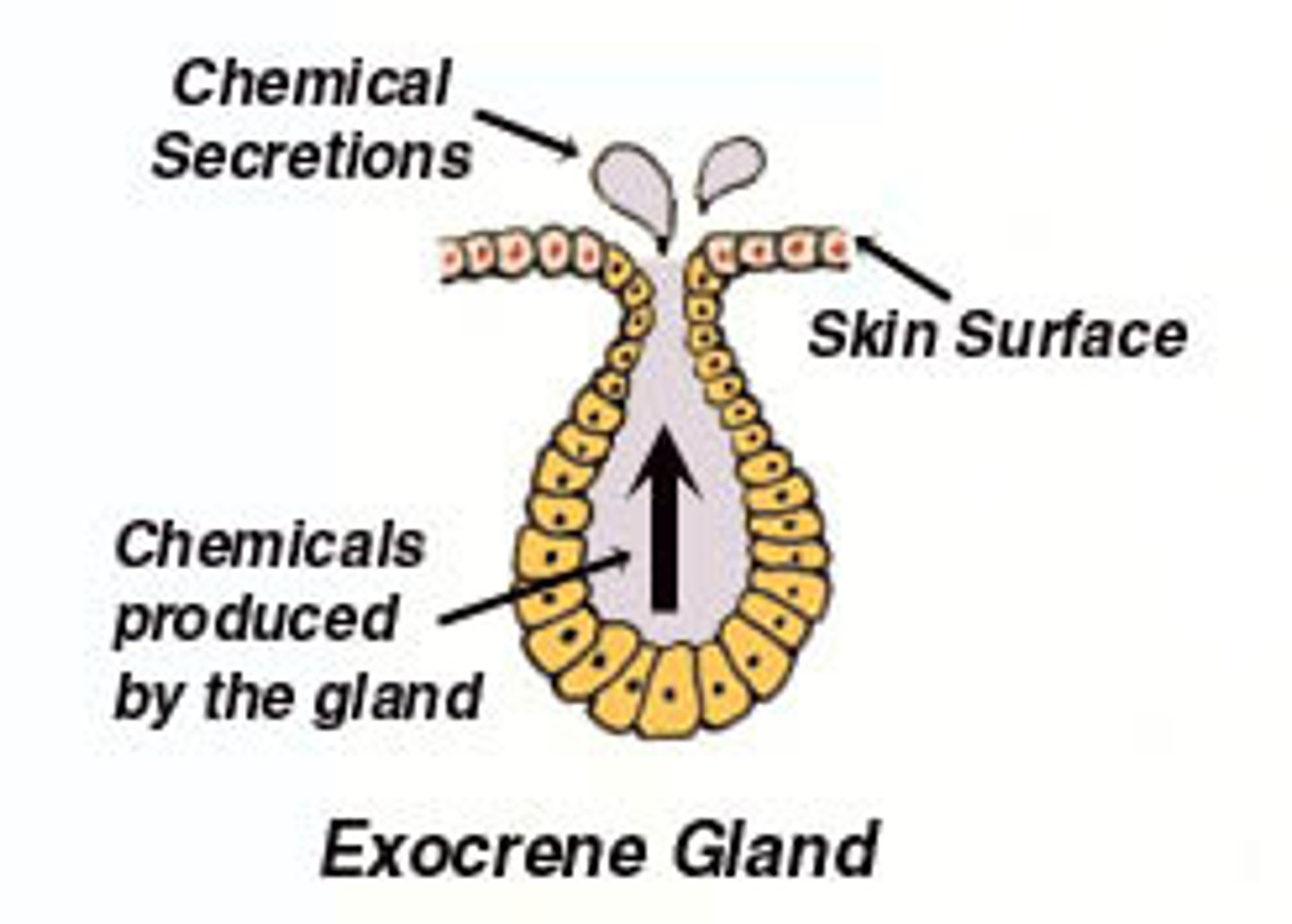
how are exocrine glands classified?
as either unicellular or multicellular
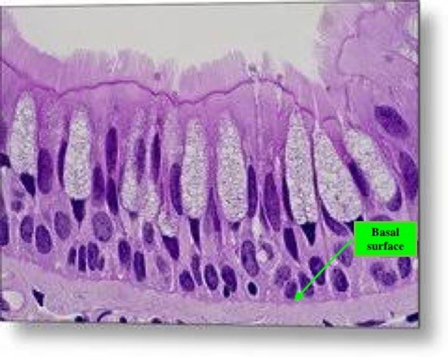
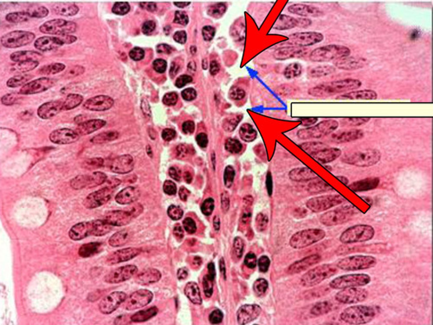
goblet cell
the only example of a unicellular gland in the body which secretes musus directly onto the surface of certain epithelial membranes
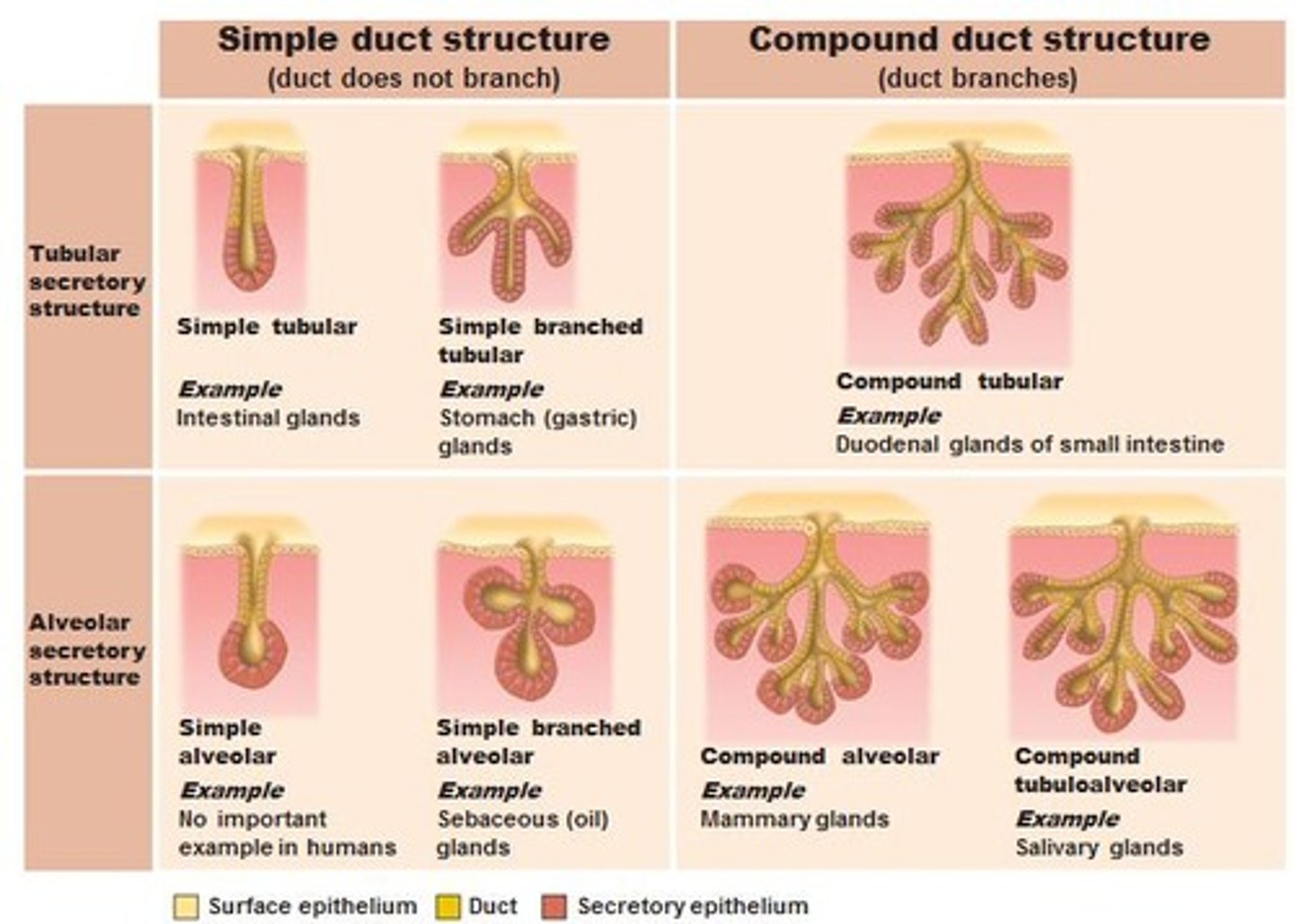
multicellular glands
expel their secretions into a tiny tube called a duct
how are multicellular glands classified?
based on the structure of the gland and the method of secretion (apocrine, holocrine, merocrine)
apocrine glands
make secretions consisting of portions of glandular cells that have broken off (ex. mammary glands that produce milk in the breasts)
holocrine glands
make secretions consisting of entire cells that have disintegrated (ex. sebaceous (oil) gland in the skin)
merocrine glands
make secretions by exocytosis and are the most common type of exocrine gland (include sweat glands, salivary glands and mammary glands)
membranous epithelium
- exists on the free surface of various organs, including the skin where it forms the epidermis
- also covers the outside of visceral organs such as the stomach and intestines, and lines their internal cavities
- are multicellular
how does membranous epithelium function?
in protection, absorption, filtration and secretion
what are the characteristics of membranous epithelium?
tightly packed cells, cells are arranged in layers, avascular, nerve supply, polarity, basement membrane, some house receptors
stratum
each layer of cells
innervated
what we call tissues that contain fibers from the nervous system
apical surface
outermost layer of cells in contact with the "free" surface
apical cells
cells in the apical surface region
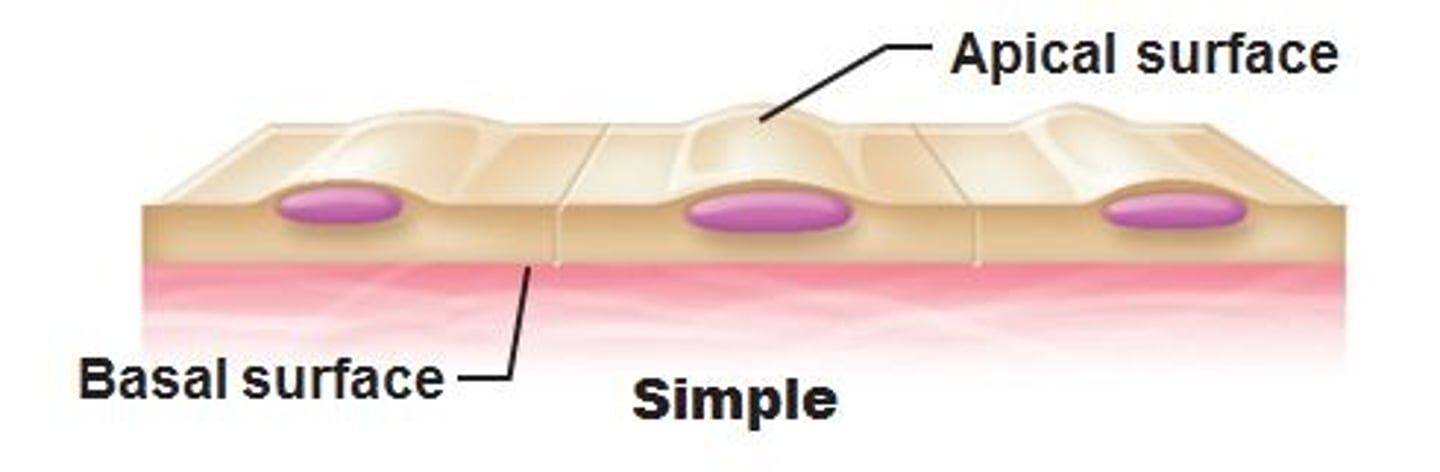
basal surface
deepest layer of cells
basal cells
cells in the basal surface region
polarity
anatomical and physiological differences between their cells located at the free surface and their cells in contact with the underlying connective tissues
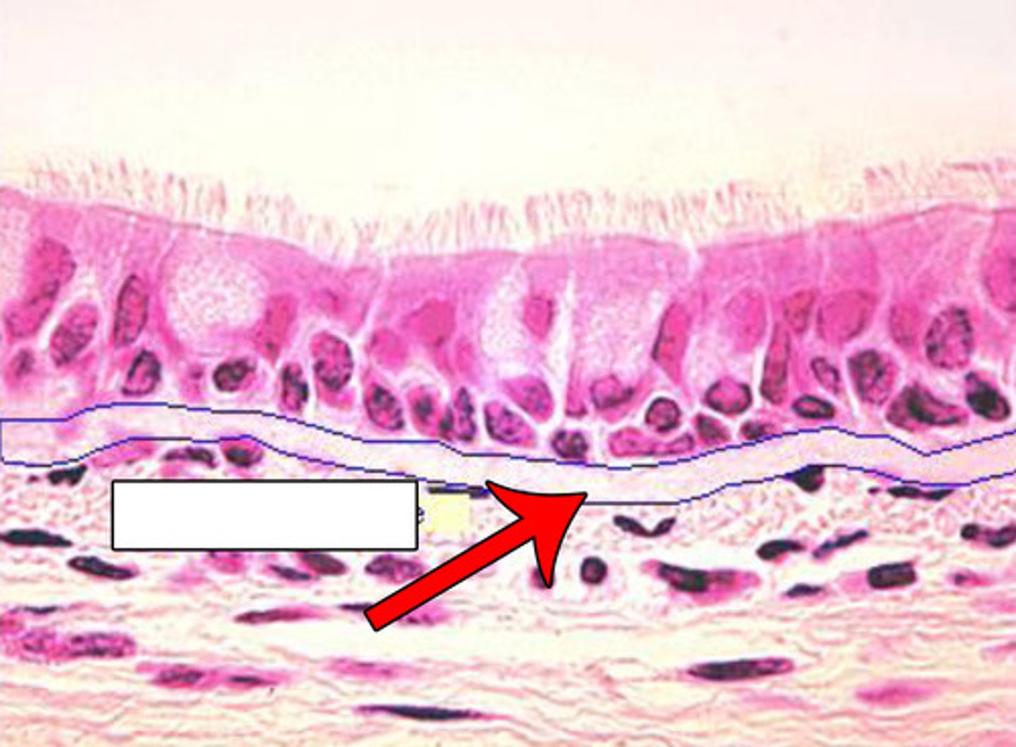
basement membrane
a thin layer of glycoproteins and collagen fibers
basal lamina
the glycoprotein component of the basement membrane that is secreted from the basal cells of the epithelium
reticular lamina
the collagen protein component of the basement membrane that is secreted from connective tissue cells lying beneath the epithelium; collagen fibers in this region intertwine to form a net-like pattern
receptors
allow the body to detect changes in the internal and external environment
how is membranous epithelium classified?
by cell shape, cell arrangement and location
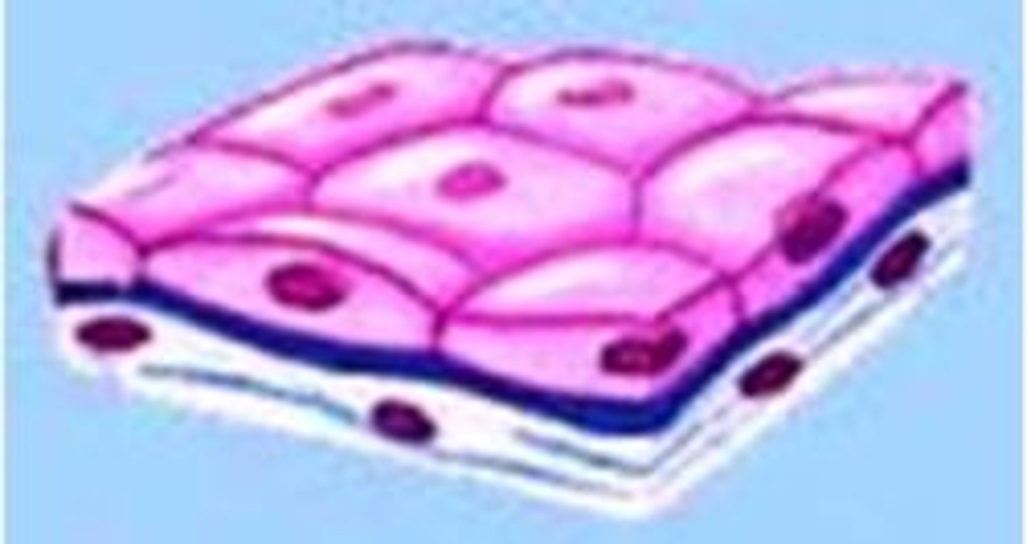
squamous
cells are flat and resemble scales on a fish (ex. outer part of your skin)
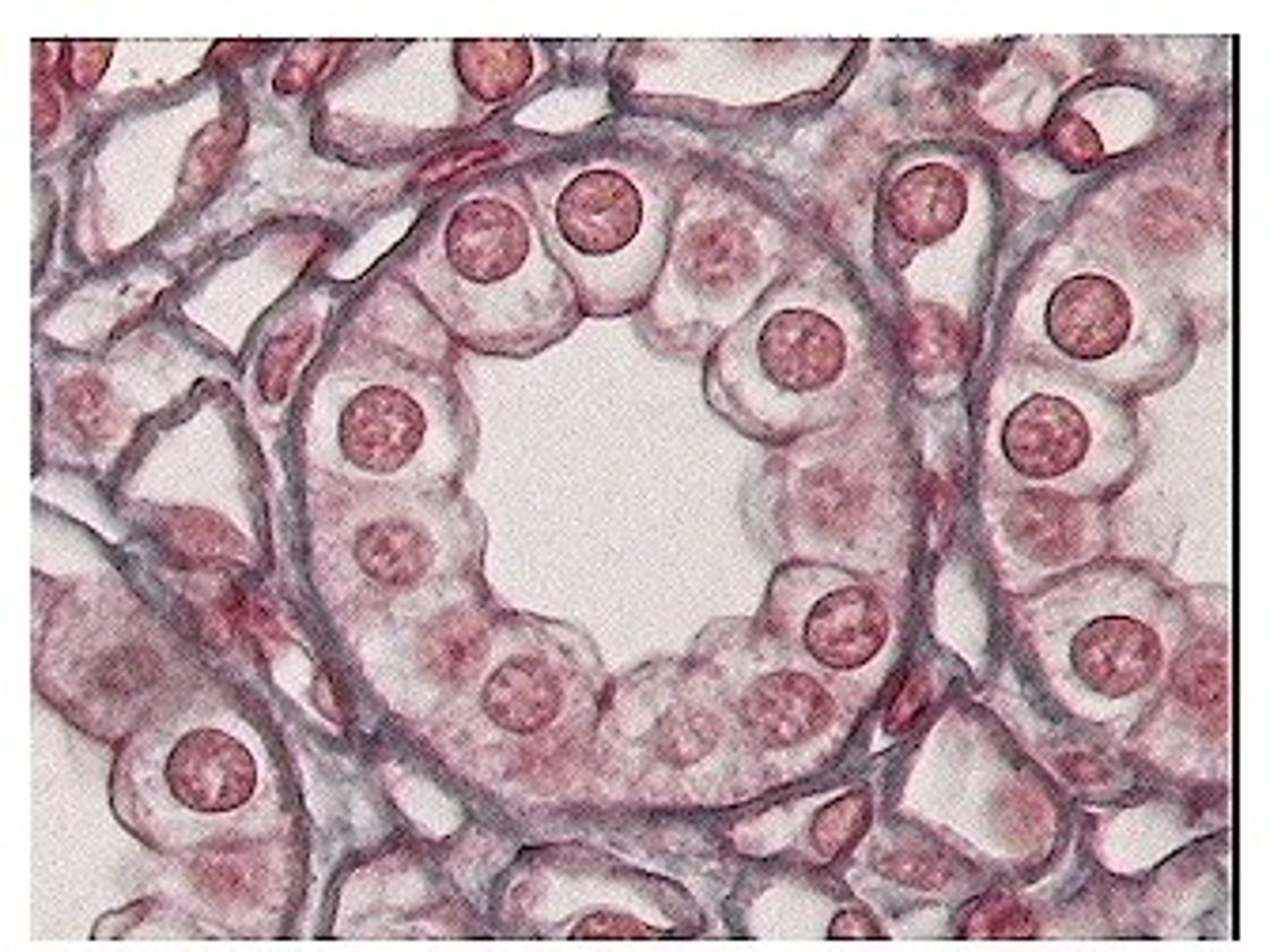
cuboidal
cells are cube shaped (ex. lining of tiny tubes in the kidneys)
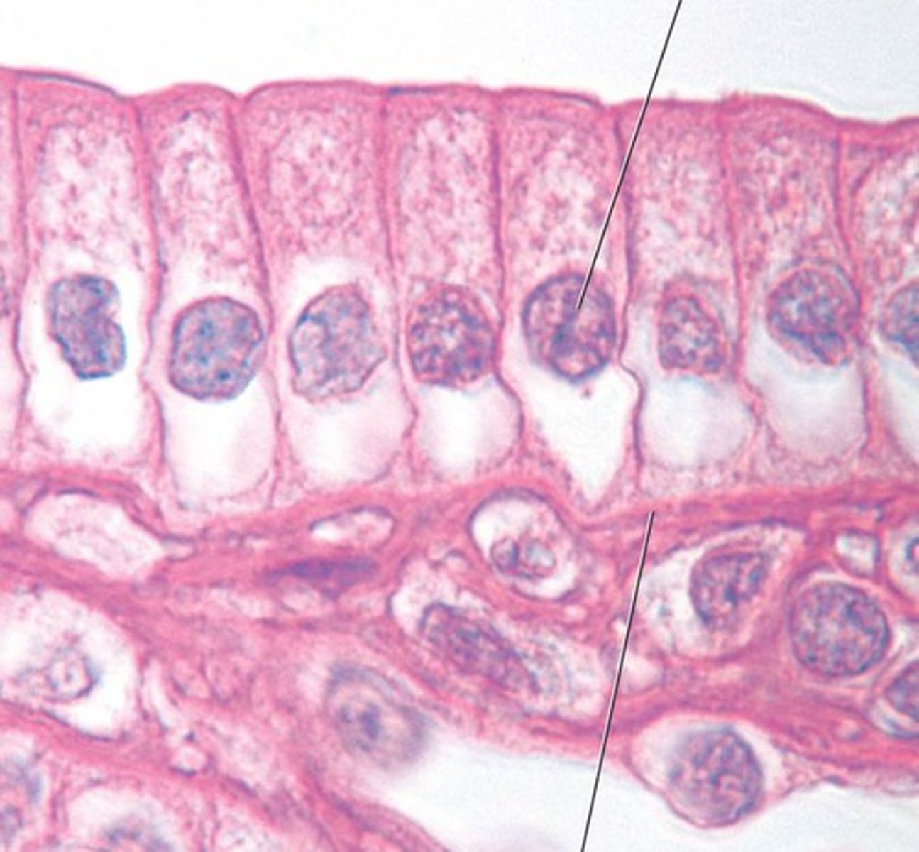
columnar
cells are column-shaped; they are taller than they are wide (ex. various glands of the body)
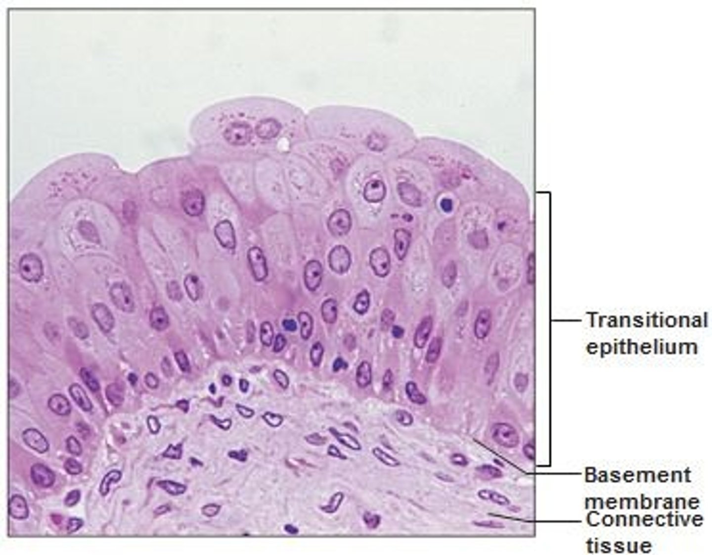
transitional
cells can change shape from cuboidal to squamous and back to cuboidal, depending on the amount of compression exerted on the cells (ex. line the inside of the urinary bladder)
simple
has only one layer of cells
stratified
has two or more layers of cells

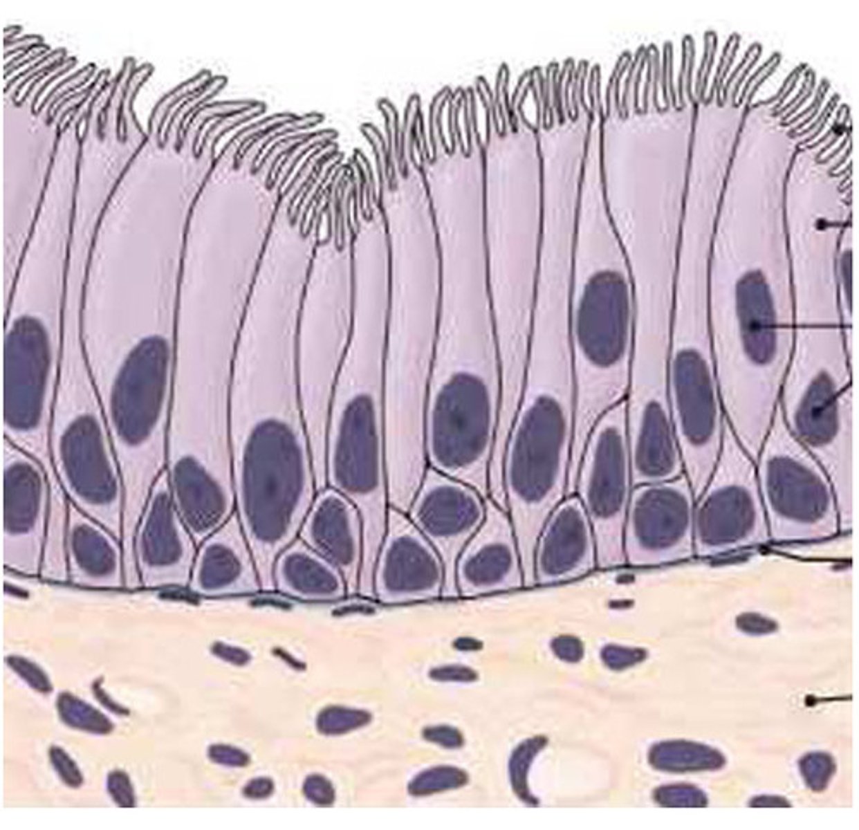
pseudostratified
is actually a simple epithelium but it looks stratified
endothelium
simple squamous epithelium that lines the inside of blood vessels and lymph vessels
serous membranes (mesothelium)
simple squamous epithelia that cover visceral organs and line major body cavities that do not have connections with the outside of the body (include the peritoneal, pleural and pericardial membranes)
serous fluid
watery fluid secreted from serous membranes that reduces friction during movement
mucous membranes
can be simple columnar, pseudostratified, or stratified squamous epithelium but all line cavities that have a connection to the outside of the body
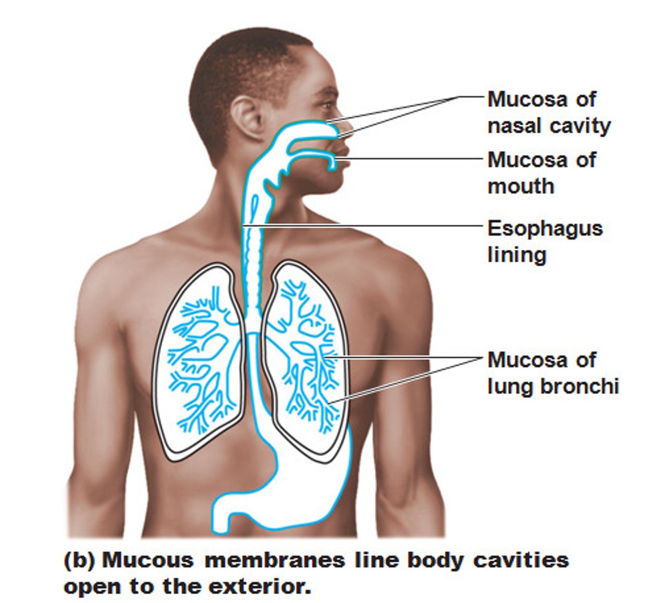
mucus
secretion secreted from goblet cells in mucous membranes
mucin
protein that gives mucus its thick viscous nature
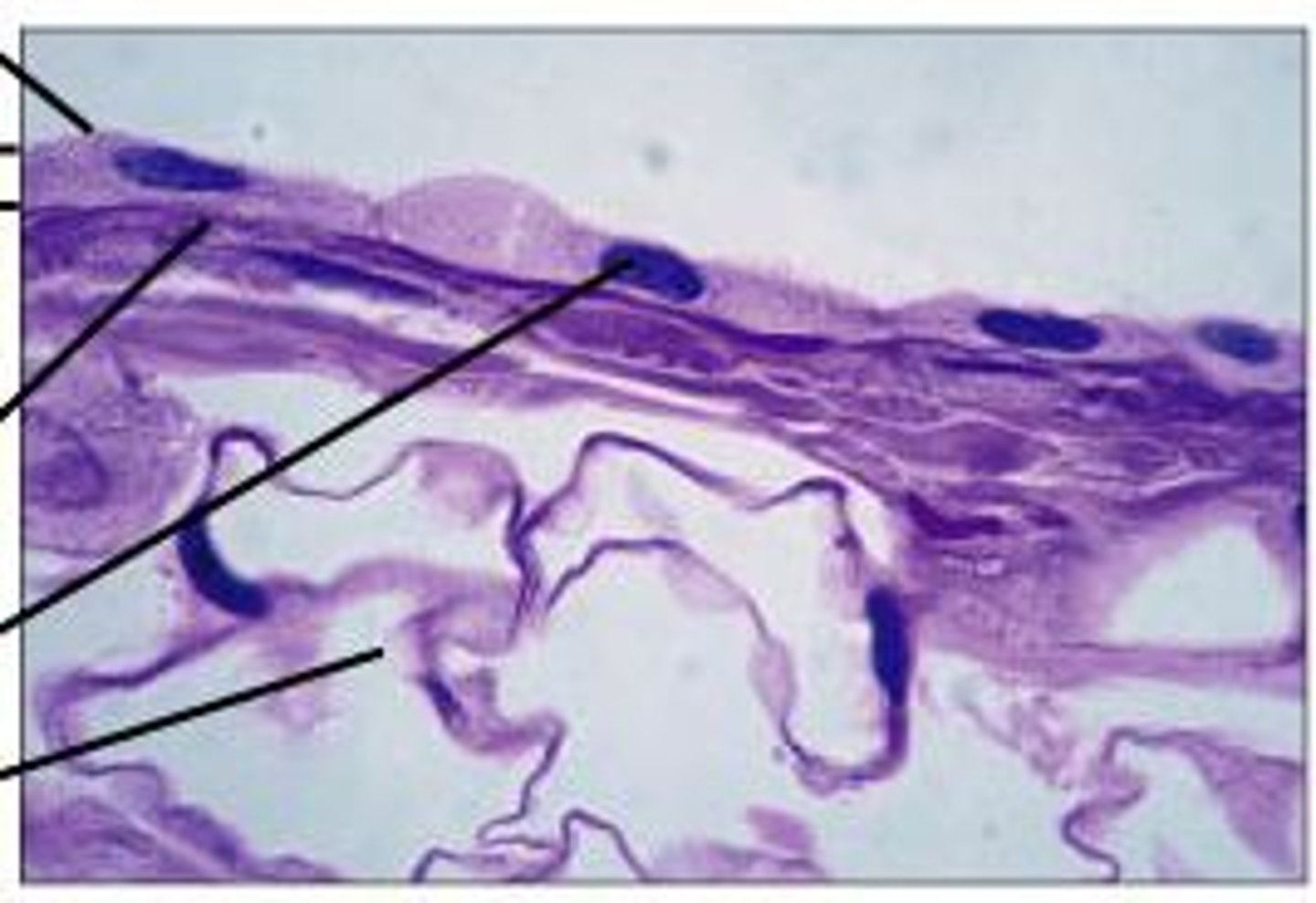
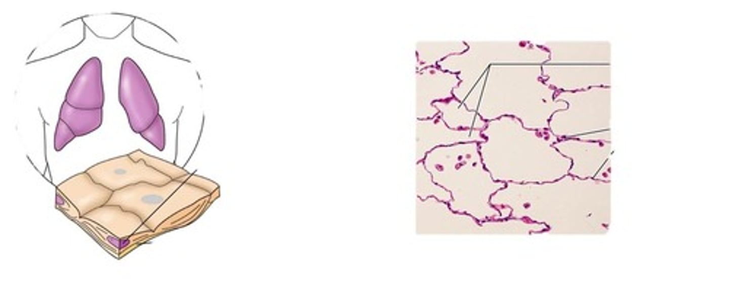
simple squamous epithelium
- forms serous membranes, small sacs within the lungs, filtering devices within the kidneys, and endothelium
- protects underlying tissues by reducing friction (in the case of serous membrane) and allows diffusion of gases through the walls of capillaries and tiny sacs in the lungs
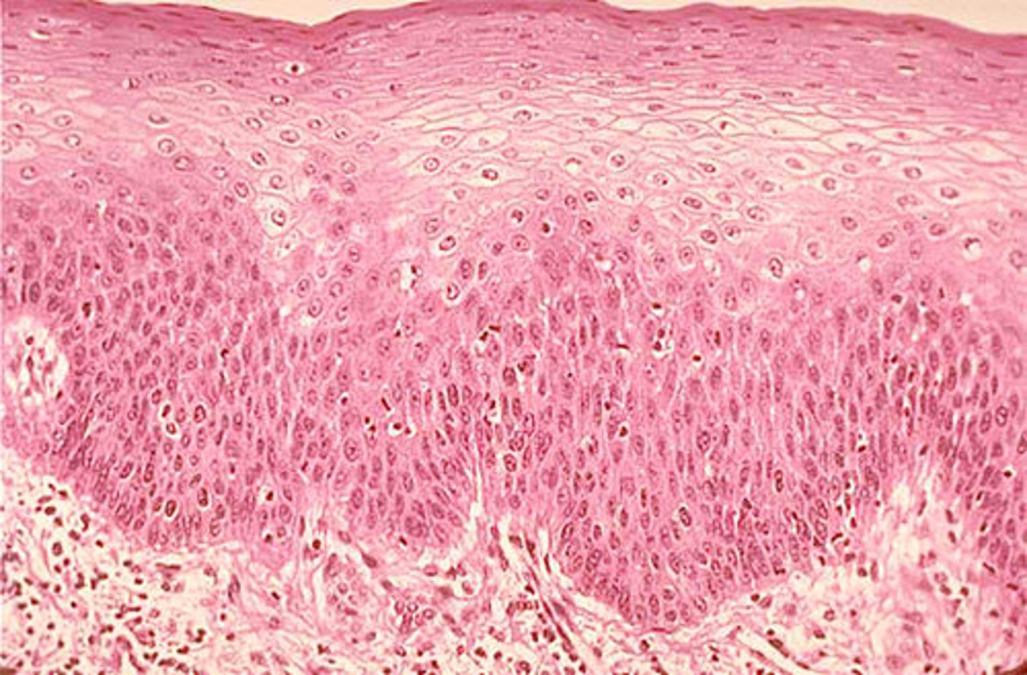
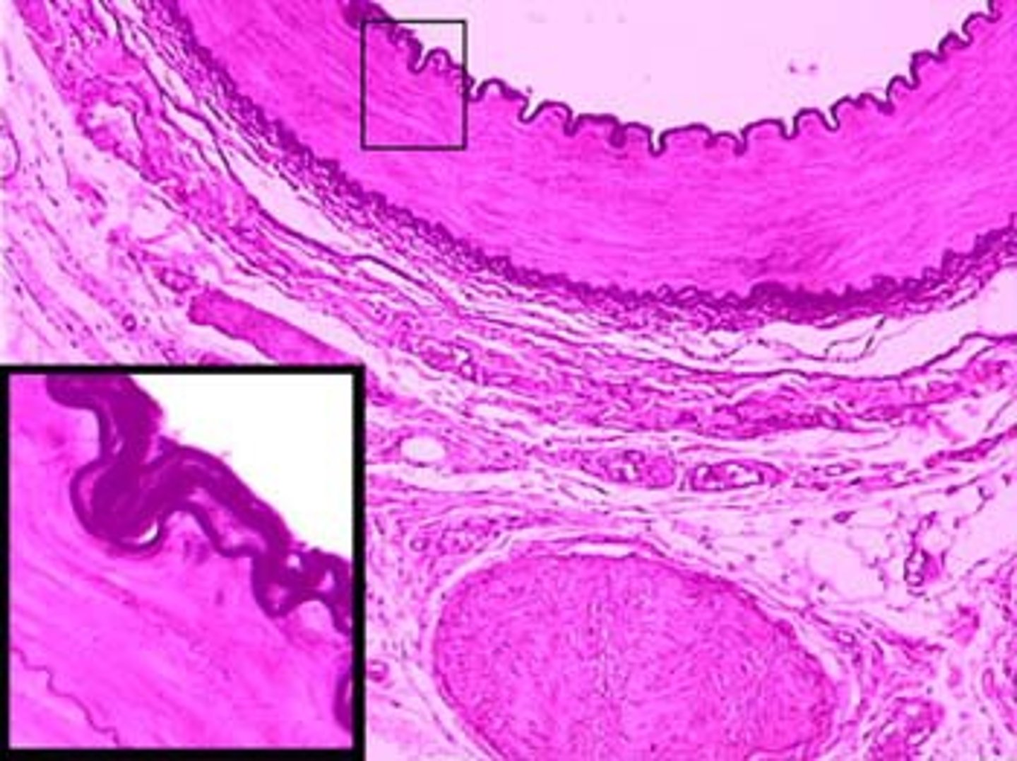
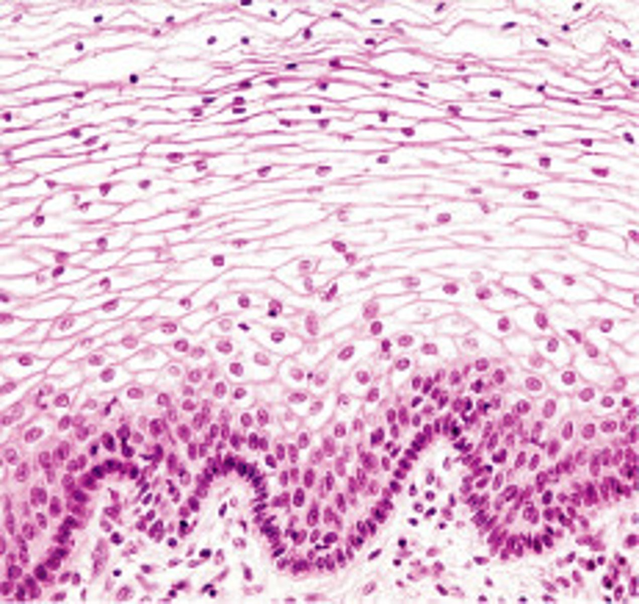
stratified squamous epithelium
- exists on the surface of the skin and the inner linings of the mouth, esophagus, and vagina
- provides protection to underlying tissues
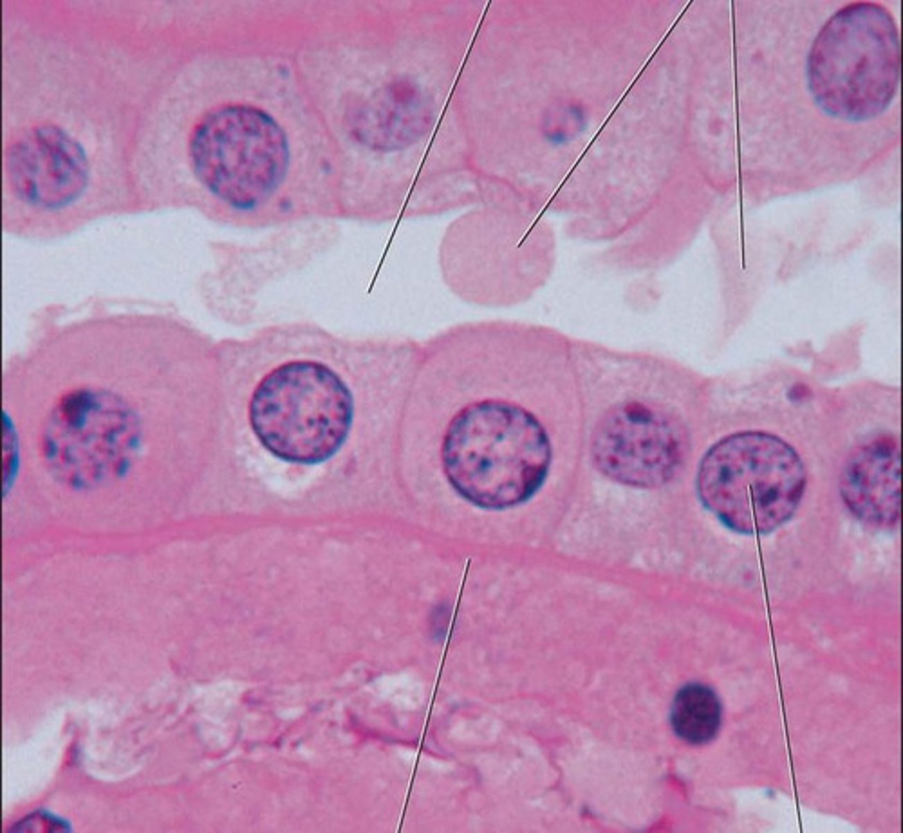
simple cuboidal epithelium
- exists in tiny tubes of the kidney and certain glands and on the surface of the ovary
- protects underlying tissues and allows passage of material between the blood and kidney fluid destines to become urine
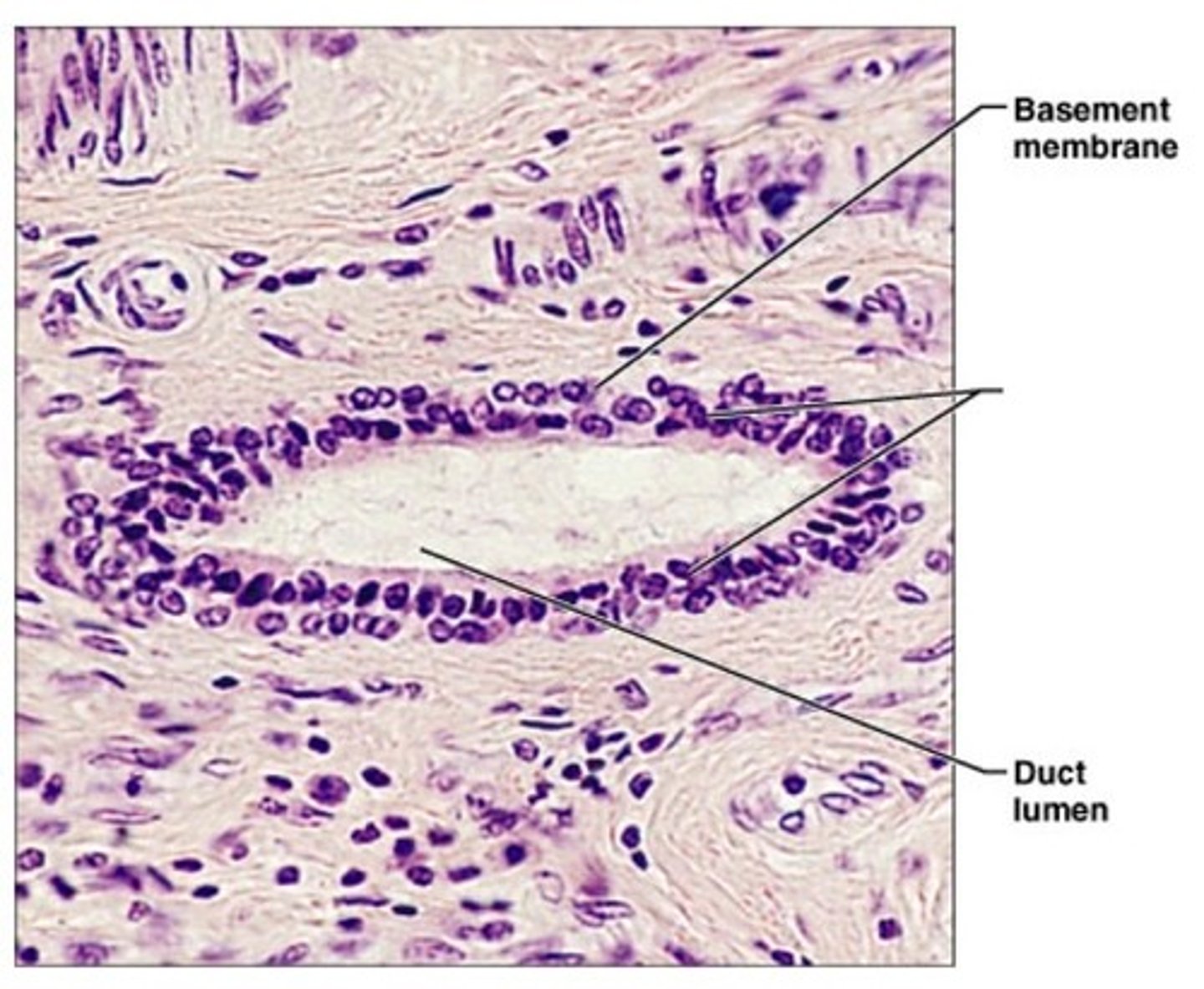
stratified cuboidal epithelium
- exists in the ducts of certain glands
- primary function is protection of underlying tissues
simple columnar epithelium
- nonciliated type makes up the inner lining of the digestive tract where it allows absorption of digested food molecules
- ciliated type lines the oviducts and the small tubes in the lungs, where it moves mucus that traps dust and other particles
stratified columnar epithelium
- lines the male's urethra and ducts of some glands
- protects underlying tissues
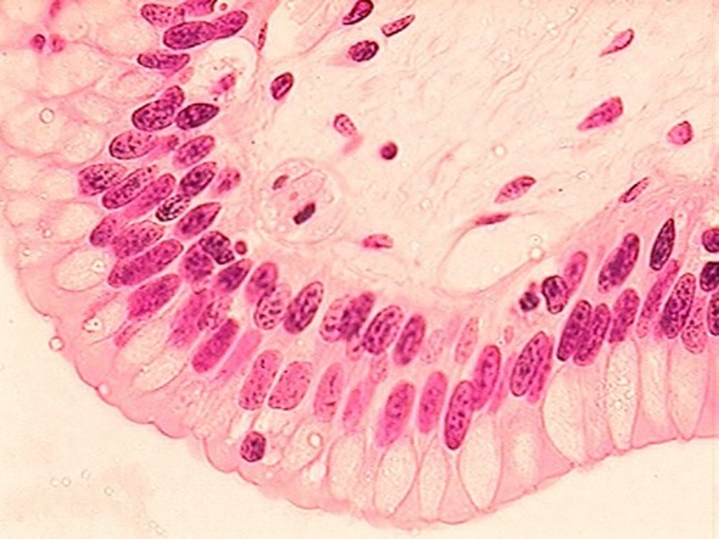
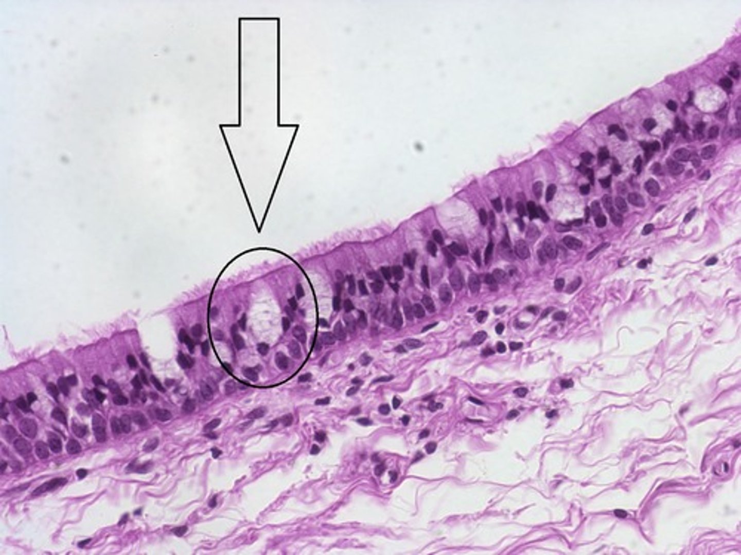
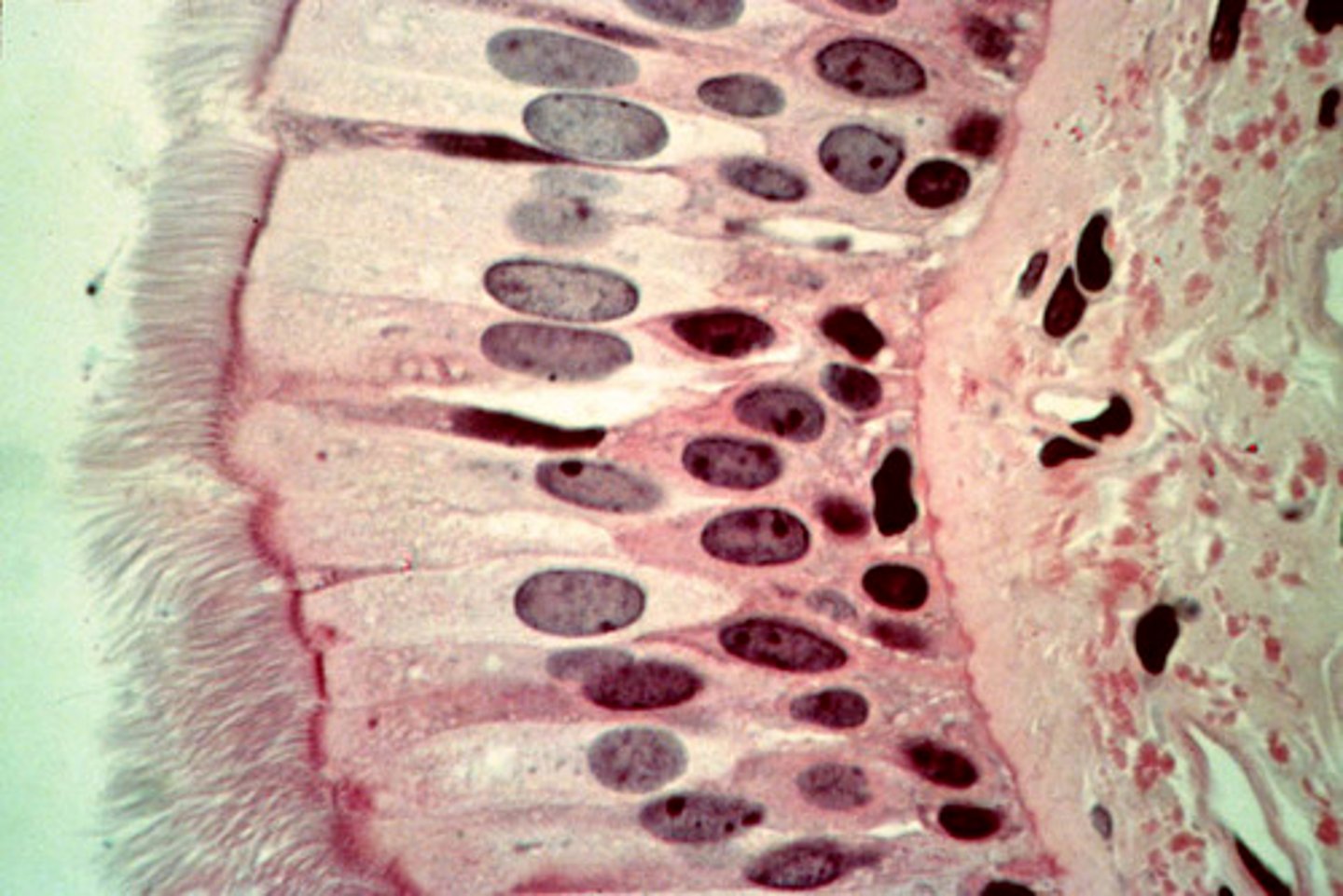
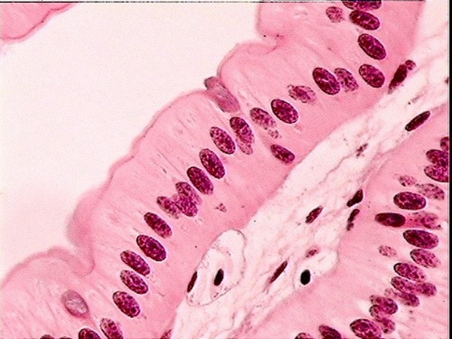
pseudostratified ciliated columnar epithelium
- contains ciliated cells and goblet cells and lines the nasal cavity, trachea, and part of the male's urethra
- protects underlying tissues by secreting and moving mucus
transitional epithelium
- exists in the lining of the urinary bladder, ureters, and part of the urethra
- allows organs to expand

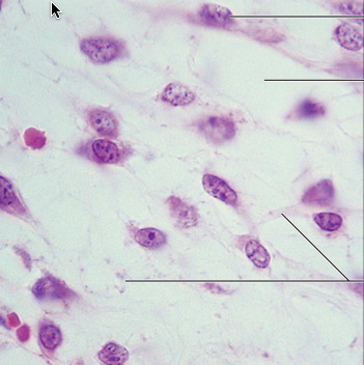
connective tissue
most abundant tissue in the body and serves primarily to bind structures together
what are the functions of connective tissue?
support, protection, insulation, movement, shock absorption, transport of nutrients and metabolites, and storage
mesenchyme
an undifferentiated, embryonic tissue that all connective tissue arises from
what are the major structural characteristics of connective tissue?
vascularization and an extensive matrix
vascularization
supplied with blood vessels (all connective tissue expect cartilage)
2 components of connective tissue matrix
ground substance and extracellular fibers
ground substance
the fluid component of the matrix and contains water, ions, nutrients, metabolities, adhesion proteins and proteoglycans
adhesion proteins
connect cells to extracellular fibers and the ground substance
fibronectin
most common adhesion protein
proteoglycans
central "wire" consists of a filamentous protein while the "bristles" include a variety of polysaccharides
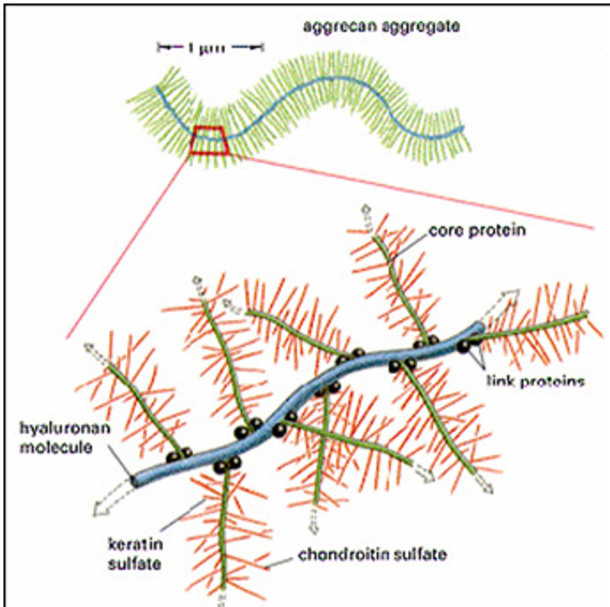
2 common polysaccharides associated with proteoglycans
hyaluronic acid and chondroitin sulfate
hyaluronic acid
makes ground substance very slippery and aids in lubrication, especially in joints
chondroitin sulfate
makes the ground substance more viscous and helps hold the matrix together, especially in cartilage and bone tissue
extracellular fibers
- abundant in most connective tissues and exists throughout the ground substance of the matrix
- provide structural support and strength for the connective tissue
3 major types of protein fibers in various connective tissues
collagenous fibers, reticular fibers and elastic fibers
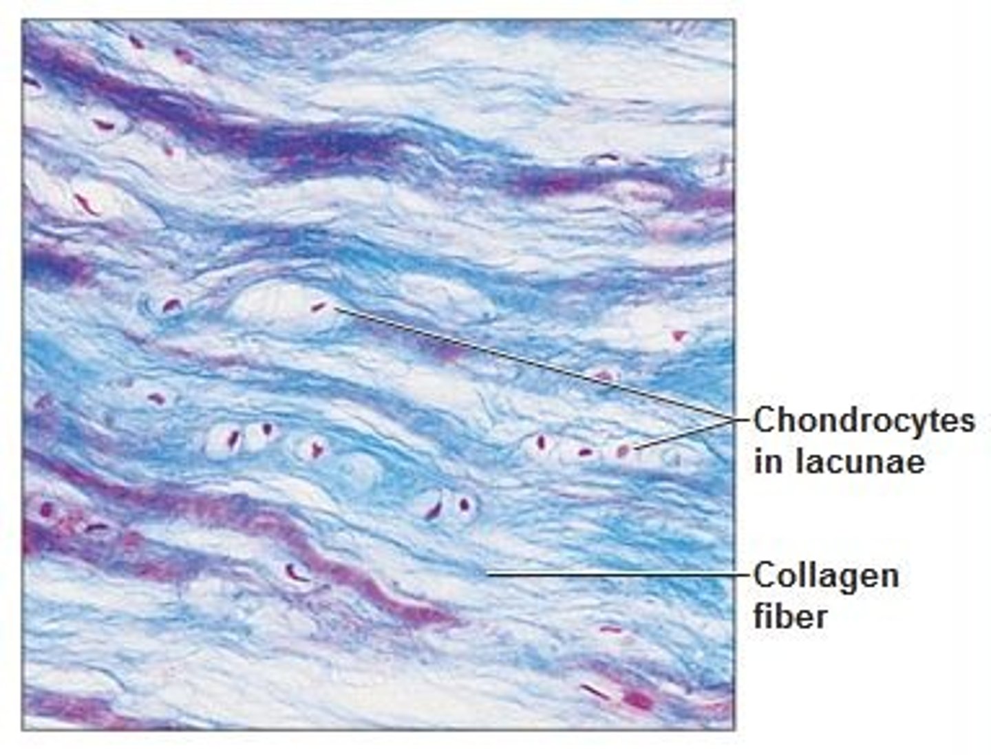
collagenous fibers
- made of a protein collagen and appear as white fibers in the matrix
- offer the connective tissue a great amount of resistance to tension
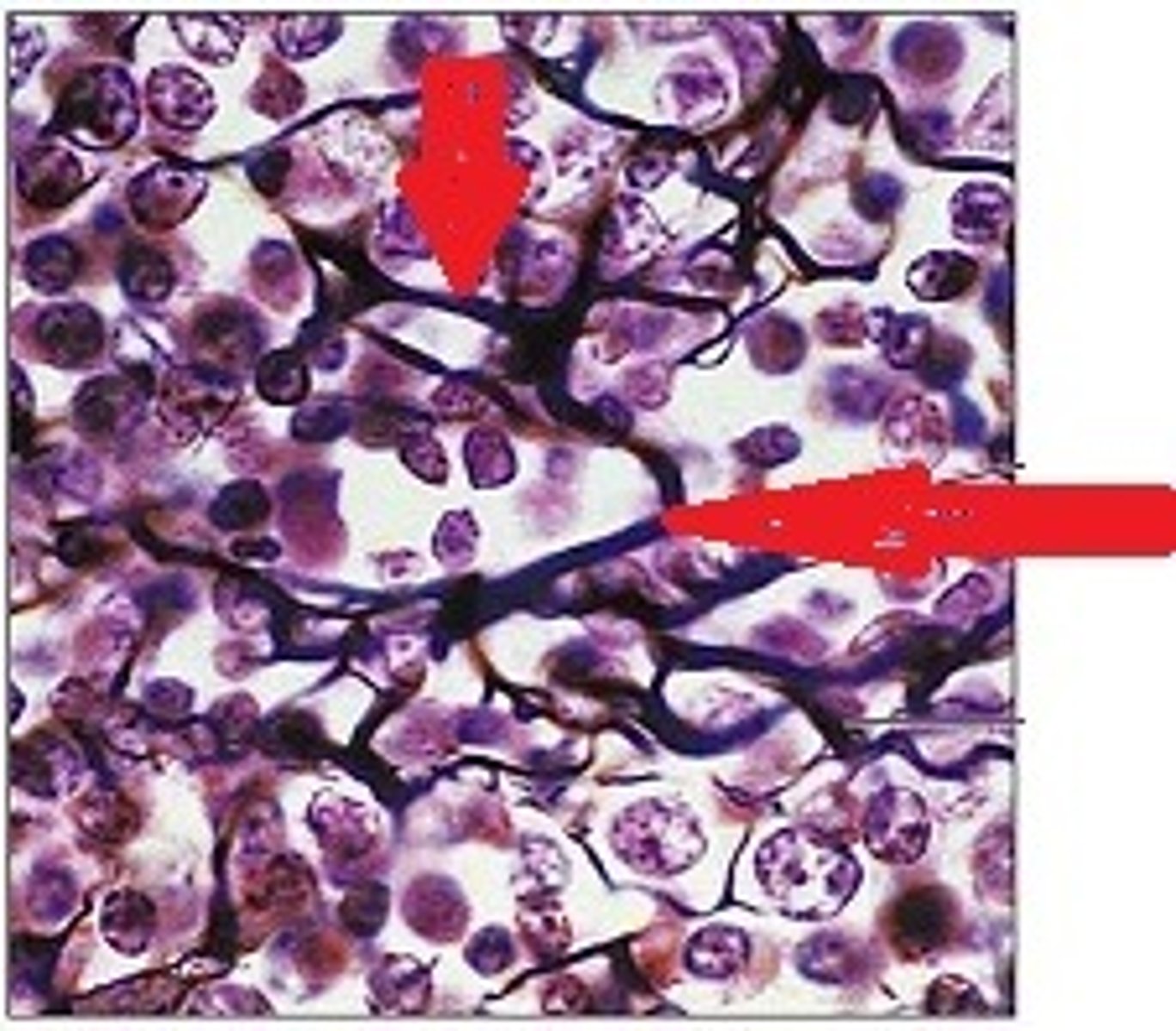
reticular fibers
very thin, highly branched collagenous fibers that give the matrix of certain connective tissues a net-like appearance
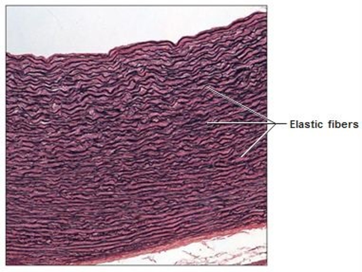
elastic fibers
- contain elastin protein and appear as yellow fibers
- allows certain connective tissue to recoil after stretching
fibroblasts
produce the matrix of most connective tissues
fibrocytes
what fibroblasts become for the most part
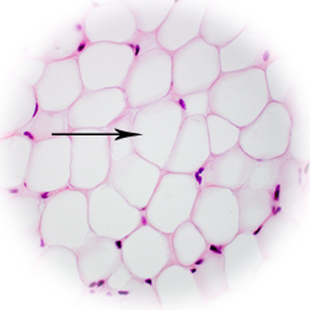
adipocytes
what fibroblasts become in adipose (fat) tissue
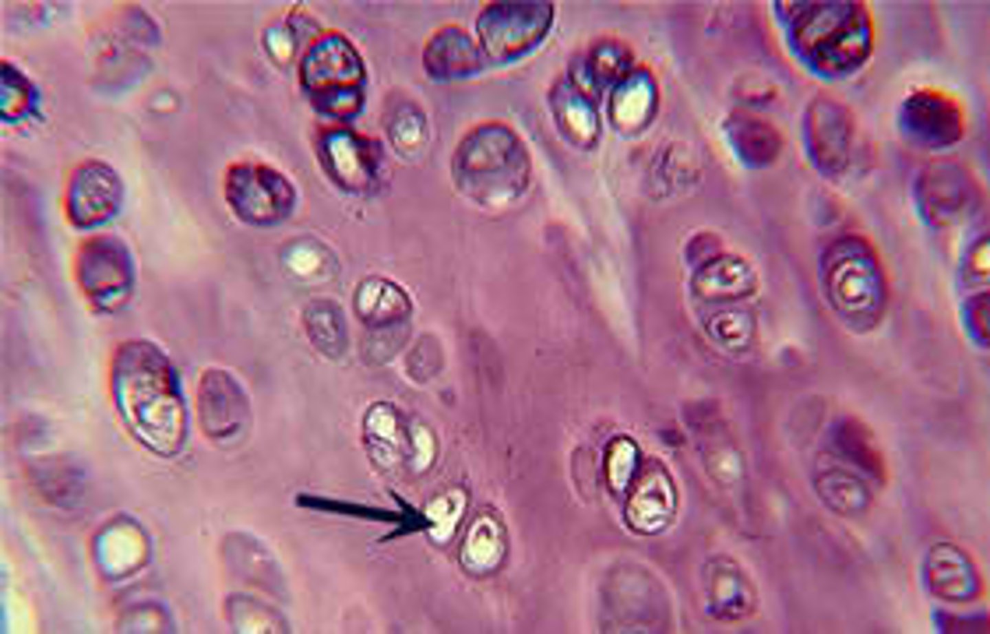
chondroblasts
produce the matrix of cartilage and eventually become chondrocytes
osteoblasts
produce the matrix of bone and eventually become osteocytes
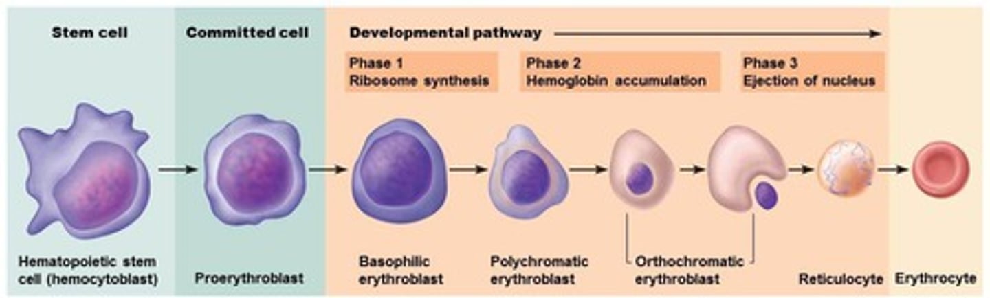
hemocytoblasts
cells that give rise to different blood cells but do not produce the liquid matrix, or plasma, of blood
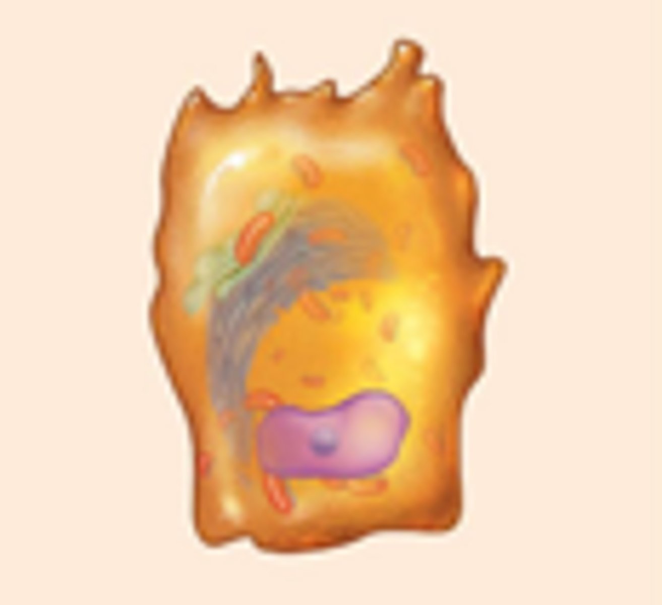
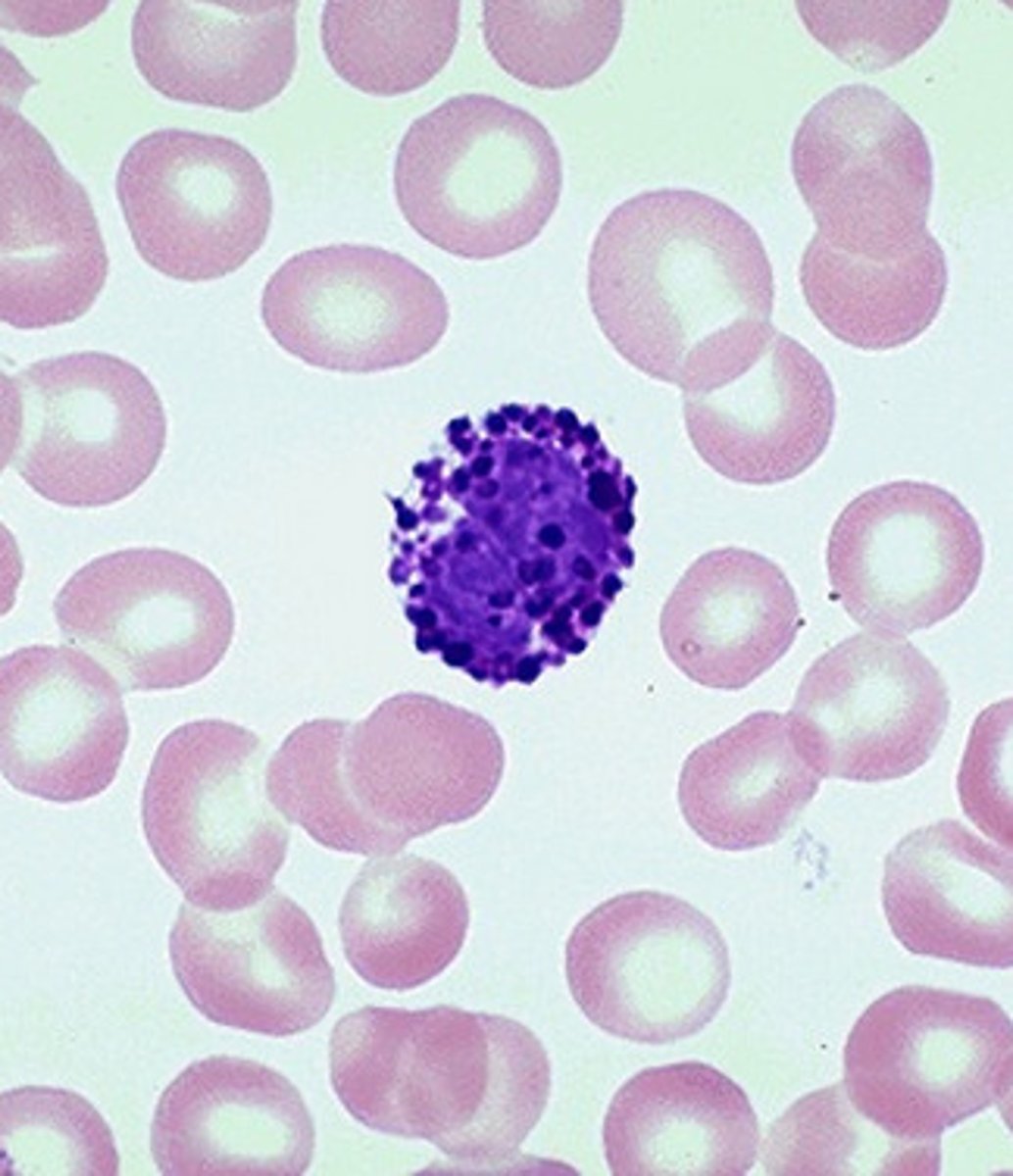
mast cells
congregate around blood vessels and when stimulated by a variety of chemicals release the chemical histamine
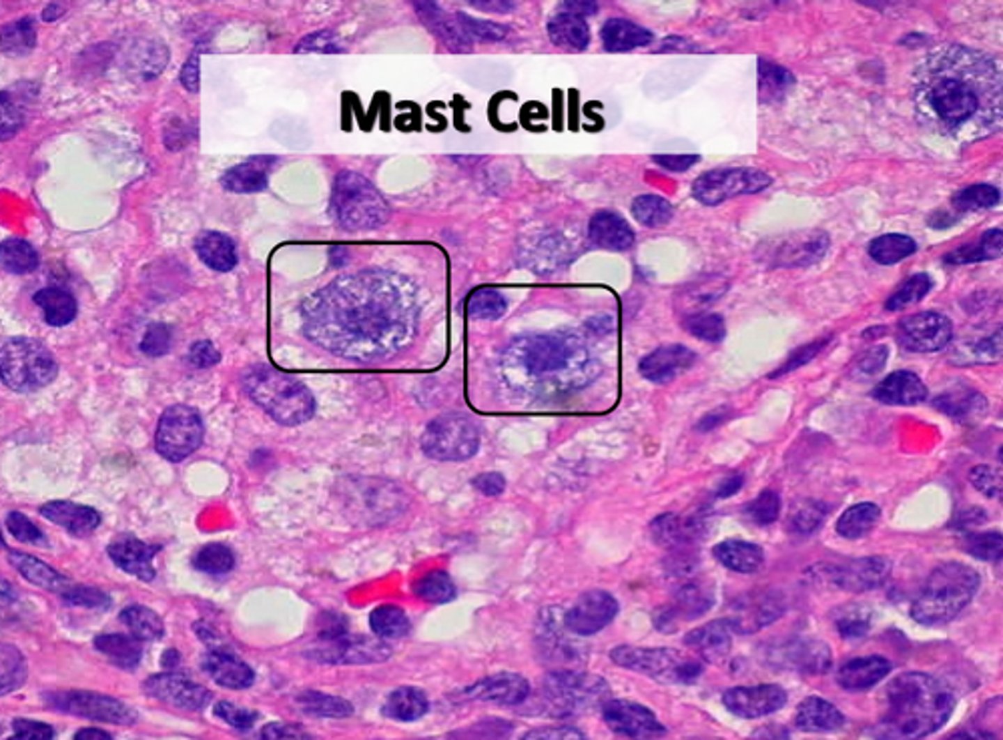
histamine
causes the blood vessels to dilate and become "leaky"
macrophages
- specialized white blood cells that once lived in the blood but squeezed out of the blood vessel into the surrounding tissues
- crawl around engulfing foreign particles and dead or damaged body cells
connective tissue proper
includes all connective tissues except bone, cartilage and blood
how is connective tissue proper subdivided?
loose connective tissue and dense connective tissue
loose connective tissue
have loosely packed intercellular fibers and include areolar, adipose and reticular tissue
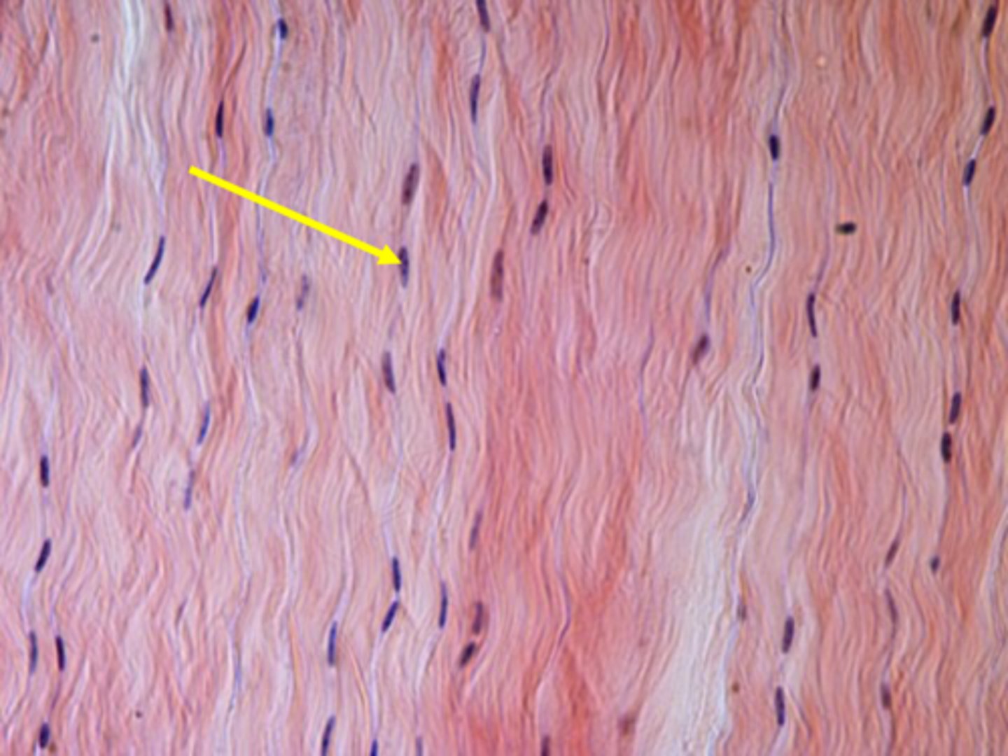
areolar tissue
has a gel-like matrix and contains collagenous, reticular, and elastic fibers produced by fibroblasts
lamina propria
areolar tissue beneath a mucous membrane
adipose tissue
a loose tissue containing adipocytes
functions of adipose tissue
energy storage, insulation, protection, and heat generation
adipose (fat) tissue consists in what 2 forms?
yellow fat and brown fat
yellow fat
- gets its characteristic yellow color from pigments such as carotene.
- most common fat in adults and is abundant just below the skin of the abdomen, buttocks and thighs