Anatomy and Physiology II Lab Practical II
1/75
There's no tags or description
Looks like no tags are added yet.
Name | Mastery | Learn | Test | Matching | Spaced | Call with Kai |
|---|
No analytics yet
Send a link to your students to track their progress
76 Terms
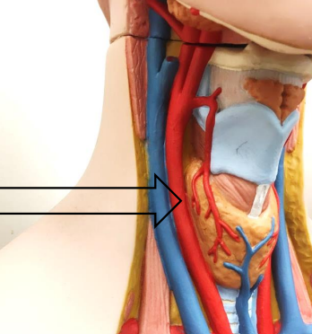
What is this?
Right Common Carotid Artery
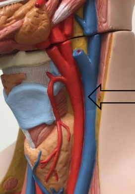
What is this?
Left Common Carotid Artery
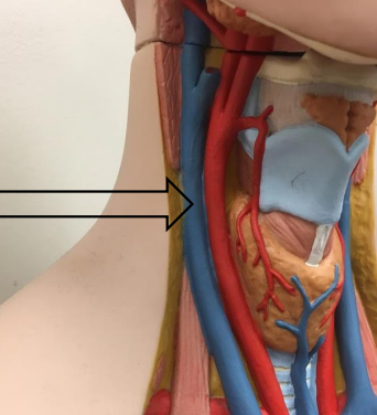
What is this?
Right Internal Jugular Vein
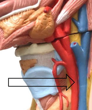
What is this?
Left Internal Jugular Vein
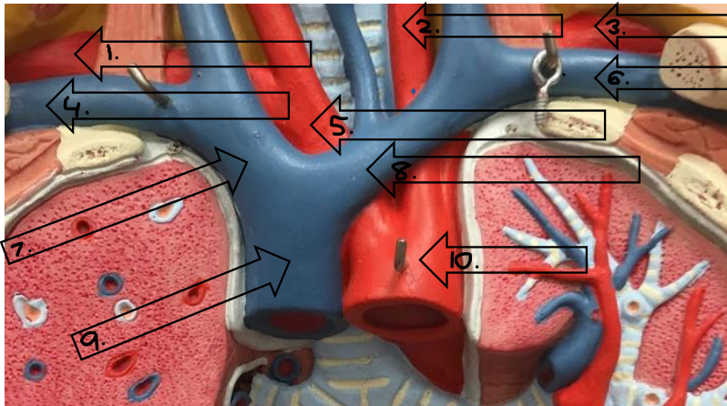
Label this image.
1. Right subclavian artery, 2. Left common carotid, 3. Left subclavian artery, 4. Right subclavian vein, 5. Brachiocephalic artery, 6. Left subclavian vein, 7. Right brachiocephalic vein, Left brachiocephalic vein, 9. Superior vena cava, 10. Aorta
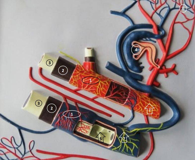
Label this image.
1. Tunica adventitia, 2. Tunica media, 3. Tunica intima, 4. Arteriovenosus anastomoses, 5. Valves in vein, 6. Capillaries, 7. Nerves
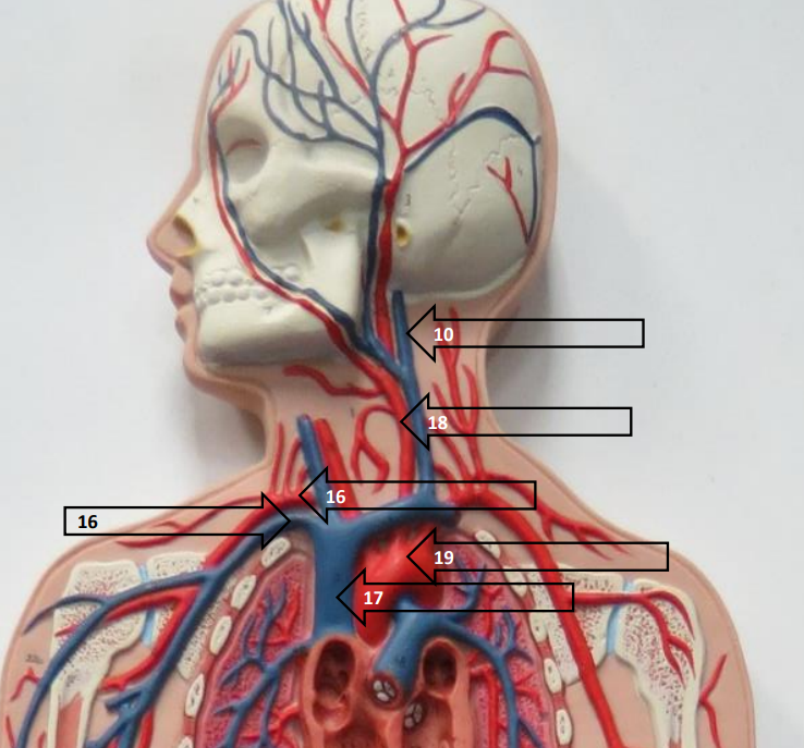
Label this image.
10 . Internal jugular vein, 18. Left common carotid, 16. (center) Right subclavan artery, 16. (right) RIght subclavian vein, 19. Aortic arch, 17. Superior vena cava
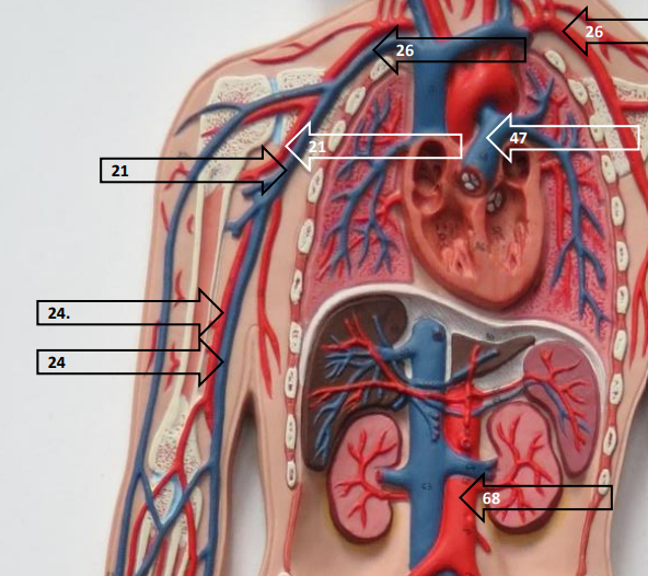
Label this image.
26. (left) Left subclavian artery, 16 (right) Right subclavian vein, 47. Pulmonary artery, 21. (center) Right axillary artery, 21. (right) Right axillary vein, 24. (top) Right brachial artery, 24 (bottom) Right brachial vein, 68. Abdominal aorta
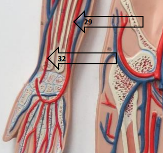
Label this image.
29. Right ulnar artery, 32. Right radial artery
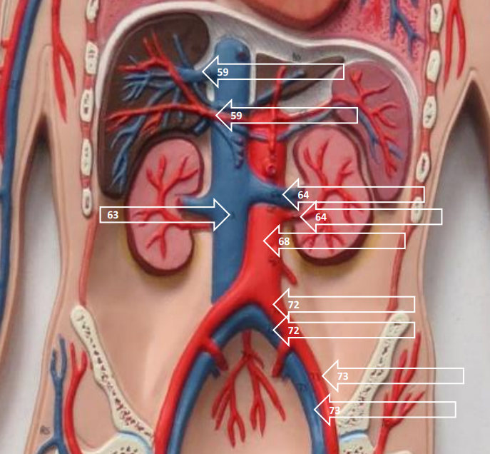
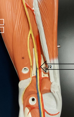
Label this image.
59 . (top) Hepatic vein, 59. (bottom) Hepatic artery, 64. (top) Left renal vein, 64. (bottom) Left renal artery, 63. Inferior vena cava, 68. Abdominal aorta, 72. (top) Left common iliac artery, 72. (bottom) Left common iliac vein, 73. (top) External iliac artery, 73. (bottom) External iliac vein
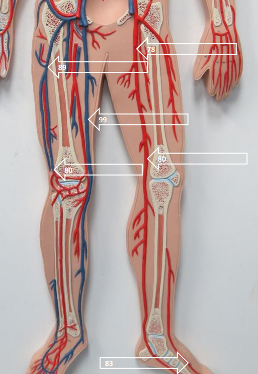
Label this image.
78. Femoral artery, 89. Femoral vein, 99. Greater saphenous vein, 80. (left) Popliteal vein, 80. (right) Popliteal artery, 83. Dorsalis pedis artery
What is this?
Axillary artery.
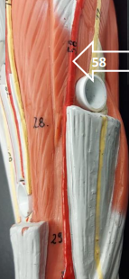
What is this?
Brachial artery.
What is this?
Radial artery.
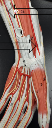
What is this?
Top arrow: Ulnar artery, Bottom arrow: Superficial palmar branch of radial artery
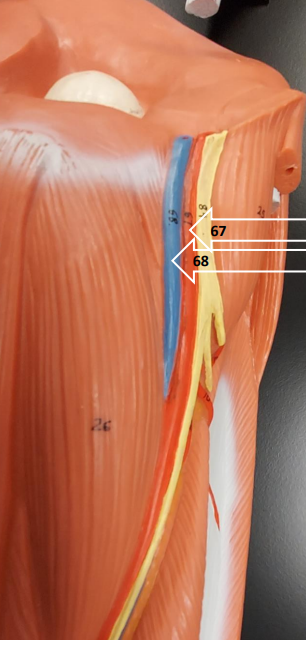
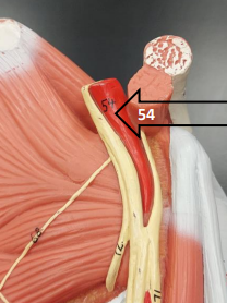
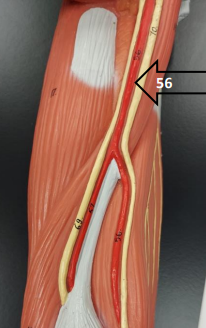
What is this?
67. Femoral artery, 68. Femoral vein
What is this?
Popliteal artery.
What is the tunica media made of?
Smooth muscle.
What is the tunica intima made of?
Simple squamous epithelium (makes up endothelium).
What is the arteriovenosus anastomoses?
A shunt (2/more blood vessels that come together to make something larger than a capillary).
Where is respiratory epithelium and what is it made of?
Conducting zone; consists of pseudostratified ciliated columnar epithelium.
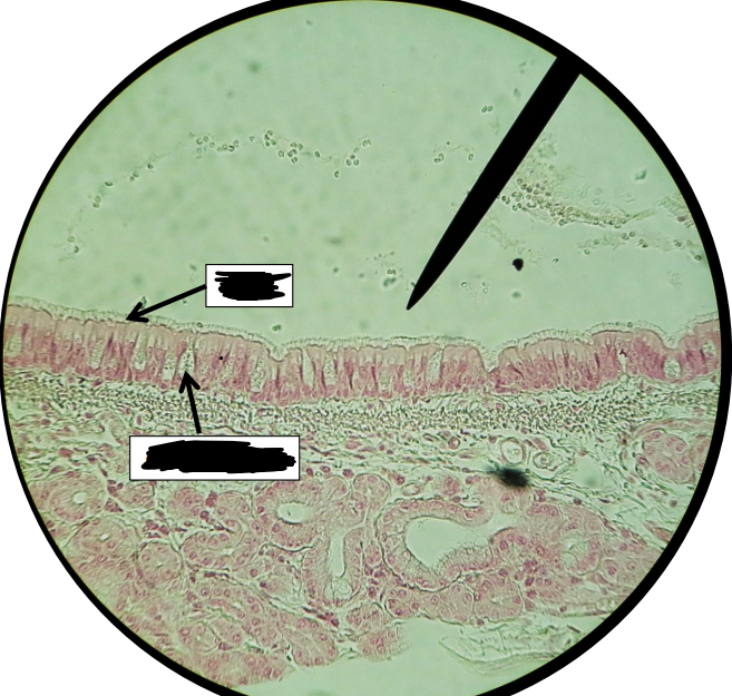
What is this?
Respiratory epithelium; top box is cilia, bottom box are goblet cells.

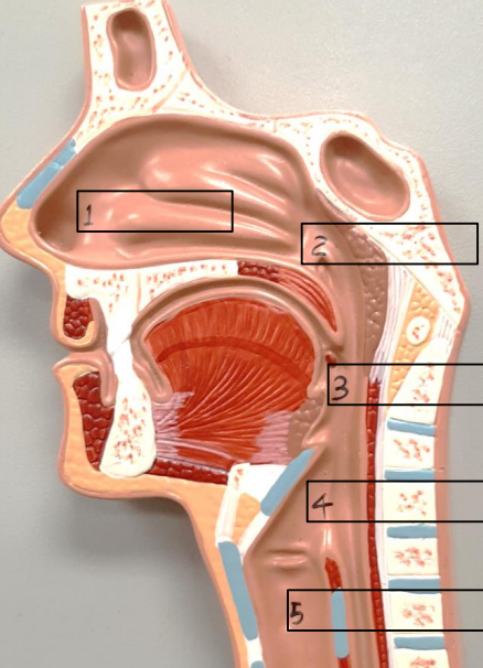
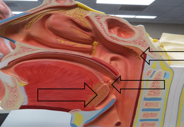
Label this image (numbers only).
1 . Nasal conchae (superior, middle, inferior), 2. Pharyngeal tonsil, 3. Hard palate, 4. Soft palate, 5. Uvula, 6. Palatine tonsil, 7. Lingual tonsil, 8. Epiglottis
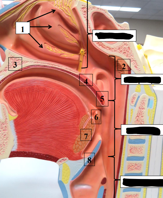
Label this image (black boxes only).
Top to bottom: Nasal cavity, Nasopharyx, Oropharynx, Layngopharynx
Why is the hard palate hard and the soft palate soft?
The hard palate is bone and the soft palate is not.
How many palatine tonsils are there?
Two.
What does the epiglottis do?
Closes to protect the trachea.
What are the parts of the pharynx?
Nasopharynx, oropharynx, laryngopharyx.
What does the nasal conchae do?
Helps to humidify the air going in.
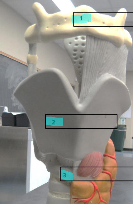
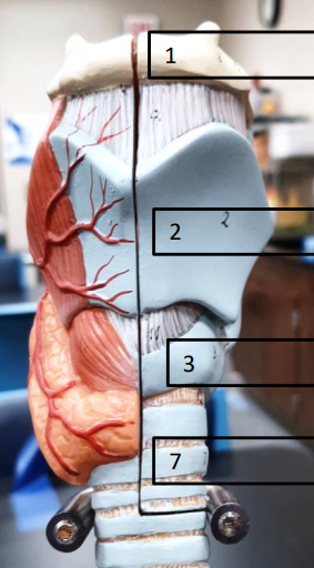
Label this image,
1. Hyoid bone, 2. Thyroid cartilage, 3. Cricoid cartilage
What is this?
Epiglottis.
What are these and what do they do?
Trachea cartilage rings; keeps windpipe open. Do not completely circle the trachea because of the esophagus’ need to expand.
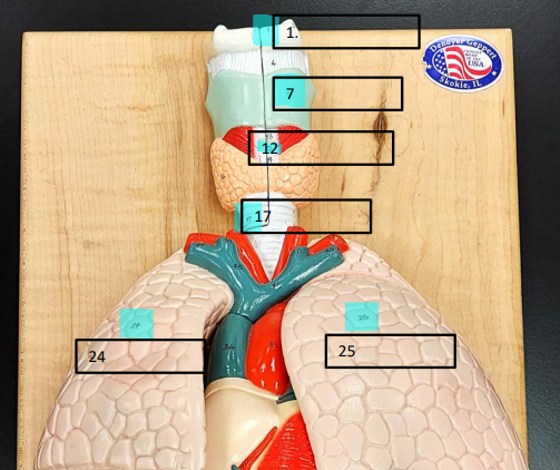
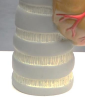
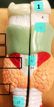
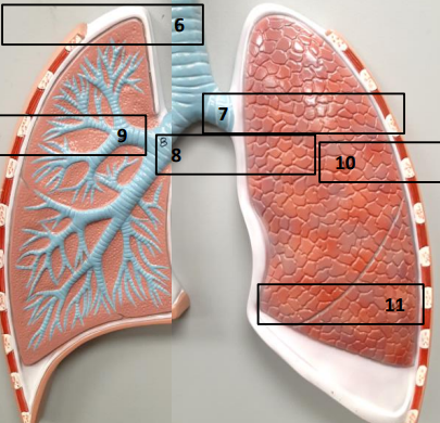
Label this image.
1 . Hyoid bone, 7. Thyroid cartilage, 12. Cricoid cartilage, 17. Tracheal cartilage, 24. Right lung, 25. Left lung
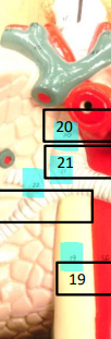
Label this image.
20. Trachea, 21. Bifurcation, Empty Box: Right bronchus, 19. Esophagus
Label this image.
1. Hyoid bone, 7. Thyroid cartilage, 12. Cricoid cartilage, Empty Box: Left bronchus, 17. Tracheal cartilage
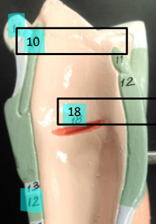
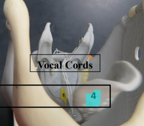
Label this image.
10. Epiglottis, 18. Vocal cords, 12. Cricoid cartilage
Label this image.
1 . Nasal cavity, 2. Nasopharynx, 3. Oropharynx, 4. Laryngopharyx, 5. Laryngeal cavity
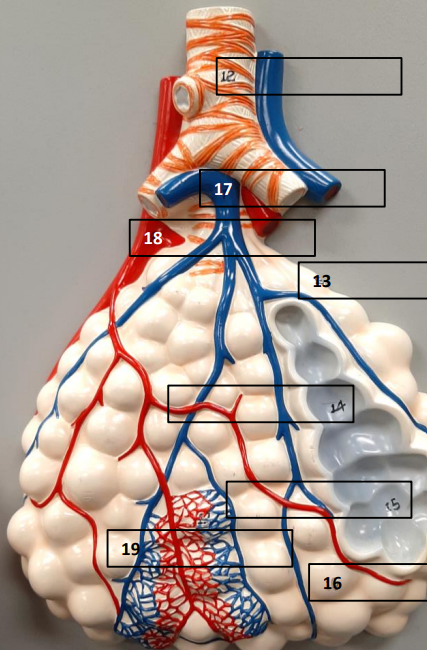
Label this image.
12. Terminal bronchiole, 17. Pulmonary artery, 18. Pulmonary vein, 13. Respiratory Bronchiole, 14. Alevolar duct, 15. Alveolar sac, 19. Capillary plexus, 16. Pulmonary alveoli
Label this image.
6. Trachea, 7. Left main stem bronchus (thicker), 8. Right main stem bronchus (thinner), 9. Secondary bronchus, 10. Left lung (superior lobe), 11. Left lung (inferior lobe)
Label this image.
1. Hyoid bone, 2. Thyroid cartilage, 3. Cricoid cartilage, 7. Tracheal cartilage
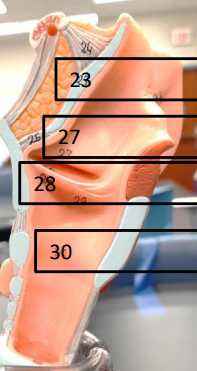
Label this image.
23. Epiglottis, 27. Vestibular fold, 28. Vocal cord, 30. Trachea
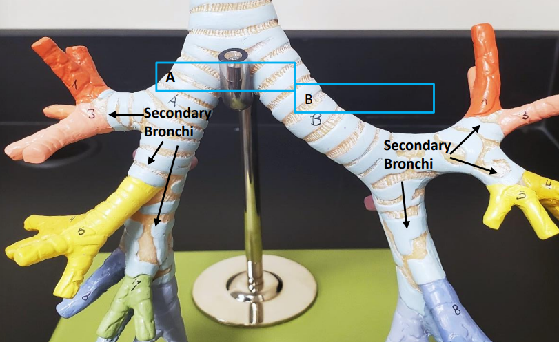
Label this image.
A. Right main bronchus, B. Left main bronchus, 1-10: Tertiary/Segmental Bronchi
What is capacity?
A combination of two or more volumes.
What is functional residual capacity?
Volume of air in lungs at the transition point.
What is tidal volume?
Amount of air moved in and out during quiet breathing.
What is inspiratory reserve volume?
Extra air inhaled above tidal volume.
What is expiratory reserve volume?
Extra air exhaled above tidal volume.
What is vital capacity?
Total amount of air that can be moved in one breath with maximum inhalation and exhalation.
What is residual volume?
Air left in the anatomical dead space.
What is total lung capacity?
Entire volume of air the lungs can hold.
What is the equation for vital capacity?
VC = TV + IRV + ERV
What is the equation for total lung capacity?
TLC = VC + RV
What is the lymphatic system?
A series of organs, tissues, and vessels in the body and help the immune system and return excess fluid to the bloodstream.
What is this?
Inguinal lymph nodes (A lymph node cluster).
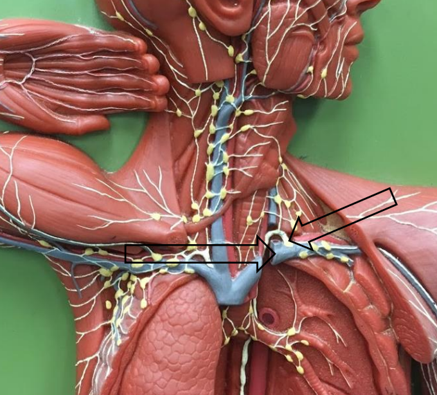
Label this image.
Top arrow: cervical lymph node cluster, Bottom arrow: axillary lymph node cluster.
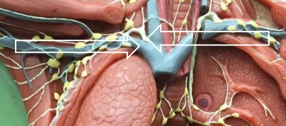
Label this image.
Right arrow: Right subclavian vein, Left arrow: Right lymphatic duct
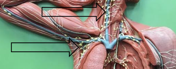
Label this image.
Right arrow: Left subclavian vein, Left arrow: Thoracic duct
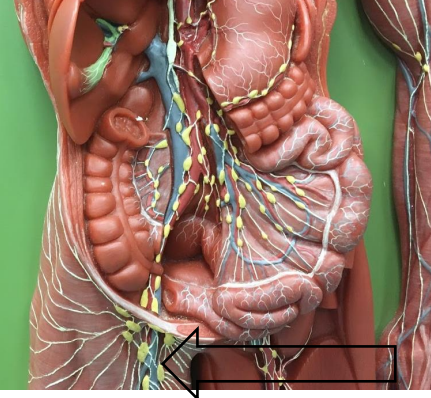
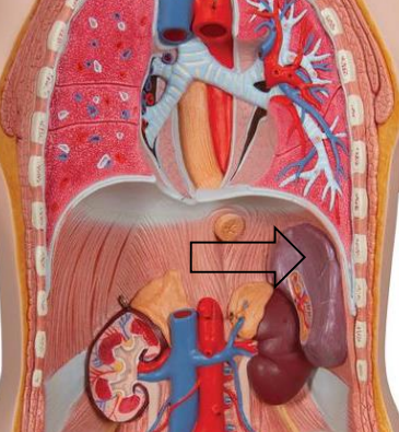
What is this? What does it do?
Cisterna chyli (entire white line), collects lymph from abdomen and lower extremities.
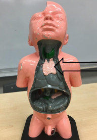
What is this?
Thymus gland.
Where is the thymus gland? What is special about it?
It’s located in the chest near the heart and is largest in puberty before becoming smaller. Immature lymphocytes go there to mature before entering the blood.
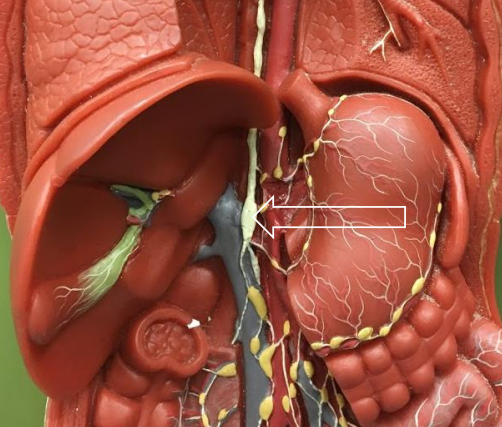
What is this?
The spleen.
What is important about the spleen?
It has red and white pulp (tissue), has a ‘standing army of immune cells that respond to bodily needs, makes RBCs in fetuses and anemic adults, and stores platelets until they’re needed to replace the ones used in hemorrhaging.
What does red pulp do?
Filters the venous blood using macrophages and lymphocytes,
What does white pulp do?
Filters arterial blood using lymphocytes and macrophages.
What do splenic macrophages do?
Break down old RBCs and remove the iron that is then sent to the liver.
What is MALT? What are the three structures that make it up?
Mucosa-Associated Lymphoid Tissue. Made up of tonsils, appendix, and Peyer’s patches.
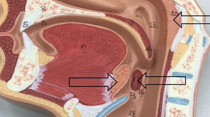
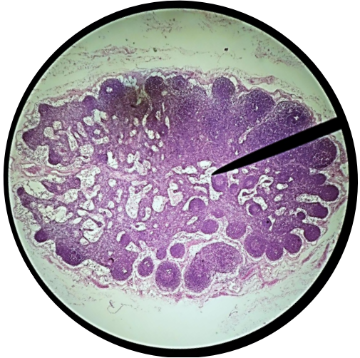
Label this image.
Top arrow: Pharyngeal tonsil, Arrow Directly Below Top Arrow: Palatine Tonsil, Other Bottom Arrow: Lingual Tonsil
Label this image.
Top Arrow: Pharyngeal tonsil, Middle Arrow: Palatine Tonsil, Bottom Arrow: Lingual Tonsil
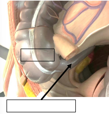
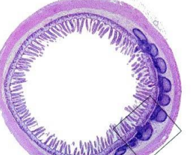
Label this image.
Top Box: Cecum, Bottom Box: Appendix
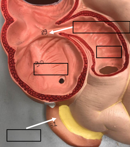
Label this image.
29 . Ileocecal valve, 30. Cecum, 31. Appendix, Unlabeled Box: Ileum
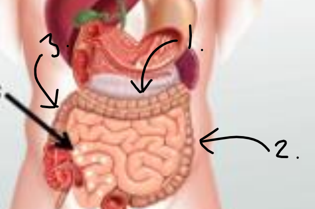
Label this image.
1 . Transverse colon, 2. Descending colon, 3. Ascending colon, Unlabeled Arrow: Peyer’s patches.
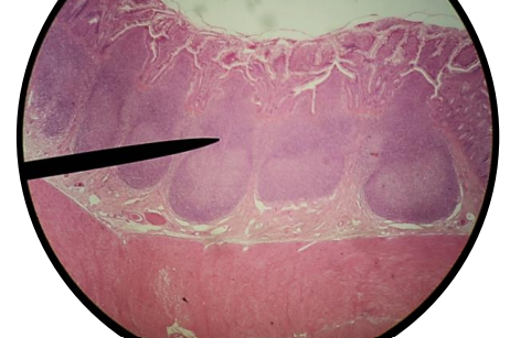
What is this?
Peyer’s patches.
What is this?
Peyer’s patches.
What is this?
Lymph node. The mesh like nature of the node which is full of macrophages and lymphocytes that help to remove and destroy any particles that are being carried in the lymph fluid before it is returned to the venous circulation.
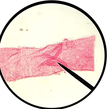
What is this?
Lymph Vessel with Valve; have a one-way flow of fluid, usually against gravity.
What is special about lymph vessels?
Lymph vessels use skeletal muscle contraction and the one-way valves to help move lymph through the lymphatic system to the R lymphatic and Thoracic duct where it is returned to the venous circulation right near the heart.