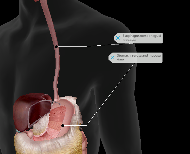
esophagus and stomach
1/16
There's no tags or description
Looks like no tags are added yet.
Name | Mastery | Learn | Test | Matching | Spaced | Call with Kai |
|---|
No analytics yet
Send a link to your students to track their progress
17 Terms
phases of food moving down espohagus
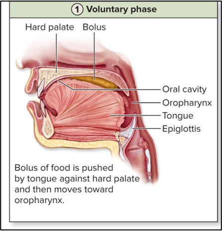
voluntary phase
pharyngeal phase
esophageal phase
voluntary phase
voluntary control
upper musculature of RT is skeletal
tongue pushes a bolus of food backwards. moves into oropharynx
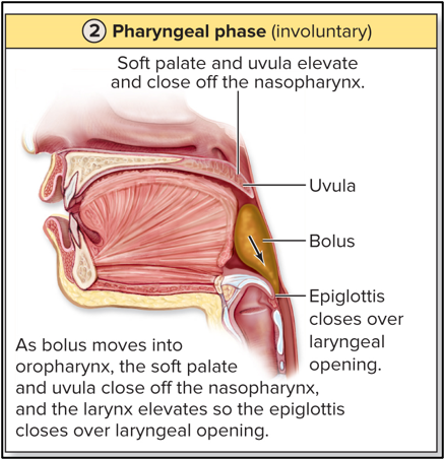
pharyngeal phase (involuntary)
swallowing draws the larynx up and propulsion of food
oral and nasal cavities are blocked
can’t breathe during this phase
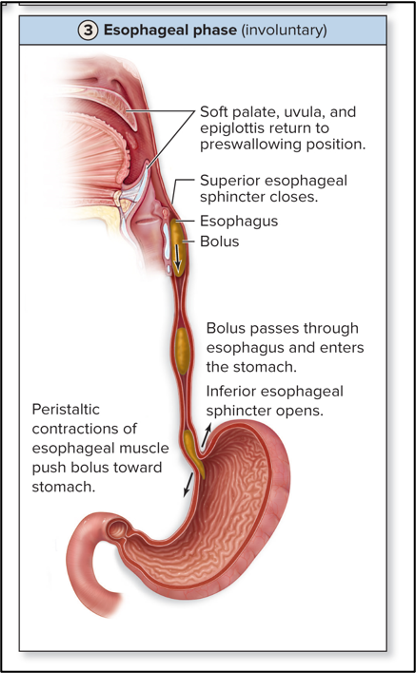
esophageal phase (involuntary)
involuntary actions; peristalsis
primary peristaltic contraction forces food down length of esophagus (pressure high behind food)
lower esophageal sphincter will then open
sphincters in the esophagus
superior esophageal sphincter: ring of circular skeletal muscle at the upper end of esophagus
inferior esophageal sphincter: ring of circular smooth muscle at the lower end of esophagus that includes part of diaphragm; physiological sphincter
physiological sphincter
contraction of diaphragm closes opeining
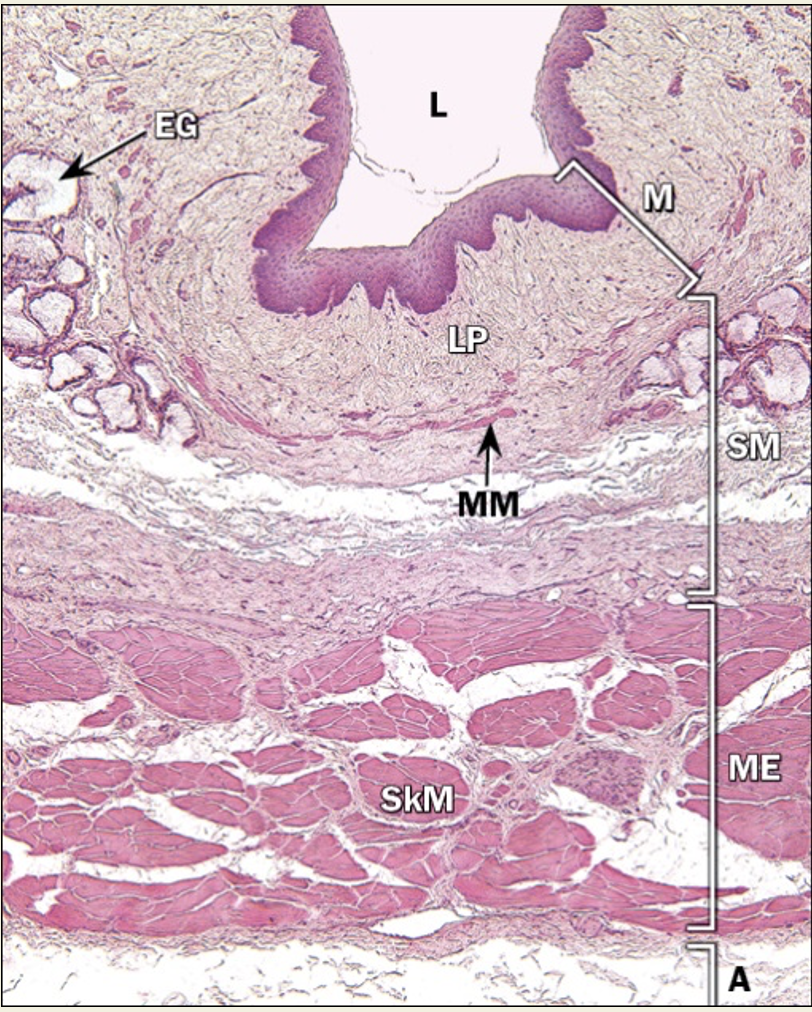
histology of esophagus
mucosa
non-keratinized stratified squamous epithelium
esophageal cardiac glands in lamina propria (near pharynx and stomach- produce mucus)
single layer of muscularis mucosae
submucosa
dense irregular CT and loose CT
esophageal glands producing mucus
submucosal plexus of ENS
muscularis
inner circular and outer longitudinal muscle
stories of thirds; proximal 1/3 is skeletal, middle 1/3 skeletal/smooth, distal 1/3 smooth
myenteric plexus
adventitia
outer dense CT for anchoring
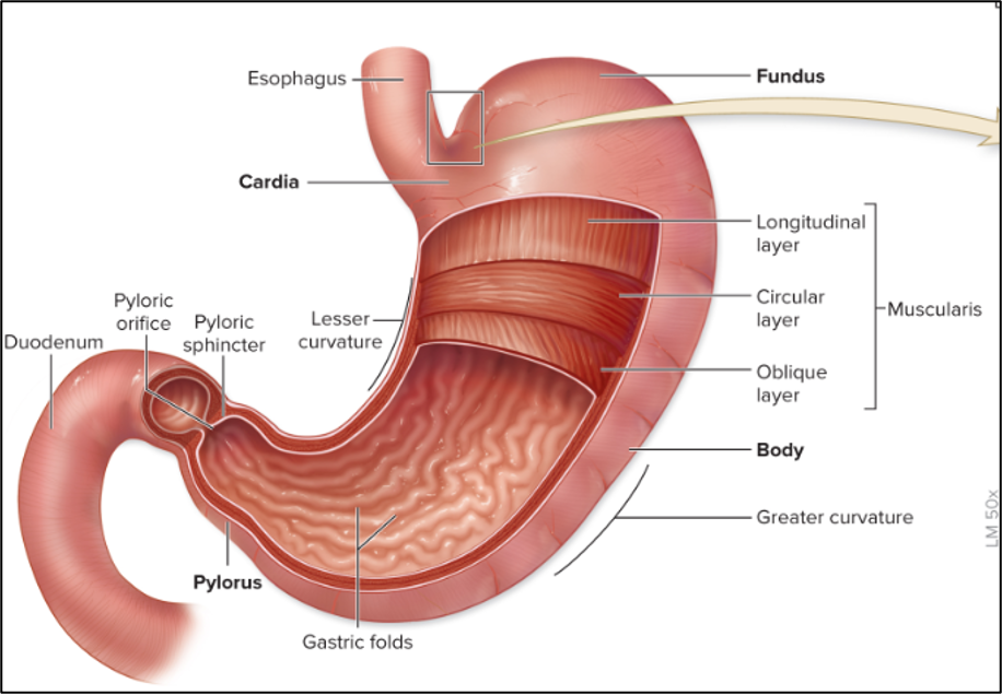
stomach
J shaped muscular organ divided into 4 regions
cardiac- GE junction
Fundic- storage of food
body- major part of stomach
pyloric- distal end, above duodenum
Curvatures of stomach
greater curvature- greater omentum (Stores fat) suspended here
lesser curvature- lesser omentum (Passageway for arteries, veins, lymphatics etc.) suspended here
sphincters of stomach
cardiac sphincter (infoerior esophageal sphincter)
pyloric sphincter

mucosa
simple columnar epithelium
gastric pits → open into gastric glands
glands have specialized secretory cells (parietal, chief, enteroendocrine)
stem cells
rugae
rugae
mucosa and submucosa wrinkle and fold to produce these
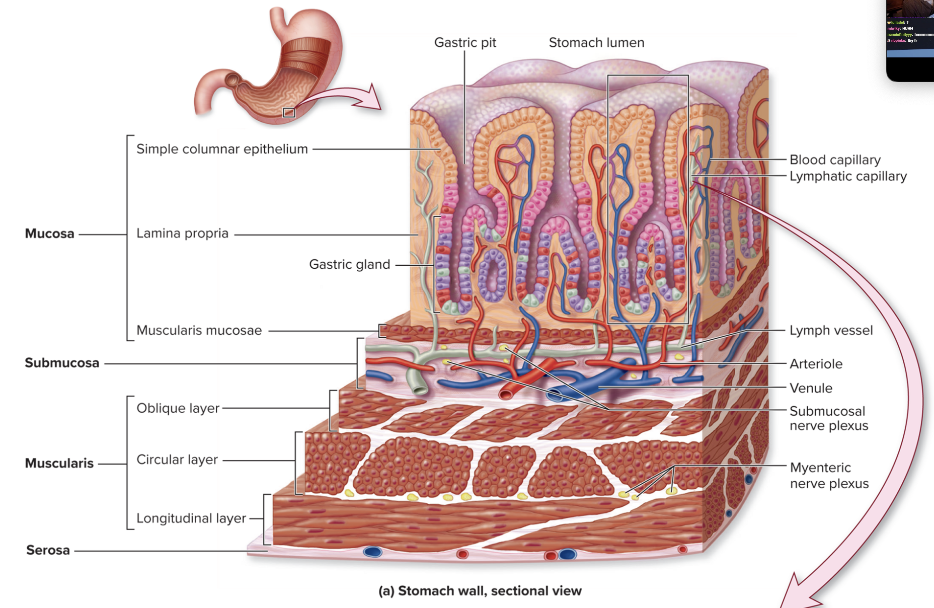
muscularis external
THREE layers or muscle
internal oblique (most well defined in cardiac region)
middle circular
outer longitudinal
cell types in stomach
Mucous and mucus neck cells
Parietal cells
Chief cells
Enteroendocrine cells
Stem cells
surface mucous and mucus neck cells
surface release basic mucus
neck cells release acidic mucus
protects mucosa from HCl and enzymes
parietal cells
release HCl and intrinsic factor
HCl activates pepsin and lingual lipase; helps liquify food
intrinsic factor enables small intestine to absorb vitamin B12 (RBC formation)
chief cells
release pepsinogen (precursor of pepsin) and gastric lipase
pepsinogen converted to pepsin which digests protein
gastric lipase digests fat
how does stomach protect itself
mucus production
tight junctions
stem cells replace differentiated cells
buffering

histology of stomach
mucosa
simple columnar epithelium (surface mucous cells) with openings (gastric pits) with mucous neck cells
pits open into glands: multiple cell types (parietal, chief, enteroendocrine, stem)
highly vascularized lamina propria
two layered mucularis mucosae
submucosa
dense irregular CT and loose CT
submucosal plexus of ENS
muscularis
inner oblique (most defined in cardiac region)
middle circular
outer longitudinal
myenteric plexus of ENS
serosa
simple epithelium
loose CT