PHYS C6
1/38
There's no tags or description
Looks like no tags are added yet.
Name | Mastery | Learn | Test | Matching | Spaced | Call with Kai |
|---|
No analytics yet
Send a link to your students to track their progress
39 Terms
Requisites for Generating X-rays
x-ray generator needs to:
▪ produce electrons
▪ accelerate electrons to high speed
▪ stop them abruptly
Diagnostic x-ray tubes
conventional Coolidge Tubes with Rotating Anodes
Thermionic Emission
Tungsten filament heated with low-voltage filament current (<10 A)
Electron excited, become loosely held
High enough temperature, electrons break free of nucleus
Increasing filament current, increases temperature, increases number of incident electrons, increases x-ray beam intensity
Space Charge
electron cloud just beyond filament where liberated electrons sit for a fraction of a second
Space charge effect
there is a limit to the number of electrons that can be in space charge before negative charge becomes strong enough to repel electrons back to filament, limiting emission
Focusing Cup
Used to concentrate emitted electrons into a tight beam towards anode target
Needed because electrons repel each other
Negatively charged concave molybdenum cup surrounding the cathode filament on 3 sides
Defines focal spot size which determines image sharpness
Dual-Focus X-ray Tube
Small and large filament parallel to each other
Only one filament energised at once
Small filament produces a narrow electron beam thus small focal spot, high spatial resolution, limited heat capacity
Large filament produces a less focused electron beam thus large focal spot, short-duration exposures, used for thicker body parts
Cold Carbon Nanotubes (CNT)
high voltage is applied to vertically aligned CNTs creating intense electric fields that extract electrons via quantum tunneling
releasing electrons without heat removes the need for cooling oil-bath and allows precise, instantaneous switching of the x-ray beam (start, stop, redirect)
have been used in portable systems, but lifetime, reliability, cost and integration challenges for universal adoption
Acceleration
Repulsion between incident electrons and focusing cups starts moving electron stream
During exposure focusing cup gets strong negative charge and anode gets strong positive charge, causing electrons to travel at high speed (high kinetic energy)
Voltage/electric potential difference
1V is the work required in Joules to move one Coulomb of charge from one point to another
Voltage formula
V = E/Q
eVs
1eV is the enrgy given to an electron accelerated through a potential difference of 1V
1eV = 1.602 × 10⁻¹⁹ J
tube voltage of 100kV will give an electron 100keV of energy
Tube current
mA
number of electrons passing from cathode to anode
X-ray tube Energy Changes/Efficiency
Electrical potential energy converted to kinetic energy then at target surface 1% is converted to x-rays, 99% to heat
transformers
Step-up transformer creates kVp
Step-down transformer heats filament
Other X-ray Generator Circuit components
Rectifier ensures unidirectional electron flow
mA Selecter controls filament current and temperature which determines tube current
Exposure Timer sets exposure duration and is used to calculate mAs
kVp
kilovoltage peak
maximum voltage applied across x-ray tube
equal to max kinetic energy of electrons
kVp Selection
technologist sets maximum voltage on console
autotransformer determines voltage applied to step-up transformer
step-up transformer boosts voltage to kV range
Relationships
kVp controls x-ray energy (thus penetration) and quantity/intensity
mA contols x-ray quantity/intensity
Target
Higher atomic number increases x-ray intensity and energy
Tungsten used because of high Z, melting point, thermal conductivity
Tungsten vaporises and is deposited onto inside of glass enclosure over
time, upsetting electrics and eventually causing tube failure
Molybdenum used for mammograhpy, producing lower energy x-rays to maximise soft tissue contrast
Stationary or rotating targets
Stationary anode
used in some portable imaging systems
Rhenium-alloyed tungsten imbedded in end of a 45° angled copper rod
Rotating Anode
Promotes cooling between exposures by evenly dispersing beam across entire surface
Enables longer scans and higher doses
Improves lifetime
a thin rhenium-alloyed tungsten focal track on top acts as target, molybdenum in middle (lightweight so allows faster spinning and thermal conductivity that pulls heat away form focal spot), graphite on bottom to absorb heat
Rotor
Cylindrical copper structure surrounding a soft iron core
Attached to the anode stem, sits inside the rotating magnetic field
Stator
electromagnetic coils arranged as a three-phase winding
supplied AC power to create roating magnetic field around the rotor’s central axis
Faraday’s Law
time-varying magnetic flux passing through a circuit induces an electromotive force (EMF) directly proportional to the rate of change of that flux
Torque
eddy currents induced in the conductive rotor and produce their own magnetic field that opposes direction of stator’s magnetic fields (lenz’s law)
this produces a torque force causing rotor to rotate
Anode Operation
~ 3000 rpm
temp can be >1000°C
Glass envelope
contains components within vacuum enclosure
X-ray tube window
thin area of enclosure(~5 cm2) through which useful part of x-ray beam is emitted
Liquid Metal Bearing (LMB)
liquid metal alloy lubricants replace ball bearings allowinf for silent operation, higher rpm, better anode cooling, and increased lifespan
Rotating envelope tubes
entire vacuum tube rotates with anode acting as part of outer wall
cooling liquid oil circulates directly against heated anode allowing heat removal via conduction rather than radiation
more efficient cooling and bearings can be on outside for easy cooling and maintanince
Types of radiation generating x-rays
bremsstrahlung (braking radiation) - continuous curve on spectrum
characteristic radiation - peaks on spectrum
Bremsstrahlung Radiation
Incident electrons attracted to positive Coulomb field around atom’s nucleus
This field causes electron to slow down and change direction
Lost kinetic energy emitted as x-ray photon
X-ray photons have continuous energy ranging from 0 to max energy of the incident electrons but most is located at ~1/3 of max kVp
Energy of x-ray photon depends on electron proximity to nucleus and energy of incident electron (tube voltage)
Major contibuter to x-ray spectrum in keV energies, sole contributer at MeV energies
Characteristic Radiation
Incident electrons eject target atom’s inner-shell electrons
incident electron keeps any excess energy that wasnt needed to overcome binding energy
outer-shell electrons fill the vacancy and emits a x-ray photon with an energy specific to energy difference between shells
Multiple peaks on x-ray spectrum because electrons that fill vacancy come from different outer shell
Instead of releasing an x-ray, energy can be transferred to another orbital electron, which is then ejected from atom as Auger electron (and can go on to cause x-ray emission)
X-ray Energy Spectrum
number of x-ray photons as a function of their energy
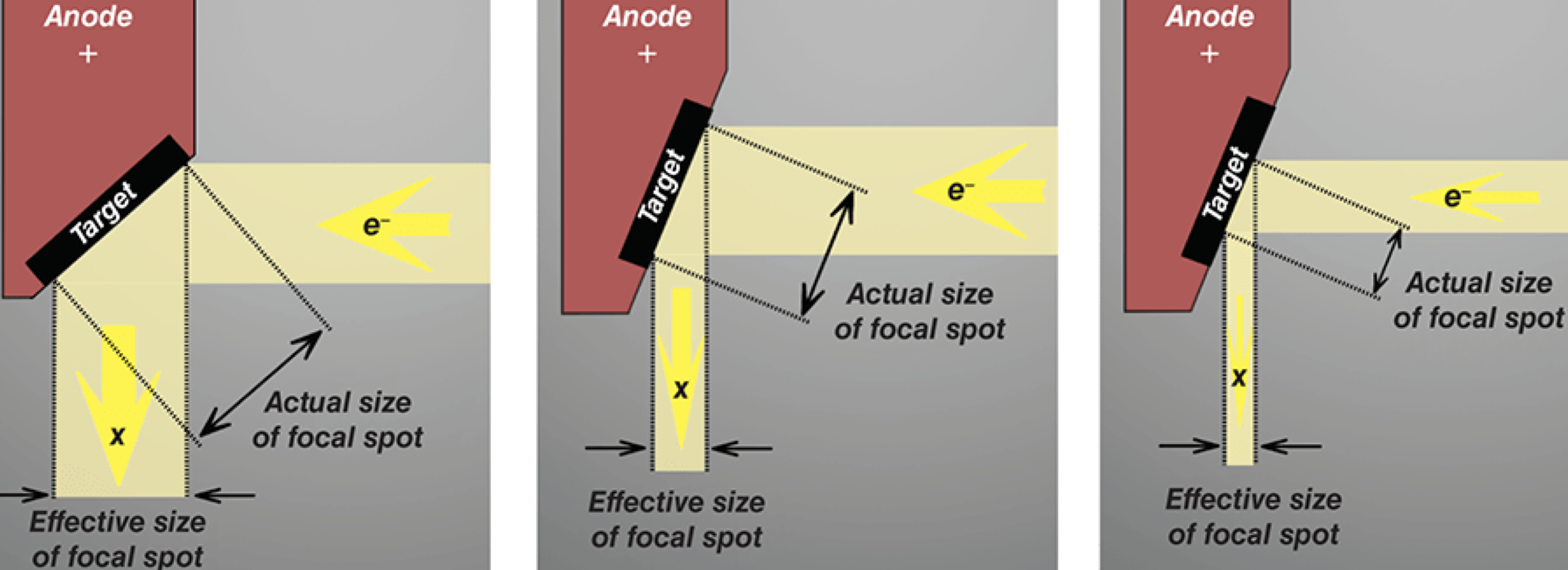
Focal Spot - def + link to image quality
Area of target where electrons strike and emit x-rays
Smaller focal spot = better spatial resolution
Line Focus Principle
Angling anode between 5 to 20 degrees (usually at 12 degrees) causes effective focal spot size to be much smaller than actual focal spot size Small effective focal spot improves spatial resolution
Large actual focal spot size allows heat to be dissipated over larger area to prevent tube melting
Smaller target angle = smaller effective focal spot size
Heel Effect
Electrons interact with target atoms at various depths in the target
x-rays emitted towards anode (target) must travel a greater distance through the target material, thus get attenuated more than x-rays emitted towards cathode
thus intensity of x-rays emitted through ‘heel’ of target is lower than (by up to 45%) compared to cathode side
Focal Effect
Effective focal spot is smaller, thus has lower radiation intensity, on anode side of x-ray field than on cathode side
So anode side produces shaper images but dissipates heat less effectively