MHS1002- Human Structure and Function (Week 7-12)
1/54
Earn XP
Description and Tags
Clinsci year 1- session 1
Name | Mastery | Learn | Test | Matching | Spaced | Call with Kai |
|---|
No analytics yet
Send a link to your students to track their progress
55 Terms
Heart- Mediastinum
Space that holds heart
Between lung
What is the position of the heart (in mediastinum) compared to diaphram?
Sits on top
What is the position of the heart (in mediastinum) compared to Sternum?
Sits posteriorly to sternum
What is the position of the heart (in mediastinum) compared to the midline
1/3rd on the right of midline
2/3rds on the left of midline
Where is the Base of the heart located?
positioned posterior and slightyl up
Anterior to thoracic spine
Where is the Apex of the heart?
points anteriorly to the left and inferiorly
Heart- Pericardium
membranous sac that encloses the heart
Pericardium- What are the two layers?
Fibrous
serous
Pericardium- What are the two layers of serous membrane
Parietal (outer)
visceral (inner)
Pericardium- What is the Pericardial cavity
space between parietal and visceral membrane
Filled with pericardium fluid
Heart wall- three layers?
epicardium (outer)
myocardium (Middle)
endocardium (Inner)
Heart Wall- What layer of the heart is made up from the pericardium?
Epicardium
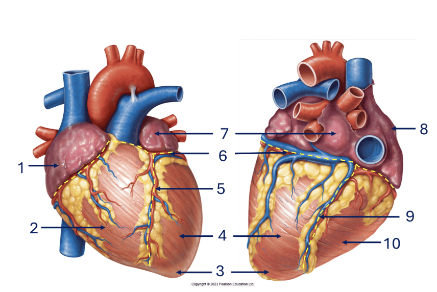
1 and 8- Right Atrium
2 and 10- Right Ventricle
3- Apex
4- Left ventricle
5- Anterior interventricular sulcus
6- Coronary sulcus
7-Left Atrium
9- Posterior interventricular sulcus
Heart- Anterior/posterior interventricular sulcus
separates left and right ventricle
Heart- Coronary sulcus
Like a crown
Separates atrium and ventricles
Heart Vessels- Superior/inferior vena cava
Deoxygenate blood arrives in right atrium through vena cava from body
Heart vessels- Pulmonary trunk/artery
Divides into right and left for lungs
sends deoxygenated blood from right ventricle to lungs
Heart Vessels- Pulmonary Veins
bring oxygenated blood from lungs to Left Atrium
One left and right
Heart Vessels- Aorta
sends oxygenated blood from left ventricle to body
Vessels delivering blood TO the heart are called:
Veins
Vessels delivering blood FROM the heart are called:
Arteries
Heart Chambers- Right atrioventricular opening
Tricupsid valce
opening between Left Atrium and left ventricle
Heart Chambers- Left atrioventricular opening
Bicuspid / mitral valve
Opening between left Atrium and left Ventricle
Heart Chamber- Papillary muscle
3 in Right ventricle, 2 in left ventricle
Contract and pulls Chordae Tendinae to open and close atrioventricular opening
Heart Chamber- Chordae Tendinae
Connects atrioventricular opening to papillary muscles
Heart Chamber- Interventricular septum
divides the ventricles
Heart Chamber- Aortic semilunar valve
valve between left ventricle and aorta
Heart Chamber- Pulmonary semilunar valve
valve between right ventricle and pulmonary trunk
What chamber of the myocardium is the thickest?
Left Ventricle because it pumps blood to the entire body
Cardiac Cycle- Atrial Diastole (Phase 1)
Heart is relaxed
blood flows from atria to ventricle
Atrioventricular valve Opens
Cardiac Cycle- Atrial Systole (phase 2)
Only Atria contract to fill ventricle
Atrioventricular valve Opens
Cardiac Cycle- Isovolumetric Contraction
Atria relaxes
Blood flow makes AV close
no valve opens
Cardiac Cycle- Ventricle systole
ventricle contracts, blood leaves heart
Semilunar valve opens
Cardiac Cycle- isovolumetric relaxation
heart is relaxed
blood flow closes semilunar valve
no valve open
Blood Vessels- Coronary sinus
vein that drains blood from heart into right atrium
Blood Vessels- coronary arteries
supply heart with blood
Pulmonary system
Cricut where deoxygenated blood leaves the heart, to the lungs, then back to the left atrium
Systemic Circuit
Circuit of blood that goes from the heart, to the body, then back to the heart
Blood vessels- What are the three layers (Tunics) of blood vessel walls
Tunica intima (innermost)
Tunic media
Tunic externa
4 diffrences between arteries and veins
Arteries:
thicker walls
more elastic (to accommodate for pressure)
Smaller lumens (round shaped)
Veins:
Thinner walls
Have valves to prevent blood flowback
Veins- Vanous valve
Large veins have valves to prevent flowback
Veins- Muscular pump
muscular muscle contract to push blood
Veins- Respiratory pump
during inhaling, pressure in thoracic increases, which increases blood pressure in veins
Veins- Vasoconstriction
decreases blood vessel room = increased blood pressure
Blood vessels- Capillaries
exchange center
connects arterioles to venules
Blood vessels- What kind of muscle tissue is in blood vessels?
smooth muscles
involuntary
regulated by autonomic nervous system
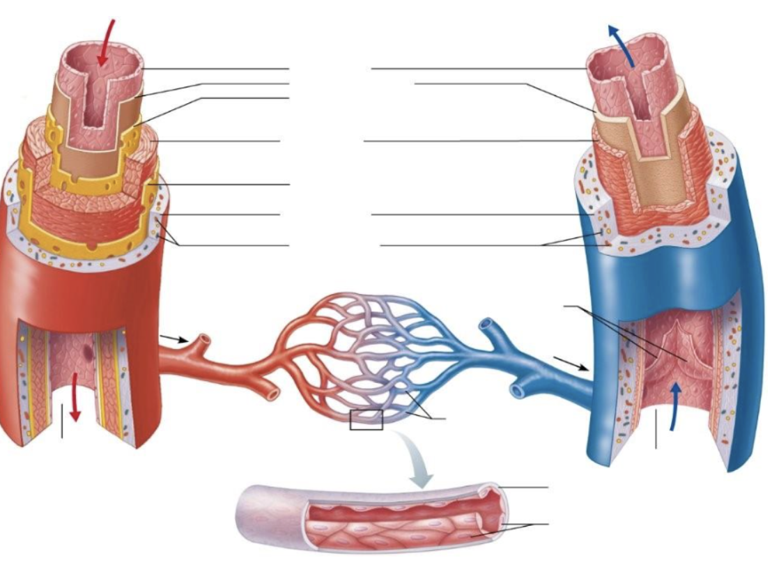
Tunica intima (BOTH)
Endothelium(BOTH)
subendothelial layer (BOTH)
Internal elastic membrane (Artery)
Tunica media (BOTH)
External elastic membrane (artery)
Tunica externa (BOTH)
vasa vasorum (BOTH)
Blood vessels- Ascending, arch, decending
3 main parts of aorta
Ascending → arch → descending
Aortic Arch- Brachiocephalic Trunk
divides into right common carotid and right subclavian
Aortic arch- Left common carotid artery
supplies blood to head / neck
divides into external and internal carotid arteries
Branches of Aortic arch- Left subclavian artery
Supplies upper limbs
extends into axillary artery
Branches of Aorta- Common iliac arteries
divide into internal and external iliac
supply pelvis and lower limbs
Arteries of upper limb
subclavian → axillary → brachial → Radial / ulnar artery
Artery of lower limbs
external iliac → femoral artery → popliteal artery → anterior / posterior tibial artery
Systemic Veins- superior vena cava