BLOCK 8- DOWN COW
1/5
There's no tags or description
Looks like no tags are added yet.
Name | Mastery | Learn | Test | Matching | Spaced | Call with Kai |
|---|
No analytics yet
Send a link to your students to track their progress
6 Terms
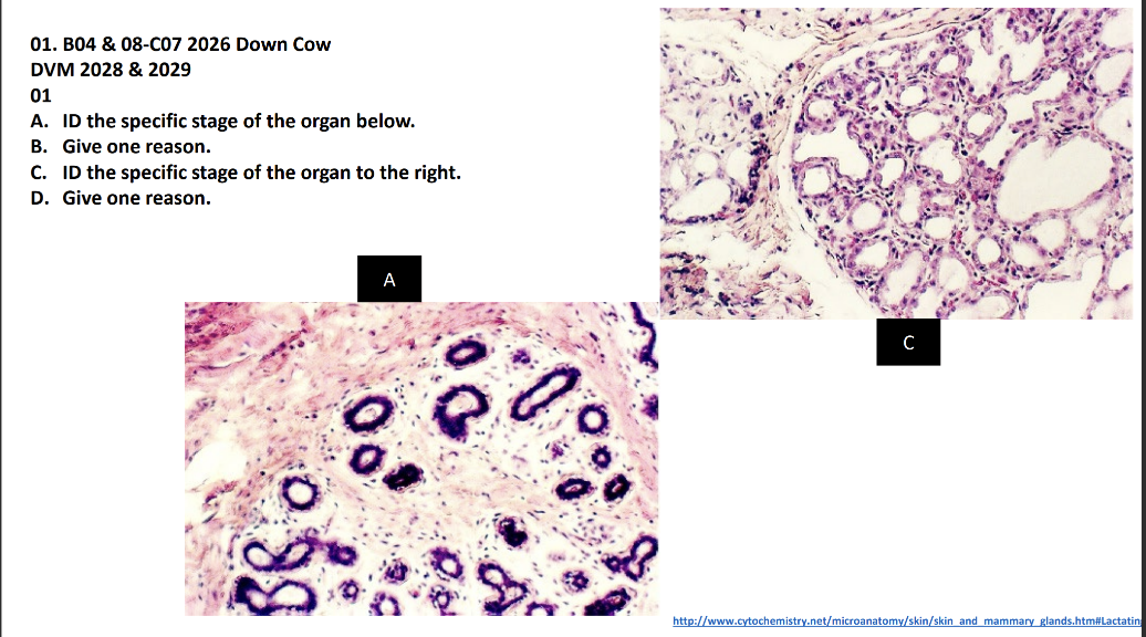
A. ID the specific stage of the organ below.
B. Give one reason.
C. ID the specific stage of the organ to the right.
D. Give one reason
A. Non lactating.
B. Few small alveoli and large amount of connective tissue.
C. Lactating mammary gland.
D. Little connective tissue and large number of alveoli.
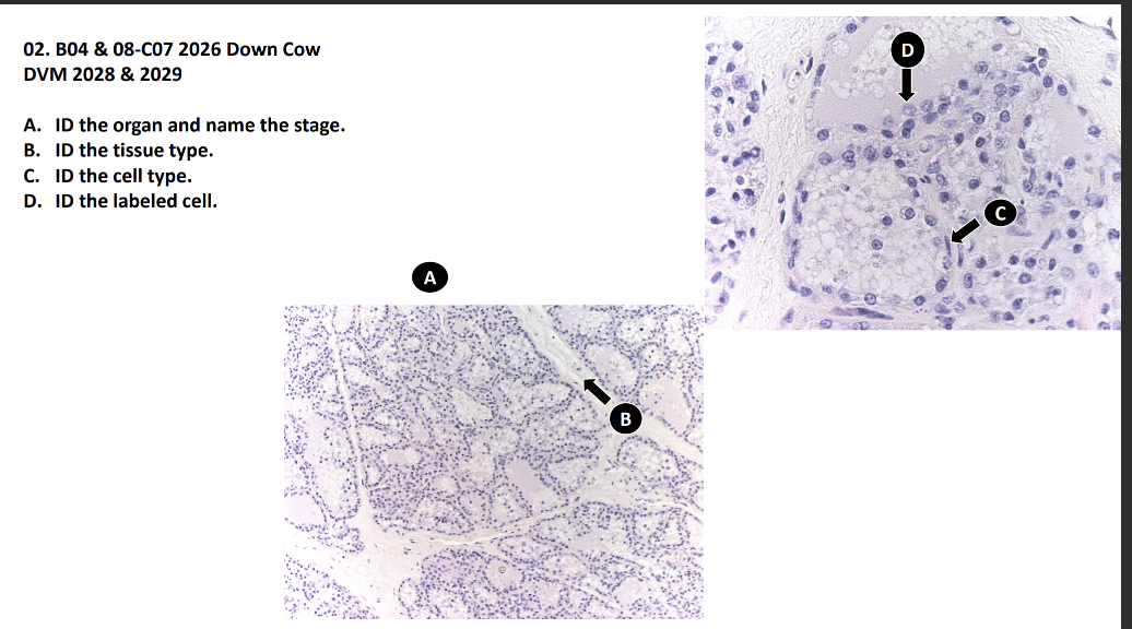
A. ID the organ and name the stage.
B. ID the tissue type.
C. ID the cell type.
D. ID the labeled cell
A. Lactating.
B. Interlobular connective tissue.
C. Myoepithelial cell.
D. Alveolar cell.
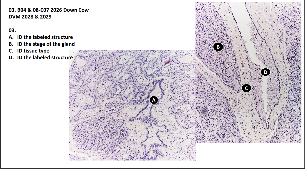
A. ID the labeled structure
B. ID the stage of the gland
C. ID tissue type
D. ID the labeled structure
A. A duct (intralobular).
B. Non lactating.
C. Interlobular connective tissue.
D. Interlobar duct.
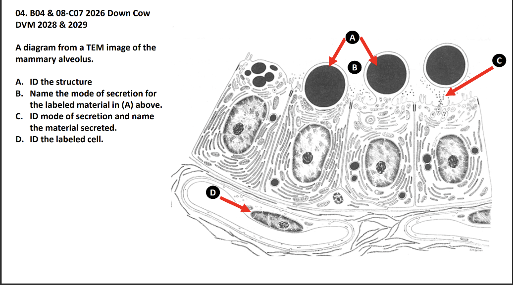
A diagram from a TEM image of the
mammary alveolus.
A. ID the structure
B. Name the mode of secretion for
the labeled material in (A) above.
C. ID mode of secretion and name
the material secreted.
D. ID the labeled cell
A. Lipid droplet.
B. Apocrine.
C. Merocrine (eccrine, exocrine), protein and sugars.
D. Myoepithelial.
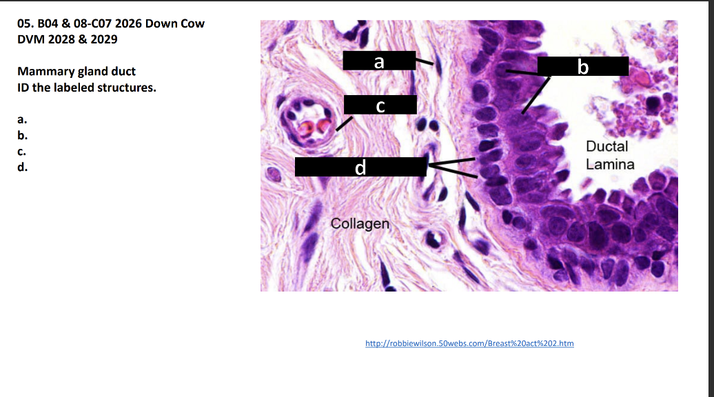
Mammary gland duct
ID the labeled structures.
a.
b.
c.
d
a. Fibrocyte
b. Ductal cell/simple cuboidal epithelium
c. Arteriole
d. Myoepithelial cells
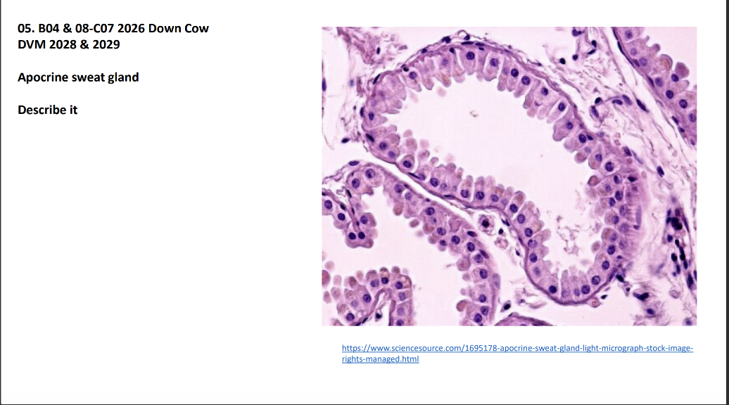
Apocrine sweat gland
Describe it
Tubular structure lined with simple cuboidal
epithelium surrounded by few myoepithelial
cells. The projections of the cells into the lumen
indicate it is apocrine type of sweat gland. It
has very little CT in between the glandular
tubules