Synaptic Transmission II
1/49
There's no tags or description
Looks like no tags are added yet.
Name | Mastery | Learn | Test | Matching | Spaced | Call with Kai |
|---|
No analytics yet
Send a link to your students to track their progress
50 Terms
When two stimuli are delivered in rapid succession, the second PSP often larger than the first: true or false
true, it involves more than summation of post synaptic potentials, it involves presynaptic facilitation/paired-pulse facilitation
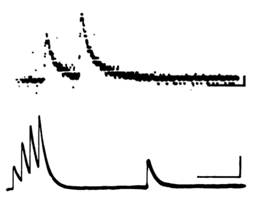
facilitation of the PSC
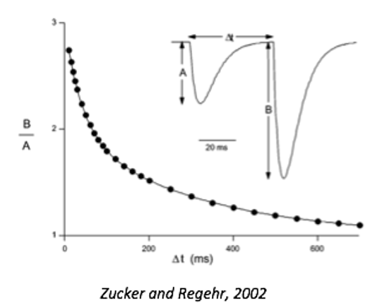
paired pulse facilitation increases the EPSC, and shows a double exponential decay.
theory before discovery of quantum
- however, the notion that neurotransmitter was released in discrete packets (called quanta) already existed—the discovery of synaptic vesicles confirmed the quantum hypothesis.
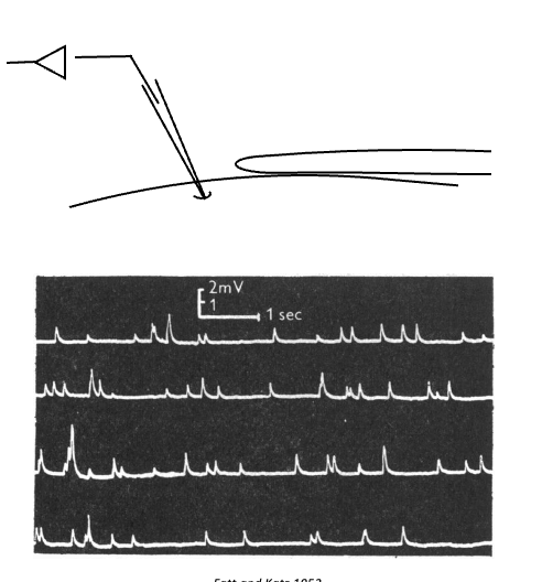
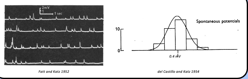
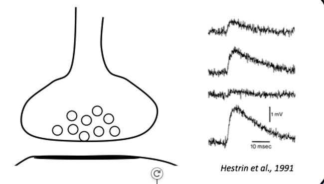
spontaneous events at the end plate
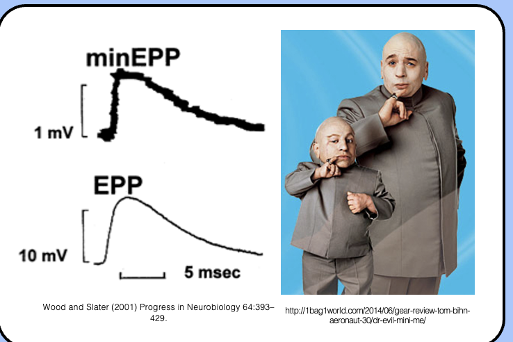
- upon close inspection, these depolarizing events looked like miniature versions of the EPP, and were called miniature EPPs (mEPPs; or just “minis”).
size of mEPP compared to EPP
it’s a scaled-down version of the EPP
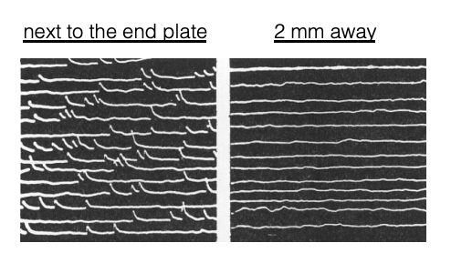
what happened when Katz moved the electrode away from the end plate?
- when Katz moved the electrode away from the endplate, the mEPPs became smaller and eventually disappeared—mEPPs seemed to be coming from the motor endplate.
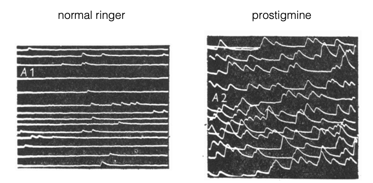
manipulating synaptic choline
- Katz manipulated the synaptic acetylcholine concentration by adding prostigmine, which blocks acetylcholinesterase.
- the resulting increased acetylcholine concentration prolongs the decay of mEPPs.
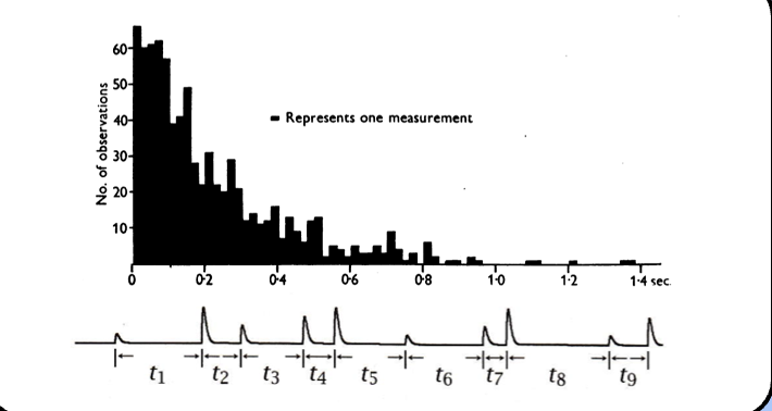
distribution of time of events of mEPPs
- they found that the time intervals between 800 serial mEPPs was exponentially distributed—this suggests that mEPPs are random events.
mEPP amplitude
- Katz also analyzed the magnitude of mEPPs by plotting the distribution of their amplitudes—they found that mEPP amplitudes were normally distributed with a mean value of 0.4 mV.
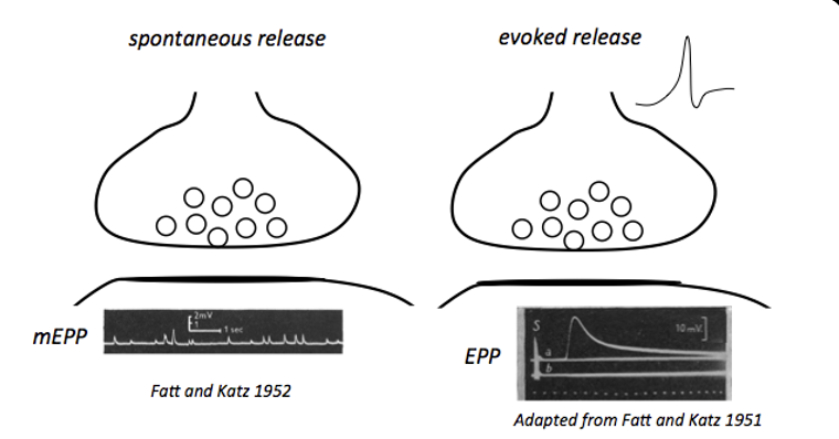
spontaneous release vs evoked release
spontaneous release = mEPP
evoked release = EPP
why did Katz have to decrease the EPP?
in order to investigate the relationship between mEPPs and EPPs
how did Katz decrease the EPP?
applying curare
manipulating the Ca/Mg ratio by lowering it
relationship between EPP and mEPP in terms of magnitude
The EPP was quantized, the amplitude of the EPPs were integer multiples of the mEPP
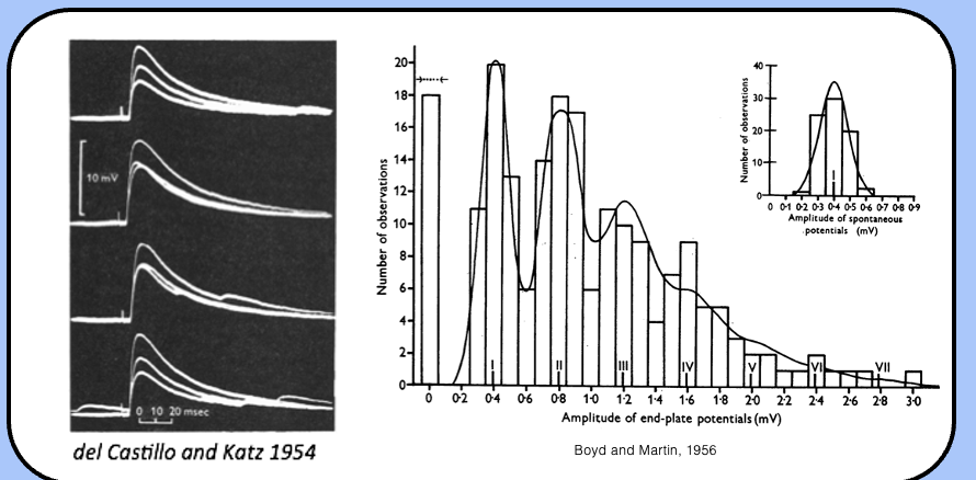
the quantum hypothesis
from these careful observations, experiments, and analyses, Katz was able to discover a fundamental aspect of synaptic transmission—neurotransmitter is released in discrete packs called quanta
parameterizing synaptic transmission
quantal analysis
assumptions of quantal analysis
release sites
many release sites
receptors are not limiting
small release probability with low concentration of Ca/Mg
independent release
transmitter released in quanta
release sites
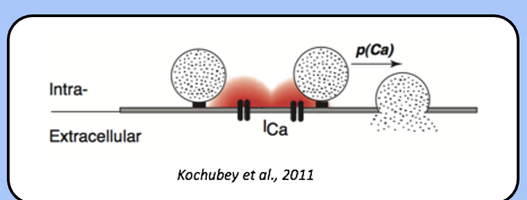
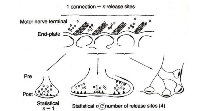
- quanta are released from discrete locations called release sites (n)—the NMJ has many release sites arranged in two rows, whereas central synapses have only one of a few release sites.
release probability
- at any given moment, the release site “flips a coin,” and determines whether or not to release its quantum.
- the “decision” to release a vesicle is determined by a parameter called the release probability (Pr)
changing the release probability
- when the membrane potential is at rest, the release probability is very low, and quanta trickle out infrequently—these are the mEPPs.
- however, when a presynaptic action potential increases the presynaptic calcium concentration, the release probably rapidly increases—this results in an EPP
low Pr versus high Pr
low Pr = spontaneous release = mEPP
high Pr = evoked release = EPP
quantal content
- the quantal content is the number of quanta that are released in response to a presynaptic stimulus—this usually refers to a presynaptic action potential.
quantal content equation
m = n*Pr
Katz and the post synaptic receptor
- neurotransmitter release evokes a postsynaptic response, which was the only signal Katz could measure—we need some parameter that incorporates the postsynaptic response.
quantal size
q
the postsynaptic response to the release of a single presynaptic vesicle—this simply corresponds to the size of the mEPP.
PSP = n*Pr*q.
three parts of quantal size
the number of neurotransmitter molecules loaded into a vesicle, (although this is actually pretty consistent)
and the number of available postsynaptic receptors.
degradation of neurotransmitters, re-uptake, diffusion when in synaptic cleft
how many molecules are in a vesicle
tends to be very consistent
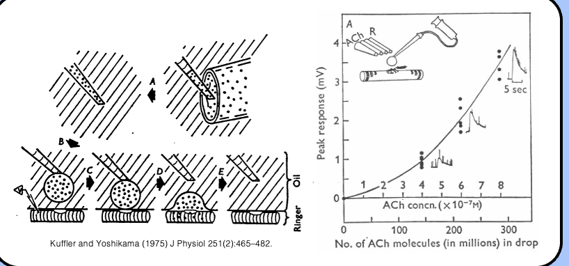
kuffler determining the number of ACh molecules
- in the mid 1970s Steven Kuffler’s group determined that about 7000 acetylcholine molecules were required to mimic a mEPP by applying water droplets filled with known concentrations of acetylcholine to muscle fibers.
spontaneous release v evoked release using quantal language
when at rest the Pr is low of the presynaptic terminal so PSP is low resulting in mEPPs, which produce a response equal to the quantal size
when an action potential invades the terminal, the Pr increases and m vesicles are release, which produces a postsynaptic potential equal to m*q
measuring presynaptic calcium
- they found that the the calcium concentration built up in the presynaptic terminal over time.
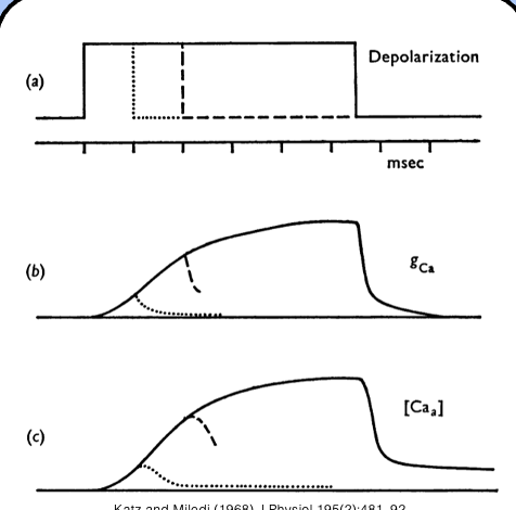
the residual calcium hypothesis
Katz and Miledi proposed that residual calcium from the first pulse remained in the presynaptic terminal for some time after the action potential.
if a second action potential arrived in the terminal, the incoming calcium would add to the calcium already present in the terminal—this became known as the residual calcium hypothesis.
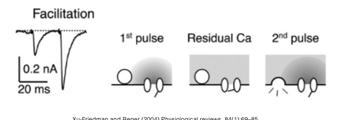
which quantal analysis parameter do you think is being affected during facilitation?
During facilitation, the parameter being affected is the probability of release (Pr) of synaptic vesicles, leading to an increased postsynaptic response.
presynaptic depression
- sometimes, sequential stimulations produce responses that decrease in magnitude—this is a form of short term plasticity called depression.

synaptic vesicle pools
- there are three physiologically distinguishable populations of presynaptic vesicles—the readily releasable pool (RRP; 1%), the recycling pool (10–15%), and the reserve pool (80–90%).
which vesicle pool is released upon stimulation
the readily releasable pool (RRP)
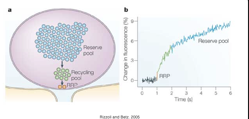
the mechanism of depression
- presynaptic depression (often called paired-pulse depression) involves the use-dependent depletion of readily-releasable pool (RRP).
- when the presynaptic terminal is allowed to recover, recycling vesicles reestablish the RRP.
which quantal analysis parameter is being affected during depression
Release probability, but by reducing the amount of vesicles it can act on- exposes the other aspect of Probability of releas (Pr)
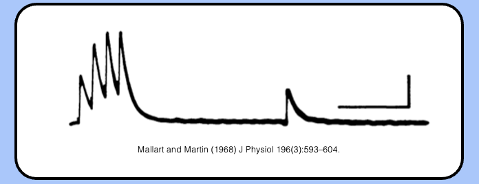
a mixture of facilitation and depression
facilitation can be followed by depression, as shown by the graph- example of short-term plasticity
additional forms of short-term plasticity
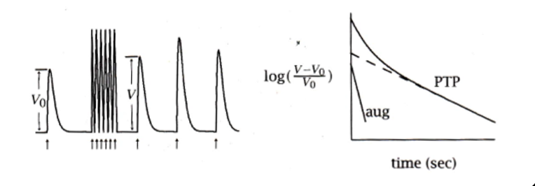
strong stimuli (such as a tetanizing shock) can induce an additional form of short-term plasticity that lasts for tens of minutes.
post -tetanic potentiation
augmentation
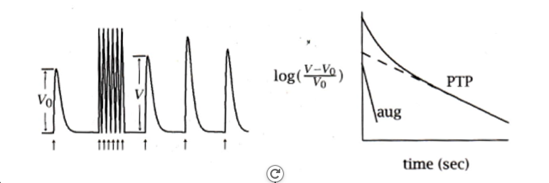
post-tetanic potentiation (PTP)
decays with two time constants—the faster decay, which takes several seconds is called augmentation, whereas the slow decay, which takes minutes, is called PTP.
both augmentation and PTP are thought to result from an increase in the quantal content through an increase in Pr.
synaptic overkill
- the NMJ is a synapse with a high safety factor—this means that a presynaptic action potential is almost always going to evoke a postsynaptic response that triggers an action potential.
quantal content of central synapses
- the NMJ might have several hundred release sites for each contact it makes with the muscle fiber (i.e., the motor end plate), which means that it can have a quantal content of a hundred vesicles or more.
- in comparison, central synapses are puny, with just one, or a handful of release sites per contact—therefore, many postsynaptic inputs must add together in order to trigger an action potential.
NMJ synapses v central synapses in terms of action potential
central synapses are very unreliable while NMJs have a high safety factor
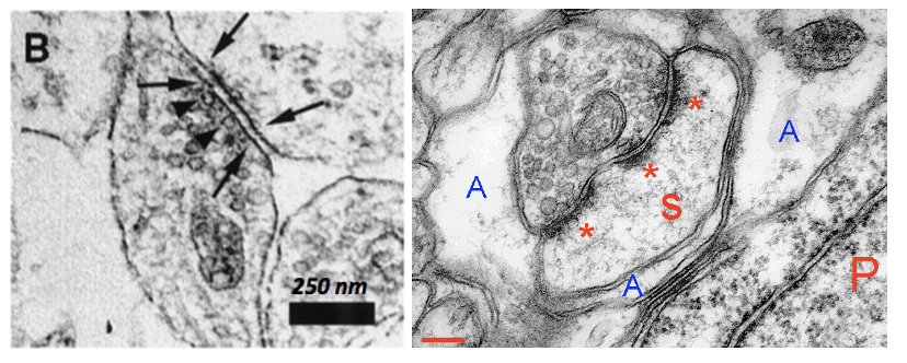
release site configurations
- central synapses can have a single release site, or several, depending on the synapse.
perforated synapse
- when multiple release sites (aka presynaptic active zones) are positioned across from their own postsynaptic density, we call this a split, or perforated synapse.
why are central synapses unreliable
- the quantal content of central synapses is so low that they produce highly variable postsynaptic responses, with a surprisingly high probability of failure.
what is the function of central synapses’ unreliability
- the low probability of release in central synapses is presumably important for forcing neurons to work together in ensembles—the brain must be wired with redundancy, so that it is fault-tolerant.
COOPERATION AND FAULT TOLERANCE
has 10000s of synapses sometimes on one neurons
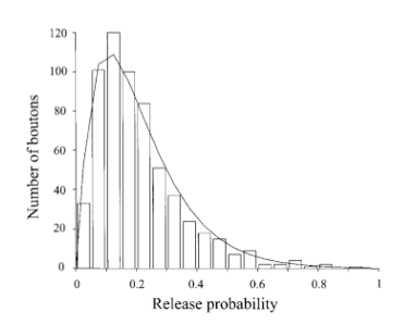
the distribution of Pr of a central synapse
- the release probability of central synapses can range from extremely high, to vanishingly low—this distribution is centered around an average Pr of ~ 0.3.
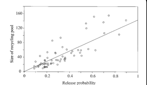
the correlation btwn release probability and the pool of synaptic vesicles in a central synapse
- interestingly, the release probability shows a strong correlation with the size of the pool of synaptic vesicles available for release.
postsynaptic receptors become limiting for central synapse
- in sharp contrast to the ocean of postsynaptic receptors waiting for a quantum to be released at the NMJ, the number of postsynaptic receptors waiting to receive a quantum from a central synapse is rather small.
- this imposes a limitation of the ability to experimentally apply quantal analysis to central synapses.
limitations of central synapse on quantal analysis
stimulation of a single axon
unitary events might be hard to measure
receptors are often limiting
distant recording site