RP2: Preparation of stained squashes of cells from plant root tips; set-up and use of an optical microscope to identify the stages of mitosis in these stained squashes and calculation of a mitotic index
1/15
There's no tags or description
Looks like no tags are added yet.
Name | Mastery | Learn | Test | Matching | Spaced | Call with Kai |
|---|
No analytics yet
Send a link to your students to track their progress
16 Terms
How is the mitotic index calculated? (1)
Mitotic index = (Number of cells undergoing mitosis) ÷ (Total number of cells)
How do you express the mitotic index as a percentage? (1)
Multiply the mitotic index by 100.
How do you calculate the number of minutes a cell spends in a particular phase of the cell cycle? (2)
(Number of cells in the phase ÷ Total number of cells) × Duration of one cell cycle
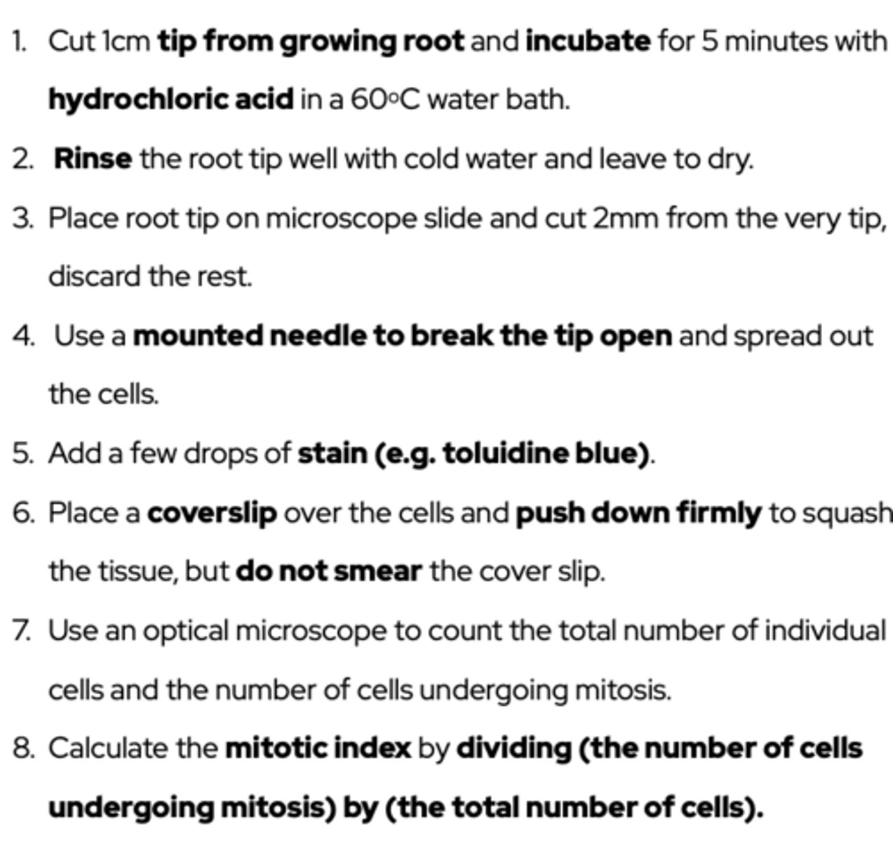
What is the method for this experiment? (8)
Why were cells from the tip of the root used? (1)
Because these cells are actively dividing
What is the purpose of incubating the root tip with hydrochloric acid? (2)
- Stops mitosis.
Breaks down links between cell walls (middle lamella)
- So that the cells can be separated easily
Why is a stain added? (1)
Stain binds to chromosomes so they become visible
Why is it important to press the coverslip down firmly, but not smear it? (2)
- Pressing down firmly spreads the cells out so they are in a single layer, allowing light to pass through the specimen.
- The coverslip mustn't be smeared from side to side because the chromosomes may be damaged.
Describe a safety concern associated with this method. (2)
- Hydrochloric acid is corrosive → wear a lab coat/safety goggles.
- The coverslip is glass → handle carefully to avoid breakages/cuts.
How can you ensure the mitotic index obtained is accurate? (3)
- Examine a large number of fields of view to ensure a representative sample.
- Repeat the count to ensure the figures are accurate.
- Count only whole cells to standardise counting.
Why do cells further away from the root tip appear longer? (1)
These cells are elongating (and no longer dividing)
Why might different students calculate different mitotic indices despite following the same method? (3)
- Root tips may have been from different plants or were a different age.
- Some students may have looked at more than one field of view and calculated a mean.
- Different fields of view are from different parts of the root tip.
Describe and explain the appearance and behaviour of the chromosomes in prophase. (3)
- Chromosomes are becoming visible because they are condensing.
- Chromosomes are arranged at random because they are not attached to spindle fibres yet.
- Chromosomes appear as two sister chromatids joined at the centromere because DNA has replicated.
Describe and explain the appearance and behaviour of the chromosomes in metaphase. (2)
- Chromosomes are lined up on the equator (metaphase plate).
- The chromosomes attach to spindle fibres by their centromeres.
Describe and explain the appearance and behaviour of the chromosomes in anaphase. (2)
- Centromeres divide & chromatids from each pair are pulled to opposite poles of the cell as the spindle fibres shorten.
- The V-shape shows that chromatids have been pulled apart at their centromeres.
Describe and explain the appearance and behaviour of the chromosomes in telophase. (1)
Chromosomes become longer/thinner because they decondense