Pleura and Lungs
1/44
There's no tags or description
Looks like no tags are added yet.
Name | Mastery | Learn | Test | Matching | Spaced | Call with Kai |
|---|
No analytics yet
Send a link to your students to track their progress
45 Terms
A double-layered membrane surrounding the lungs, consisting of the visceral pleura attached to the lungs and the parietal pleura lining the thoracic cavity. It facilitates lung movement and creates a pleural cavity filled with pleural fluid.
Pleura
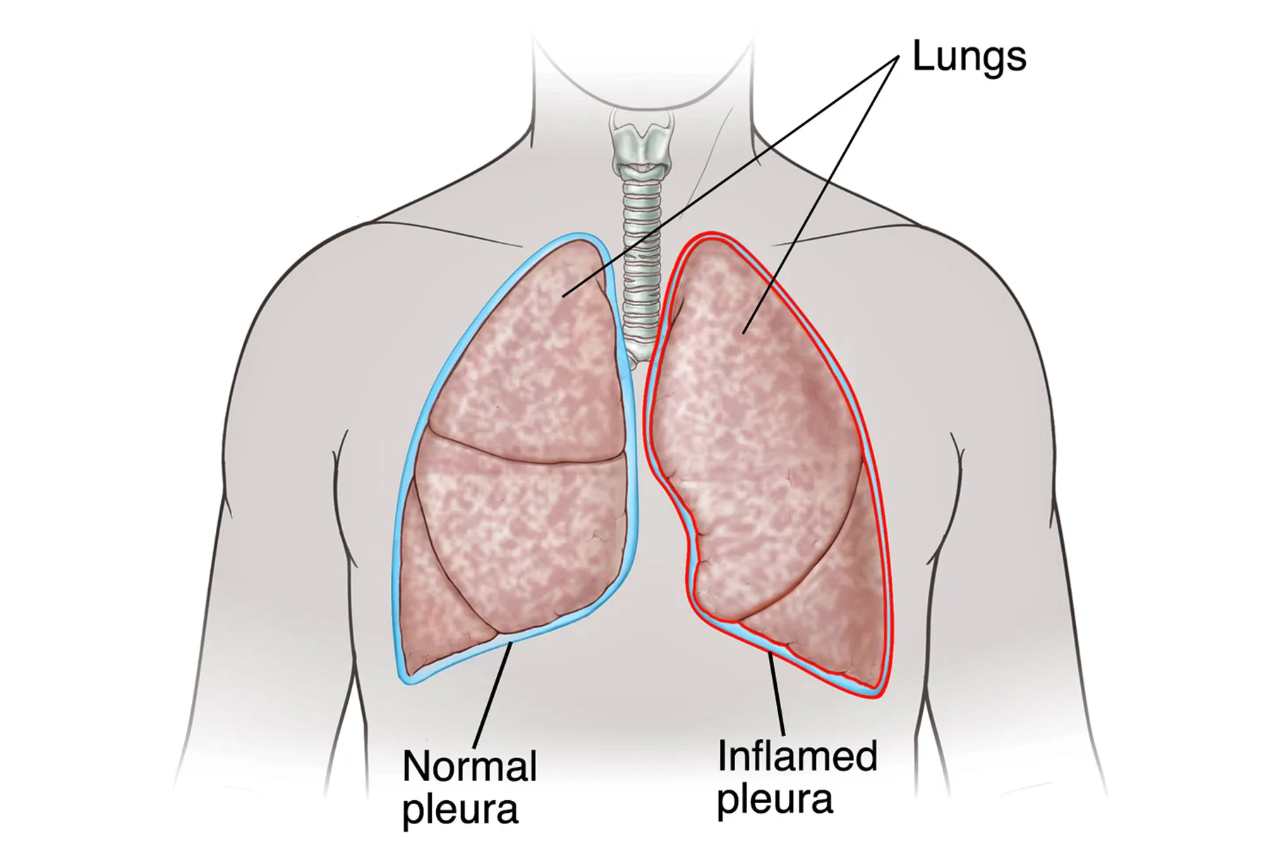
Visceral Pleura
The inner layer of the pleura that directly covers and adheres to the surface of the lungs. It provides a protective barrier and helps in reducing friction during respiration.
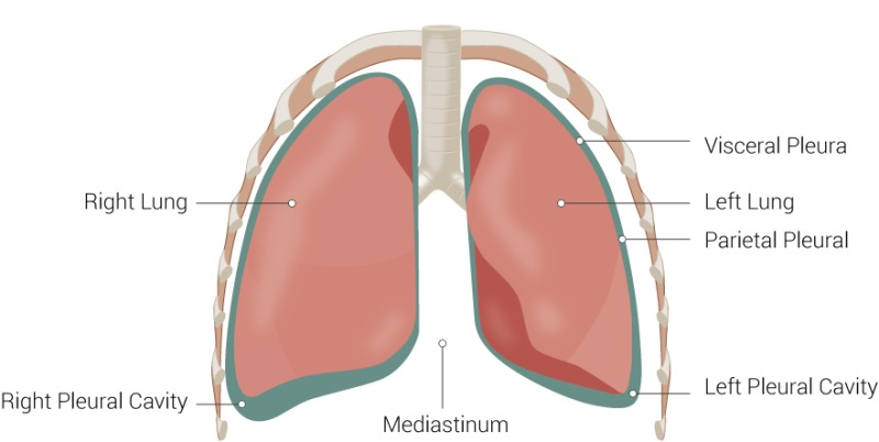
Parietal Pleura
The outer layer that lines the inner surface of the thoracic wall, diaph and mediastinum.
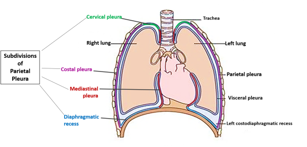
What are the subdivision of Parietal Pleura?
Cervical pleura (Extends through the superior thoracic aperture into the root of the neck)
Costal Pleura (Lines the internal surfaces of the ribs, costal cartilages, and intercostal muscles)
Mediastinal pleura (Covers the lateral aspects of the mediastinum)
Diaphragmatic pleura (Covers the superior surface of the diaphragm)
Where does Sympathetic Nerve Originates?
In the Spinal cord in the region of sympathetic trunk.
What is Sympathetic trunk?
A ganglia chain that runs alongside the vertebral column, consisting of sympathetic ganglia and nerve fibers that communicate with the spinal nerves, contributing to the autonomic nervous system.
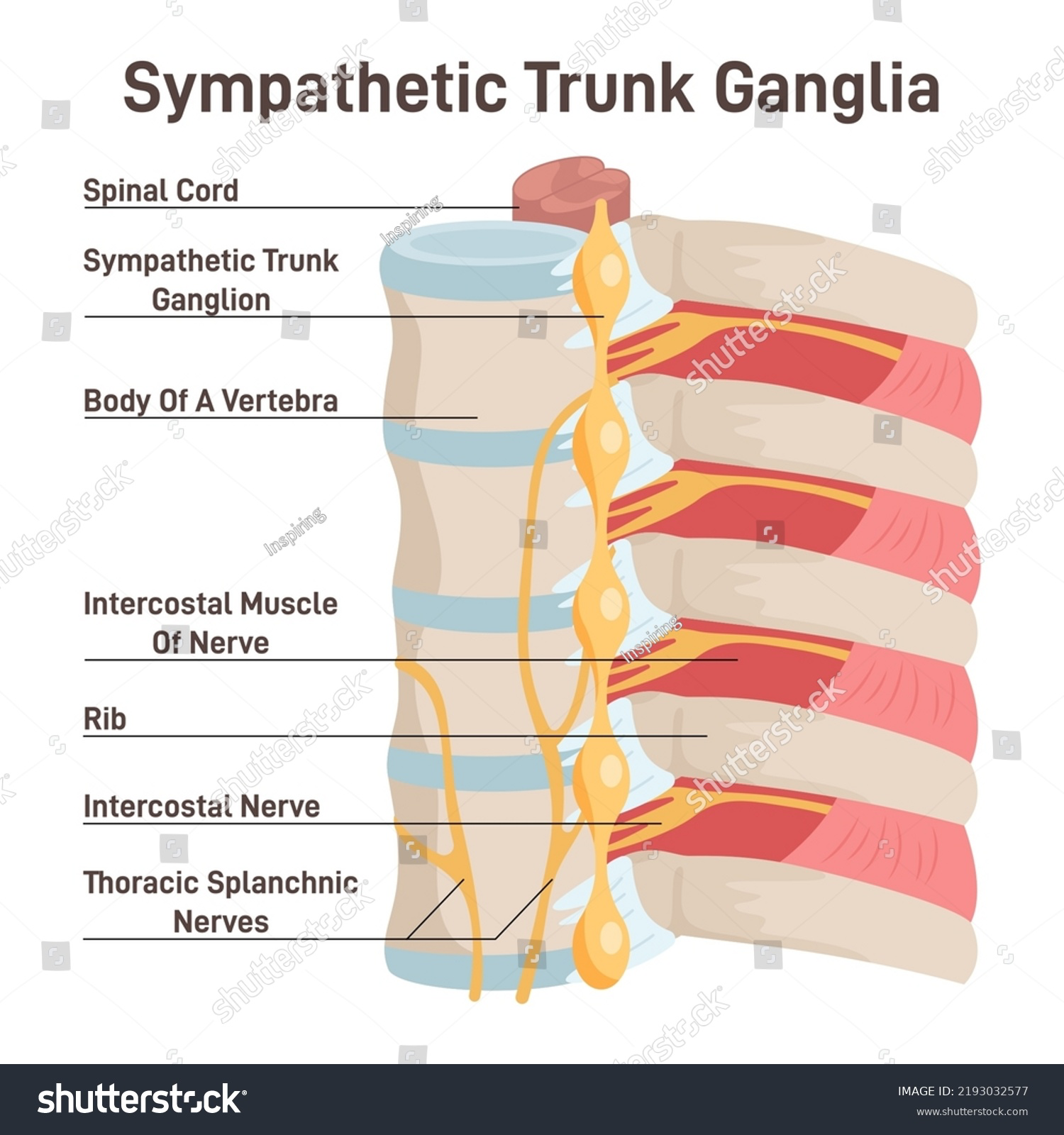
What is Cardiopulmonary splanchnic nerve?
Postganglionic fibers from the sympathetic ganglion to the heart and lungs.
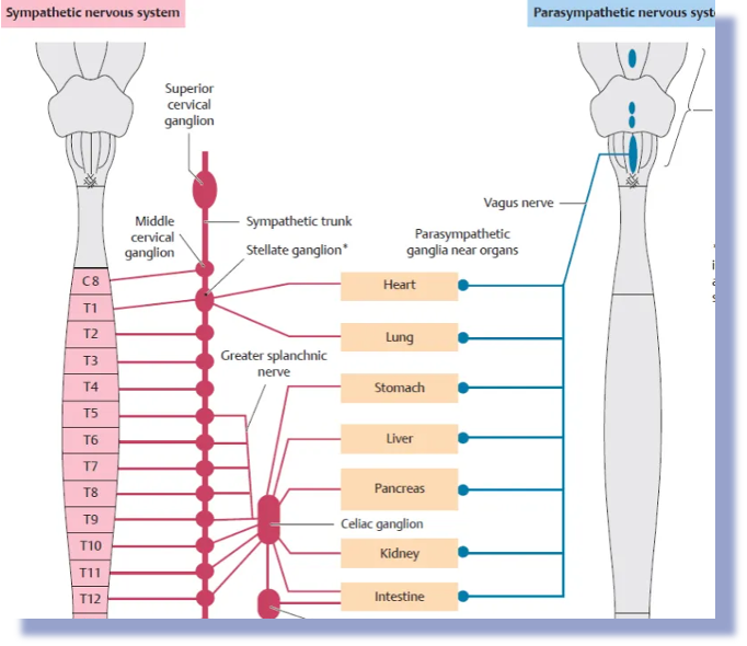
What are the effect of the Sympathetic nerve to the lungs?
Bronchodilation and reducing glandular secretions
Vasoconstriction in areas to shunt blood to muscles
Where does parasympathetic nerve originates?
Originate from the brainstem.
In the thorax, it is almost entirely controlled by the vagus nerve.
They do not synapse until they are on the right on or inside their target organ.

What are the effects of Parasympathetic nerves on Lungs?
Bronchoconstriction
No effect of vasculature

What is visceral nerve?
Supply the internal organs (heart, lungs, stomach, intestines, etc.) Carry autonomic (involuntary) signals.
Visceral pain is often:
Dull or aching
Poorly localized (hard to pinpoint)
Can be felt in a different location than the actual organ (referred pain)
What is Parietal nerve?
Supply the walls of body cavities and the parietal layers of serous membranes (such as the parietal pleura, peritoneum, and pericardium).
Carry somatic (voluntary/body wall) sensory information.
Parietal pain is:
Sharp
Well localized (easy to pinpoint)
Usually worsens with movement or pressure
What is Pleura and what are the two type?
The pleura is a double-layered membrane surrounding the lungs.
It consists of two layers:
Visceral pleura and Parietal pleura.

The outer most Layer of the lungs?
Parietal pleura: lines the inside of the chest wall, diaphragm, and mediastinum.
“Wallpaper lining the chest cavity”
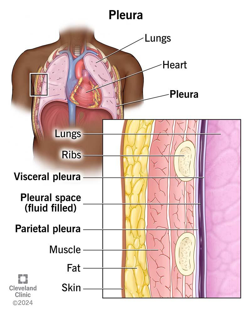
What is the innermost Layer of the lungs?
Visceral Pleura- directly covers the lung surface.
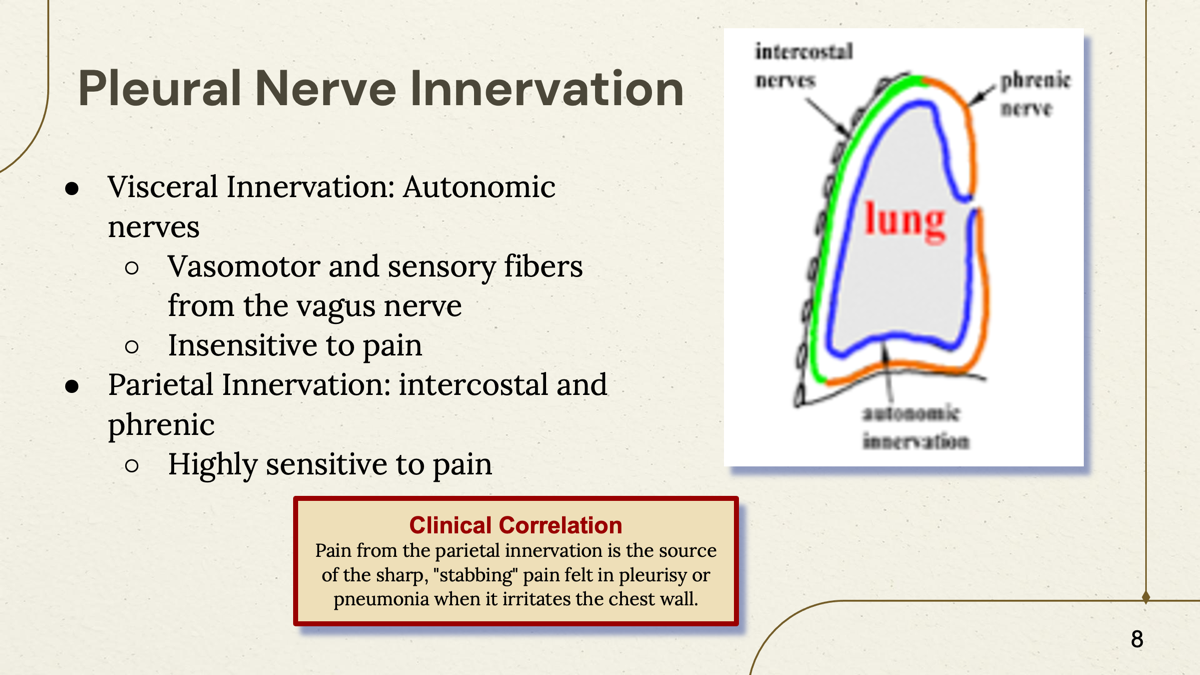
What does innervation means?
Innervation simply means the nerve supply to a structure.
When anatomy professors ask:
"What is the innervation of this muscle?"
They are asking:
"Which nerve controls or supplies this muscle?"
Where does Visceral Pleura receives its nerve fiber? (visceral innervation)
The visceral pleura receives nerve fibers from the autonomic nervous system (including fibers traveling with the vagus nerve).
The visceral pleura is insensitive to pain.

Where does Parietal pleura receive its nerve fibers? (Parietal Innervation)
The parietal pleura receives its nerve fibers from intercostal and Phrenic nerve.
Highly sensitive to pain
What is the function of lungs and is properties?
External respiration, ventilation, and has elasticity and recoil properties.
where is the Apex located in the lungs?
Superior rib (above the first rib)
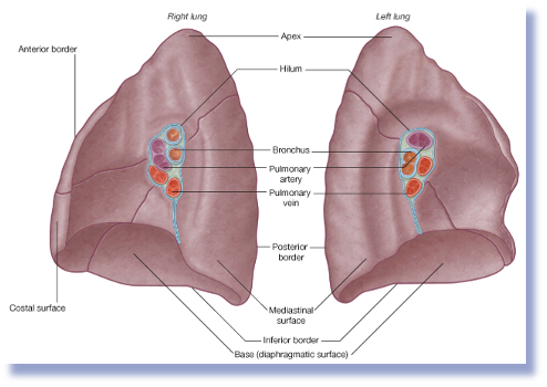
Where is the base located in lungs?
In the concave Inferior surface to the rests of the diaphragm.

The location where bronchi and neuromuscular structure enter and exit?
Hilium/Root
Everything entering or leaving the lung passes through here (like a front door)
What lung is heavier and larger
The right lung
How many lobes are in the right lung?
Three lobes- superior, middle, and inferior.
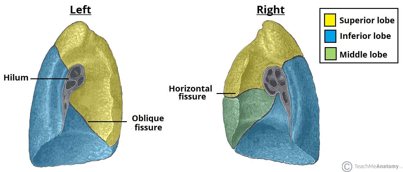
How many fissures are in the right lung?
Two- horizontal and oblique.

How many lobes are in the left lung?
Two lobes- Superior and inferior
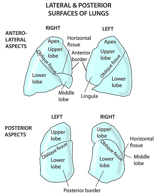
How many fissure in the left lung?
One- oblique fissure.

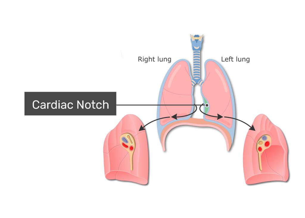
What is cardiac notch?
The cardiac notch is a dent in the left lung where the heart sits.
Think of it as the lung "making room" for the heart.
What is lingula?
The lingula is a small tongue-like projection of the superior lobe.
Bronchopulmonary segments
Smallest segments of lungs that can be surgically removed without affecting the surrounding lung tissue.
They are functionally independent and receive their own air supply from tertiary bronchi.

How many segments are for each lung?
10 segments for each lung.
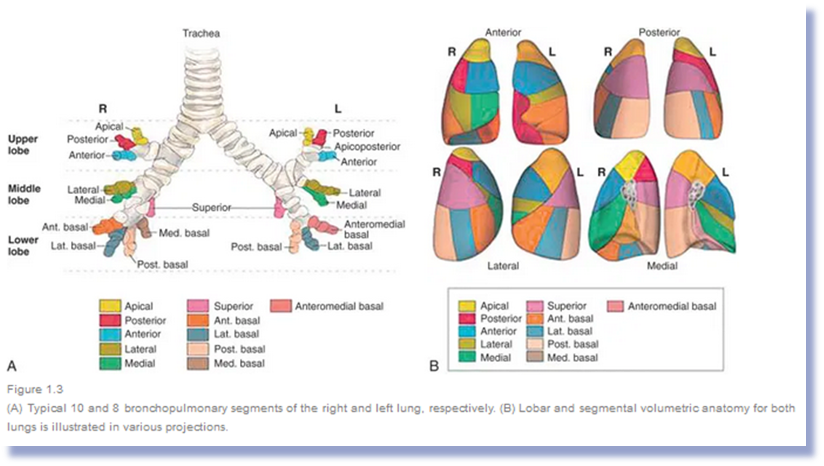
Segment of each lung are separated by?
Connective tissue that divides the bronchopulmonary segments.
-Lobes are separated by?
-Segments are seared by?
-fissures
-connective tissue
What is Bronchopulmonary tree?
Connects the larynx to the bronchi.
Trachea
|
|
(Tree Trunk)
|
Carina
/ \
/ \
Right Left
Bronchus Bronchus
| |
Branches Branches

What is Bronchial Carina and its location?
The carina is where the trachea splits into the right and left main bronchi.
At the level of T4-T5
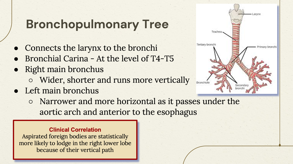
What bronchus is more wider and runs more vertically?
The right main bronchus is wider and runs more vertically than the left.
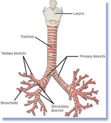
What main bronchus is narrower and more horizontal?
The left main bronchus is narrower and more horizontal.

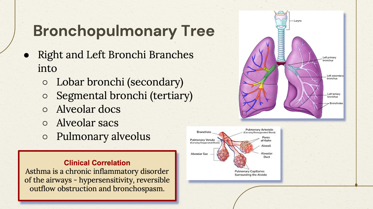
Where does the right and left bronchi branches into?
Lobar bronchi (secondary) —> segmental bronchi (tertiary) —> Alveolar docs—> Alveolar sacs—> Pulmonary alveolus
Larynx (voice box)
↓
Trachea (windpipe)
↓
Primary Bronchi
↓
Secondary (Lobar) Bronchi
↓
Tertiary (Segmental) Bronchi
↓
Bronchioles
↓
Alveolar Ducts
↓
Alveolar Sacs
↓
Alveoli
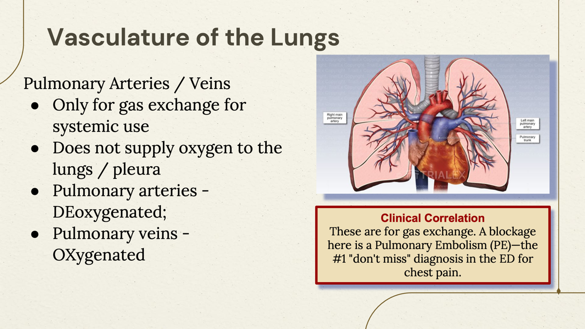
What is the job of pulmonary blood vessels (Arteries/Veins)
Only for gas exchange for systemic use.
Pick up oxygen from the lungs and remove carbon dioxide from the blood.
They don't supply Oxygen to the lungs.
Pulmonary vein carries:
Oxygenated blood.
Go from lungs → heart

pulmonary arteries carry:
Deoxygenated blood.
Go from heart → lungs
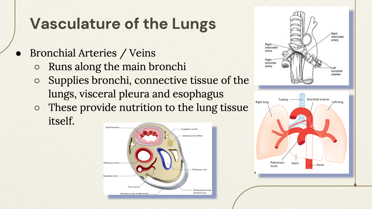
Which blood vessels supply oxygen to the lung tissue itself?
The bronchial arteries supply oxygenated blood to the lung tissue.
NOT the Pulmonary veins or arteries.
what does bronchial artery/vein supply?
Supplies bronchi, connective tissue of the lungs, visceral pleura and esophagus.
This system puts oxygen into the blood:
Pulmonary circulation system.
Job: Gas exchange
This system provides oxygen and nutrients to keep the lungs alive.
Bronchial circulation system.
Job: Feed the lung tissue
