chapter 7
1/84
There's no tags or description
Looks like no tags are added yet.
Name | Mastery | Learn | Test | Matching | Spaced | Call with Kai |
|---|
No analytics yet
Send a link to your students to track their progress
85 Terms
Carbohydrates
•aldehydes or ketones with at least two hydroxyl groups, or substances that yield such compounds on hydrolysis
•
•many carbohydrates have the empirical formula (CH2O)n
Monosaccharides
•simple sugars, consist of a single polyhydroxy aldehyde or ketone unit
–example: D-glucose
Oligosacchraides
•short chains of monosaccharide units, or residues, joined by glycosidic bonds
Disaccharides
•oligosaccharides with two monosaccharide units
example: sucrose (D-glucose and D-fructose)
Polysaccharides
•sugar polymers with 10+ monosaccharide units
–examples: cellulose (linear), glycogen (branched)
Backbones of monosaccharides
unbranched carbon chains with single bonds linking all carbon atoms
One of the carbon atoms is double bonded to an oxygen atom to form a carbonyl group
Other carbon atoms are bonded to hydroxyl group
Steroisomerism in sugars
Suagr stereoisomers arise because many of the carbon atoms to which the hydroxyl groups are attached to chiral centers
enzymes that act on sugars are stereospecific
Aldose
Carbonyl group is at he need of the carbon chain ( in an aldehyde group)
Ketone
Carbonyl group is at any other position ( in a ketone group)
Trioses
Simple set monosaccharides,three carbon backbone
Tetroses
Four carbon backbone
Pentoses
Five carbon backbone
Hexoses
Six carbon backbone
Heptoses
Seven carbon backbone
What makes sugar sweet?
•TAS1R2 and TAS1R3 encode sweet-taste receptors
•
•binding of a compatible molecule generates a “sweet” electrical signal in the brain
–requires a steric match
Fischer projection formulas
used to represent three-dimensional sugar structures on paper
bonds drawn horizontally indicate bonds that project out of the plane of the paper
bonds drawn vertically project behind the plane of the paper
D isomers
configuration at reference carbon is the same as D-glyceraldehyde
on the right (dextro) in a projection formula
most hexoses of living organisms
L isomer
configuration at reference carbon is the same as L-glyceraldehyde
on the left (levo) in a projection formula
Numbering carbons of sugar
Carbons are numbers beginningat the end of the chain near the carbonyl group
Epimers
Two sugars that differ only in the configuration around one atom
Hemiacetlas or hemiketals
derivatives formed by a general reaction between alcohols and aldehydes or ketones
product of the first alcohol molecule addition
a five- or six-membered ring forms if the —OH and carbonyl groups are on the same molecule
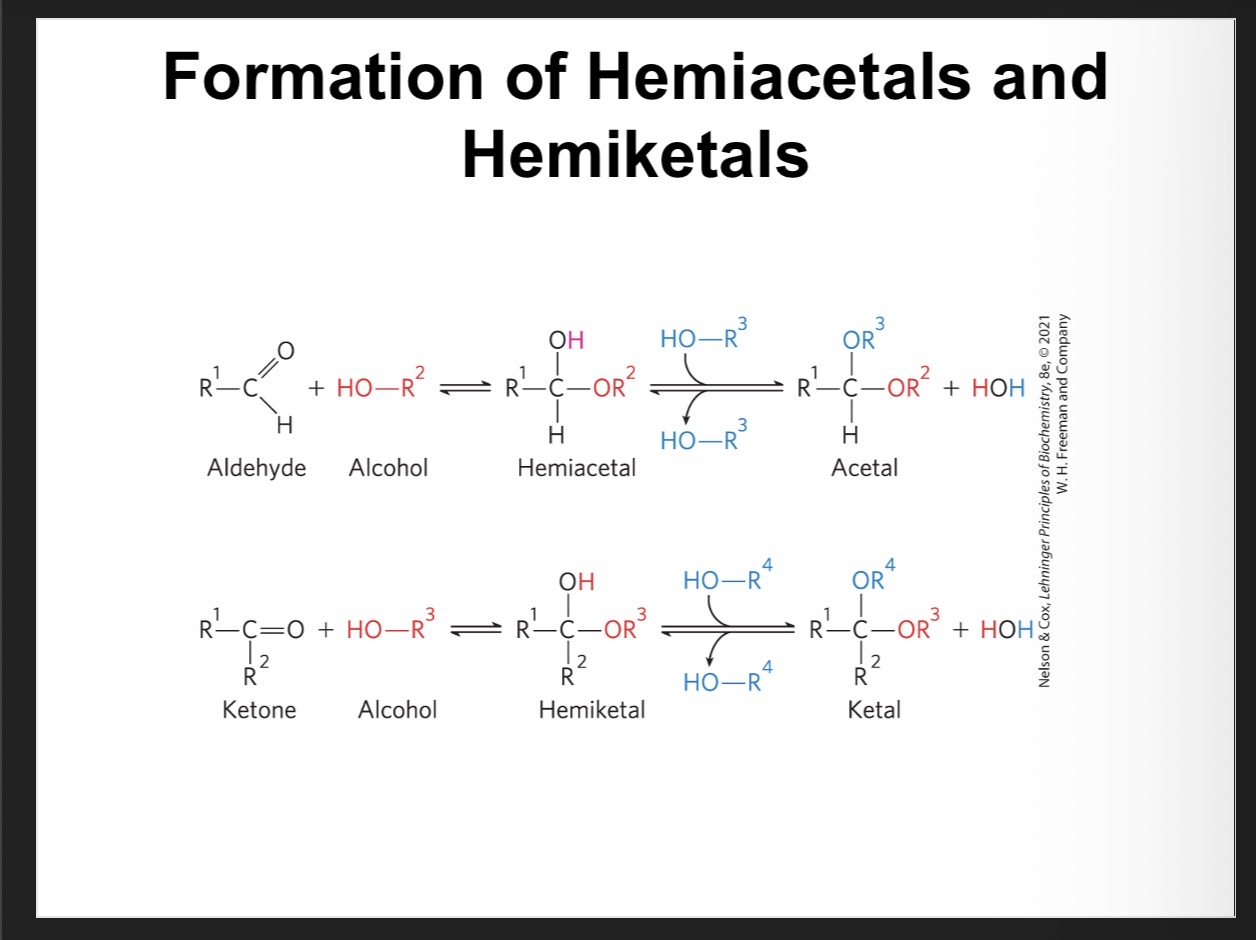
Acetal or ketal
Product of the second alcohol molecule addition
forms a glycosidic bond
Anomers
isomeric forms of monosaccharides that differ only in their configuration about the hemiacetal or hemiketal carbon atom
Formation of hte two cyclic forms of D Glucose
reaction between the aldehyde group at C-1 and the hydroxyl group at C-5 forms a hemiacetal linkage
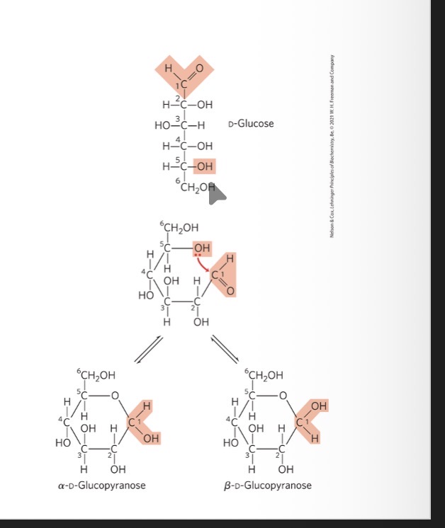
Mutarotation
The interconversion of alpha and beta anomers
Pyranoses
six-membered ring compounds
form when the hydroxyl group at C-6 reacts with the keto group at C-2
furanoses
five-membered ring compounds
form when the hydroxyl group at C-5 reacts with the keto group at C-2
Haworth perspective formulas
more accurate representation of cyclic sugar structure than Fischer projections
six-membered ring is tilted to make its plane almost perpendicular to that of the paper
bonds closest to the reader are drawn thicker than those farther away
Converting D-hexose Fischer projections to Haworth prespective formulas
step 1: draw the six-membered ring (five carbons, and one oxygen at the upper right)
step 2: number the carbons in a clockwise direction beginning with the anomeric carbon
step 3: place the hydroxyl groups
hydroxyl groups on the right in a Fischer projection are placed pointing down and those on the left are placed pointing
step 4: place the terminal —CH2OH group
projects upward for the D enantiomer, downward for the L enantiomer
step 5: place the anomeric hydroxyl group
for a β structure, the hydroxyl group is placed on the same side of the ring as C-6
for an α structure, it is placed on the opposite side
Aldonic acids
Form following the oxidation of the carbonyl carbon of aldoses
Form stable intramolecular esters called lactones
Uronic acids
Form following oxidation at C-6
Form stable intramolecluar ester called lactones
Reducing sugars
undergo a characteristic redox reaction where free aldehyde groups react with Cu2+ under alkaline condition
reduction of Cu2+ to Cu+ forms a brick-red precipitate
ketoses that can tautomerize to form aldehydes are also reducing sugars
O -glycosidic bond
covalent linkage joining two monosaccharides
formed when a hydroxyl group of one sugar molecule reacts with the anomeric carbon of the other
readily hydrolyzed by acid
Reducing end
The end of the disaccharide or polysaccharide chain with a free anomeric carbon
Name reducing oligosaccharides
step 1: with the nonreducing end on the left, give the configuration (α or β) at the anomeric carbon joining the first unit to the second
step 2: name the nonreducing residue using “furano” or “pyrano”
step 3: indicate in parentheses the two carbon atoms joined by the glycosidic bond, with an arrow connecting the two numbers
step 4: name the second residue and repeat for additional residues
Three common disaccharides
Lactose
Sucrose
Trehalose
Polysaccharides
most carbohydrates in nature occur as polysaccharides (Mr > 20,000)
also called glycans
Homopolysaccharides
contain only a single monomeric sugar species
serve as storage forms and structural elements
Heterpolysaccharides
contain 2+ kinds of monomers
provide extracellular support
Distinction between proteins and polysaccharides
The distinction between These two is the consequence of the mechanism of assembly
There is no template for polysaccharide synthesis
The program for polysaccharide synthesis is intrinsic to the enzymes that catalyze the polymerization of monomer units
Storage polysaccharides
Starch in plant cells and glycogen in animal molecules
starch and glycogen molecules are heavily hydrated because they have many exposed hydroxyl groups available to hydrogen
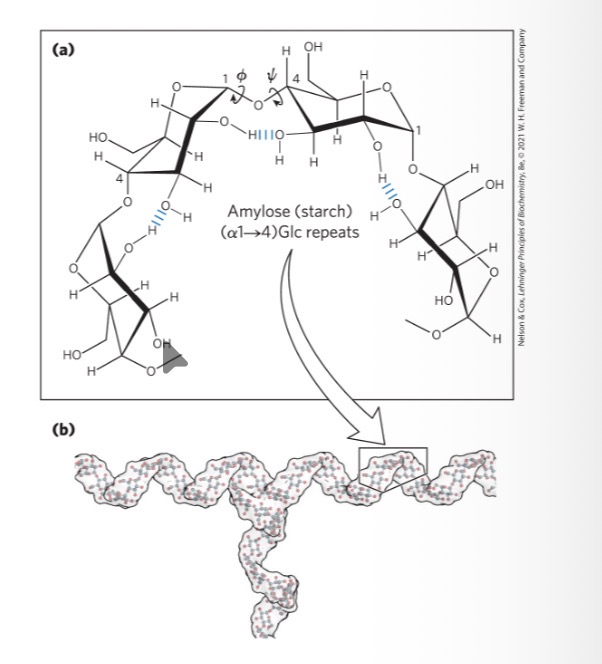
Starch
contains two types of glucose polymer, amylose and amylopectin
amylose = long, unbranched chains of D-glucose residues connected by (α1→4) linkages
amylopectin = larger than amylose with (α1→4) linkages between glucose residues and highly branched due to (α1→6) linkages
Glycogen
polymer of (α1→4)-linked glucose subunits, with (α1→6)-linked branches
more extensively branched
more compact than starch
Cellulose
tough, fibrous, water-insoluble substance
Linear, unbranched homopolysaccharide, - -consisting of 10,000 to 15,000 D-glucose units
glucose residues have the β configuration
Linked by (β1→4) glycosidic bonds
animals do not have the enzyme to hydrolyze (β1→4) glycosidic bonds
Chitin
Linear homopolysaccharide composed of N- acetylglucosamine residues in ( beta 1 —4) linkage
-Acetylated amino group makes chitin more hydrophobic and water resistant then cellulose
Helical structure of starch and glycogen
most stable three-dimensional structure for the (α1→4)-linked chains of starch and glycogen
six residues/turn
Linear structure of cellulose
most stable conformation is a straight, extended chain
each chair is turned 180° relative to its neighbors

Peptiodolglycan
rigid component of bacterial cell walls
heteropolymer of alternating (β1→4)-linked N-acetylglucosamine and N-acetylmuramic acid residues
cross-linked by short peptides
Extra cellular matrix (ECM)
gel like material in the extracellular space of tissues that hold cells together and provides a porous pathway for nutrient and O2 diffusion
Composed of interlocking mesh work of heteropolysaccharides(ground substance) and fibrous proteins
-basement membrane(specialized ECM) also contain heteropolysaccharides
Glycosaminoglycans
heteropolysaccharides in ECM
linear polymers composed of repeating disaccharide units
one monosaccharide is always either N-acetylglucosamine or N-acetylgalactosamine and the other is usually a uronic acid
unique to animals and bacteria
some contain esterified sulfate groups
Hyaluronan(hyaluronic acid)-
alternating residues of D-glucuronic acid and N-acetylglucosamine
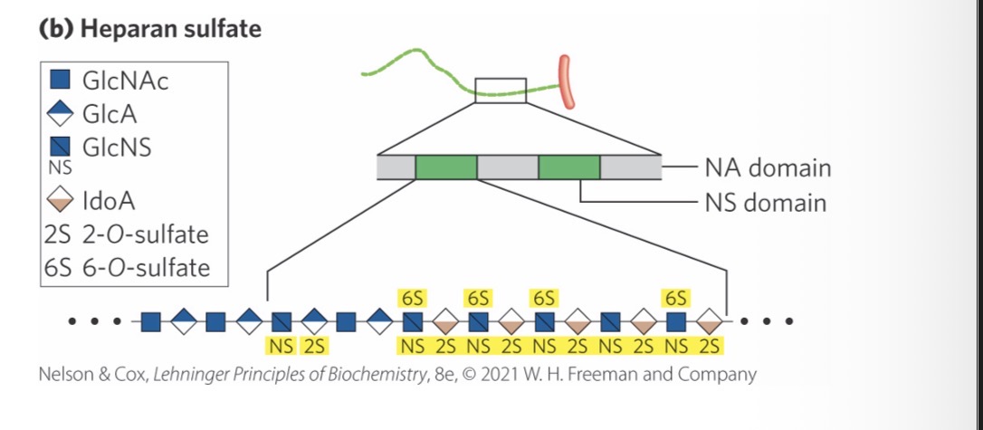
Heparin sulfate
contains variable, nonrandom arrangements of sulfated and nonsulfated sugars
sulfated residues gives the molecule the ability to interact specifically with proteins
Heparan
highly sulfated, intracellular form of heparan sulfate produced primarily by mast cells
used as a therapeutic agent to inhibit coagulation of blood through its capacity to bind the protease inhibitor antithrombin
Glycoconjugate
Biologically active molecule consisting of an informational carbohydrate joined to a protein or lipid
Proteoglycans
Macromolecules of the cell surface or ECM consisting of 1 + sufated glycosaminoglycan chain(s) joined covalently to a membrane or protein or secreted protein
major component of extracellular matrices
Glycoproteins
have one or several oligosaccharides joined covalently to a protein
- found ont he outer face of plasma membrane in ECM, in blood and in organelles( golgi complexes, secretory granules and lysosomes )
Oligosaccharides portions are heterogenous and rich in information
Glycolipids
Plasma membrane components in which the hydrophilic head groups are oligosaccharides
Gylcosphingolipids
Class of glycolipids with specific backbone structure
neurons are rich in glycosphingolipids
Play a role in transduction
Proteoglycan unit
“Core protein” with covalently attached glycosaminoglycan(s)
Tetrasaccharide linker
Connects to glycosaminoglycan to Ser residue of the protein
Syndecans
Single trans membrane domain and extracellular domain bearing 3-5 chains of heparan sulfate and chondroitin
Glypicans
attached to the membrane by a GPI anchor (a glycosylated derivative of the membrane lipid phosphatidylinositol)
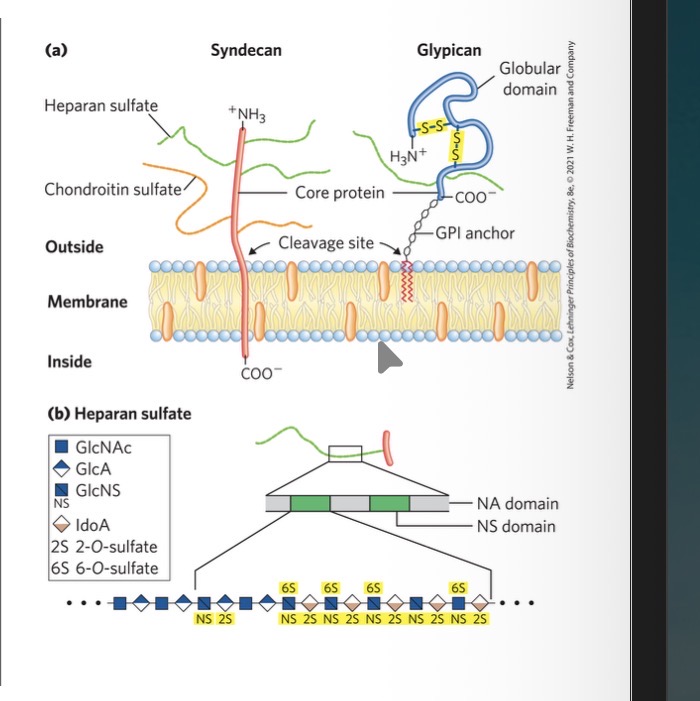
NS Domian
highly sulfated domains that alternate with domains having unmodified GlcNAc and GlcA residues
Poroteoglycan aggregates
supramolecular assemblies of many core proteins all bound to a single molecule of hyaluronan
aggrecan interacts strongly with collagen in the ECM of cartilage
Thrombin and Antithrombin
antithrombin binds to and inhibits the protease thrombin only in the presence of heparan sulfate
both proteins are rich in Arg and Lys residues
interact electrostatically with the sulfates of the glycosaminoglycans
How does chondroitin sulfate, dermatan sulfate, keratan sulfate, and heparan sulfate differ from hyaluronan in three respects:
generally much shorter polymers
covalently linked to specific proteins (proteoglycans)
one or both monomer units differ from hyaluronan
Fibronectin
has separate domains to bind fibrin, heparan sulfate, and collagen
contain the conserved RGD sequence (Arg–Gly–Asp) to bind integrins
Integrins
Mediate signaling between cell interior and ECM molecules
What is the purpose of interactions between cells and the ECM?
anchor cells tot he ECM providing strength and elasticity of skin and joints
Provide paths that direct the migration of cell interior developing tissue
Convey information in both directions across the plasma membrane
O linked -(type of attachment)
a glycoside bond joins the anomeric carbon of a carbohydrate to the —OH of a Ser or Thr residue
N-LInked ( type of attachment )
an N-glycosyl bond joins the anomeric carbon of a sugar to the amide nitrogen of an Asn residue
Mucins( ex of glycoproteins)
secreted or membrane glycoproteins
can contain large numbers of O-linked oligosaccharide chains
present in most secretions
Ex of glycoprotiens( protiens of blood )
examples: immunoglobulins (antibodies), follicle-stimulating hormone, luteinizing hormone, and thyroid-stimulating hormone
Ex of glycoproteins- milk protiens
example: major whey protein α-lactalbumin
Glycomics
= the systematic characterization of all carbohydrate components of a given cell or tissue, including those attached to proteins and to lipids
What are the advantages of adding oligosaccharides to proteins?
covalently attached oligosaccharides:
influence the folding and stability of the proteins
provide critical information about the targeting of newly synthesized proteins
allow specific recognition by other proteins
Gangliosides
membrane lipids of eukaryotic cells in which the polar head group is a complex oligosaccharide containing a sialic acid and other monosaccharide residues
Lipopolysaccharides
dominant surface feature of the outer membrane of gram-negative bacteria
Glycobiology
the study of the structure and function of the challenge is to understand how cells use specific oligosaccharides to encode information about:
intracellular targeting of proteins
cell-cell interactions
cell differentiation and tissue development
extracellular signals
Lectins
bind carbohydrates with high specificity and with moderate to high affinity
-function :cell-cel recondition, signaling,adhesion ,intracellular targeting of newly synthesized proteins
Selections
family of plasma membrane lectins that mediate cell-cell recognition and adhesion in a wide range of cellular processes
move immune cells through the capillary wall
mediate inflammatory responses
mediate the rejection of transplanted organs
Methods of carbohydrate analysis
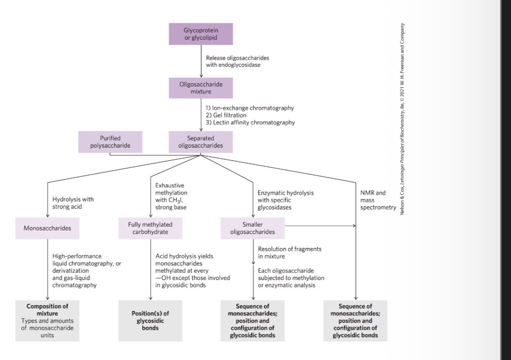
How can you determine oligosaccharides and polysaccharide structures?
more complex than protein and nucleic acid analysis
can employ a variety of methods to determine sequence, configuration at anomeric and other carbons, and positions of glycosidic bonds:
traditional chemical and enzymatic approaches
mass spectrometry
high-resolution NMR spectroscopy
Solid phase synthetic methods
carbohydrate chemists can synthesize short segments of almost any glycosaminoglycan
solid-phase oligosaccharide synthesis:
based on the same principles as peptide synthesis
yields defined oligosaccharides
useful in exploring lectin-oligosaccharide interactions