1.6. Digestive system
1/77
There's no tags or description
Looks like no tags are added yet.
Name | Mastery | Learn | Test | Matching | Spaced | Call with Kai |
|---|
No analytics yet
Send a link to your students to track their progress
78 Terms
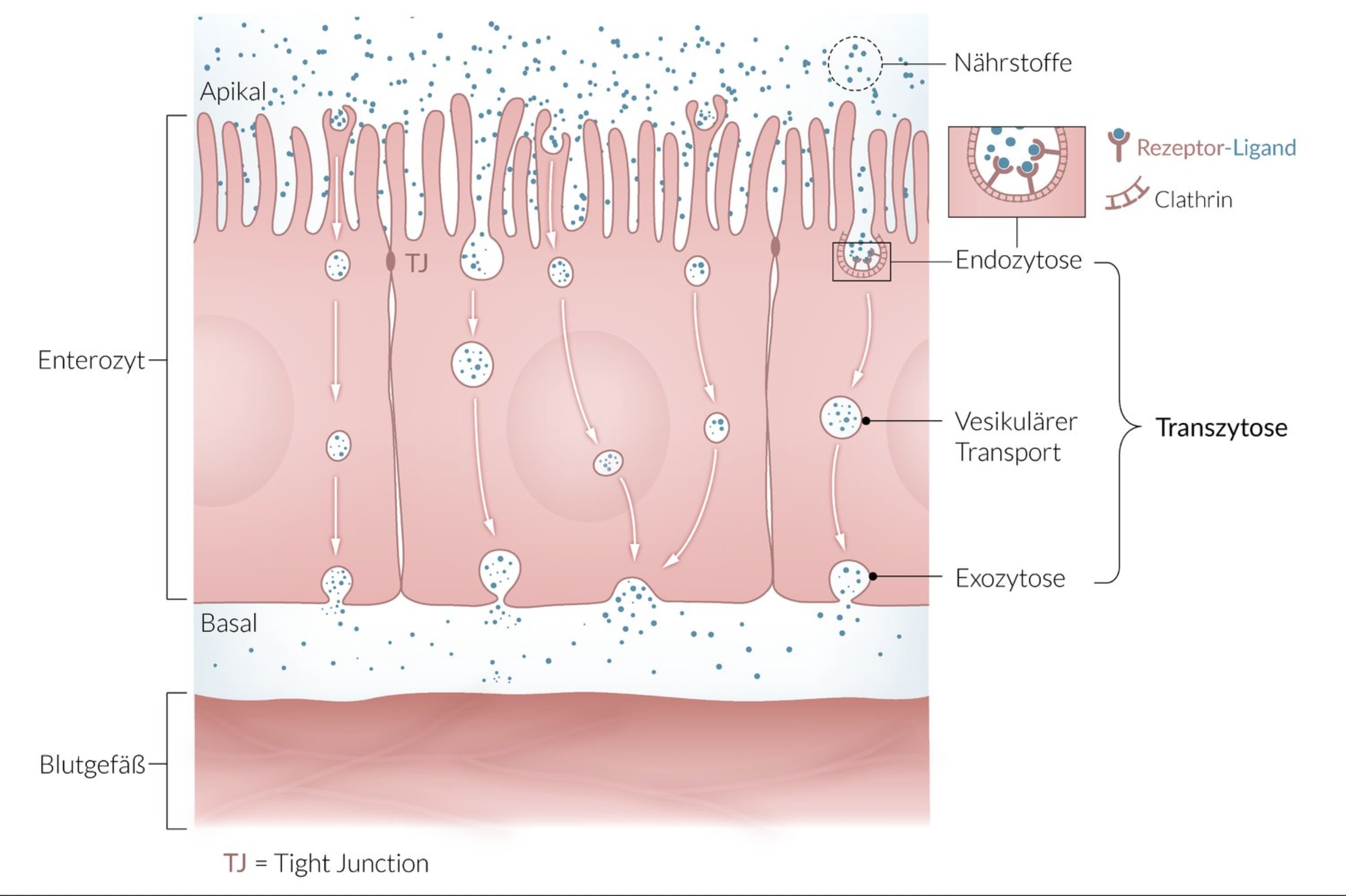
1. When vesicle-packaged macromolecules are transported through the cells of a barrier-forming epithelium or endothelium, this process is called:
A. Apoptosis
B. Endomitosis
C. Phagocytosis
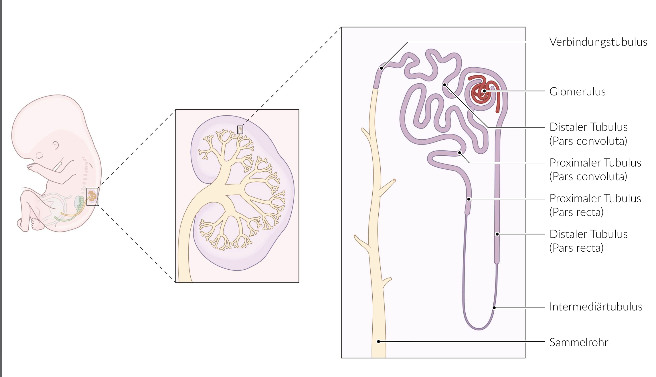
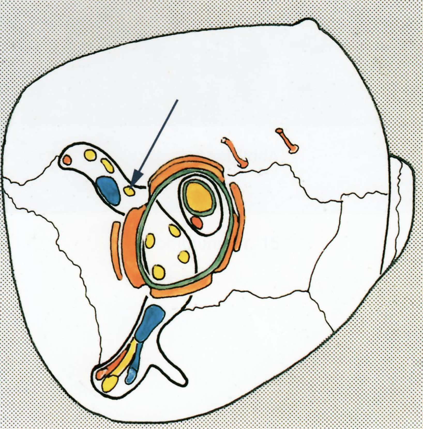
D. Pinocytosis
E. Transcytosis
E. Transcytosis
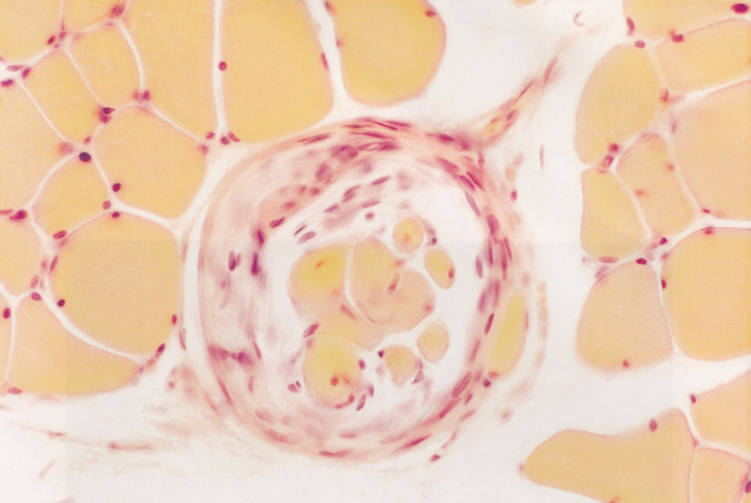
2. Which vessel is an artery of the elastic type?
A. Brachial artery (A. brachialis)
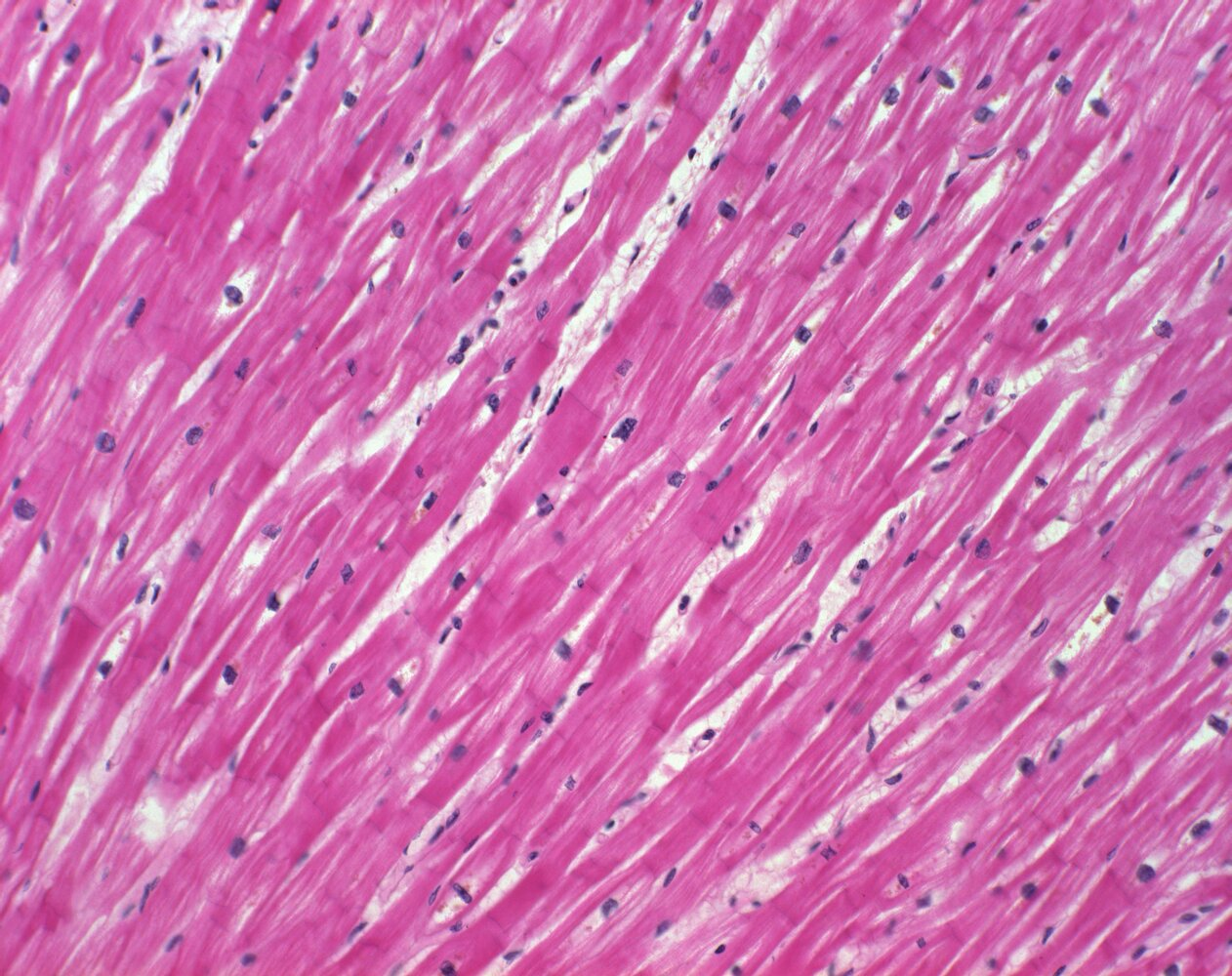
B. Common carotid artery (A. carotis communis)
C. Femoral artery (A. femoralis)
D. Maxillary artery (A. maxillaris)
E. Posterior tibial artery (A. tibialis posterior)
B. Common carotid artery (A. carotis communis)
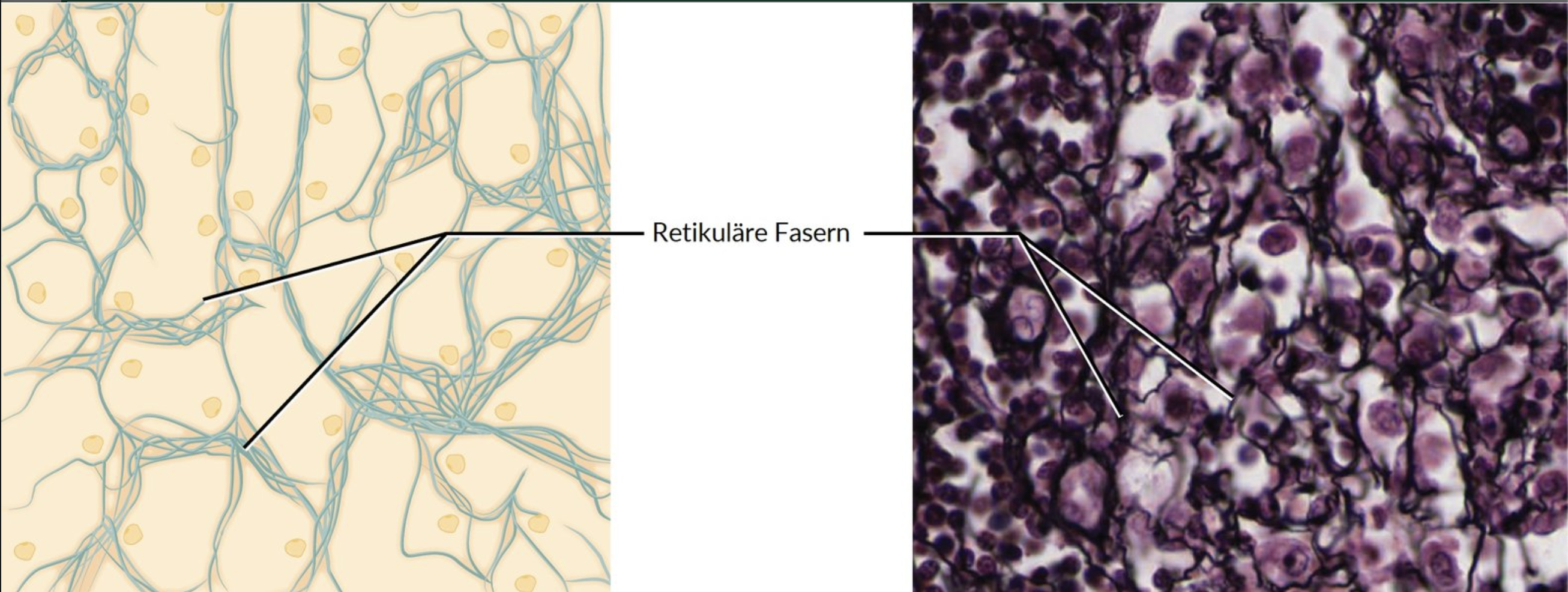
3. Which statement about reticulocytes is NOT true?
A. They develop into mature erythrocytes.
B. They produce reticular fibers.
C. They contain RNA.
D. They are found in red bone marrow.
E. They are without a nucleus.
B. They produce reticular fibers.
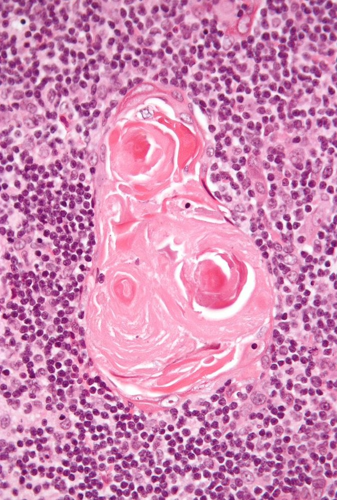
4. Which of the following histological features is found only in the thymus among the lymphatic organs?"
A. Hassall's corpuscles
B. Hilum
C. Marginal sinus (Randsinus)
D. Cortex and medulla (Rinde und Mark)
E. Secondary follicles
A. Hassall's corpuscles
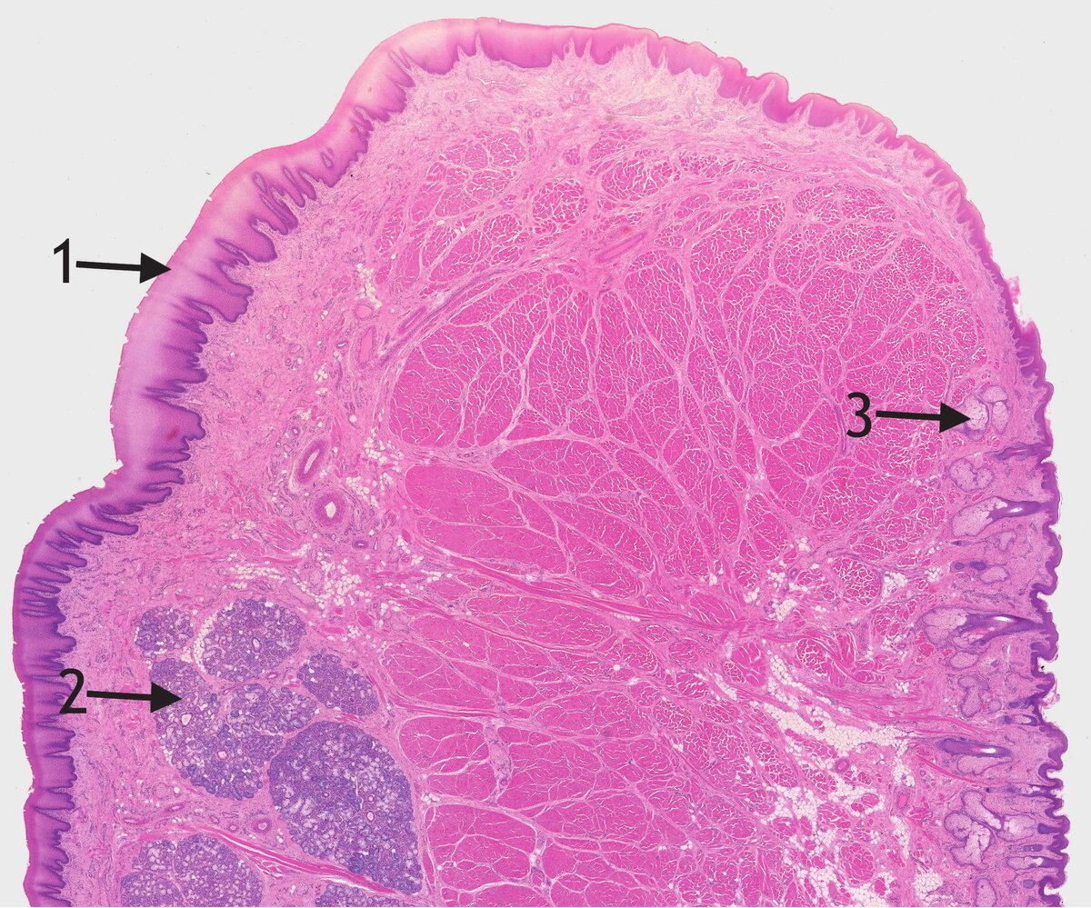
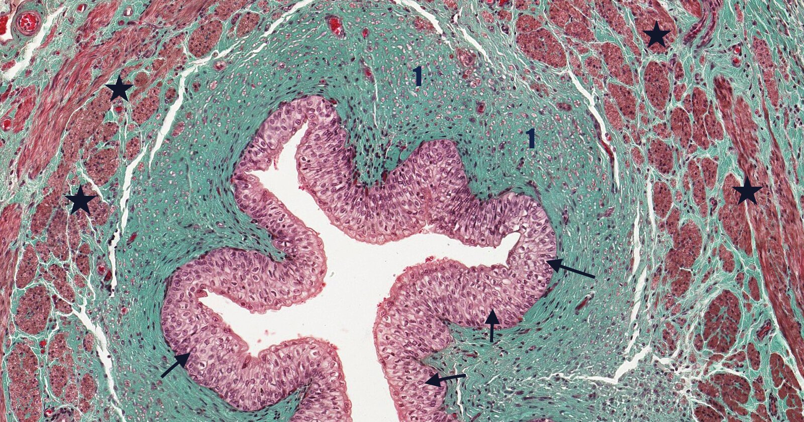
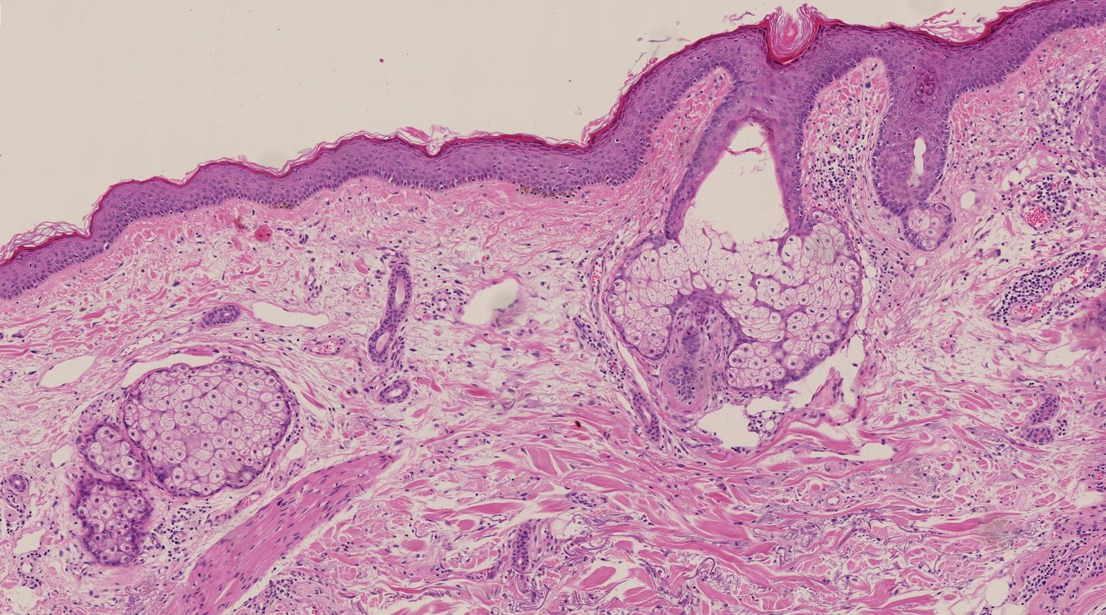
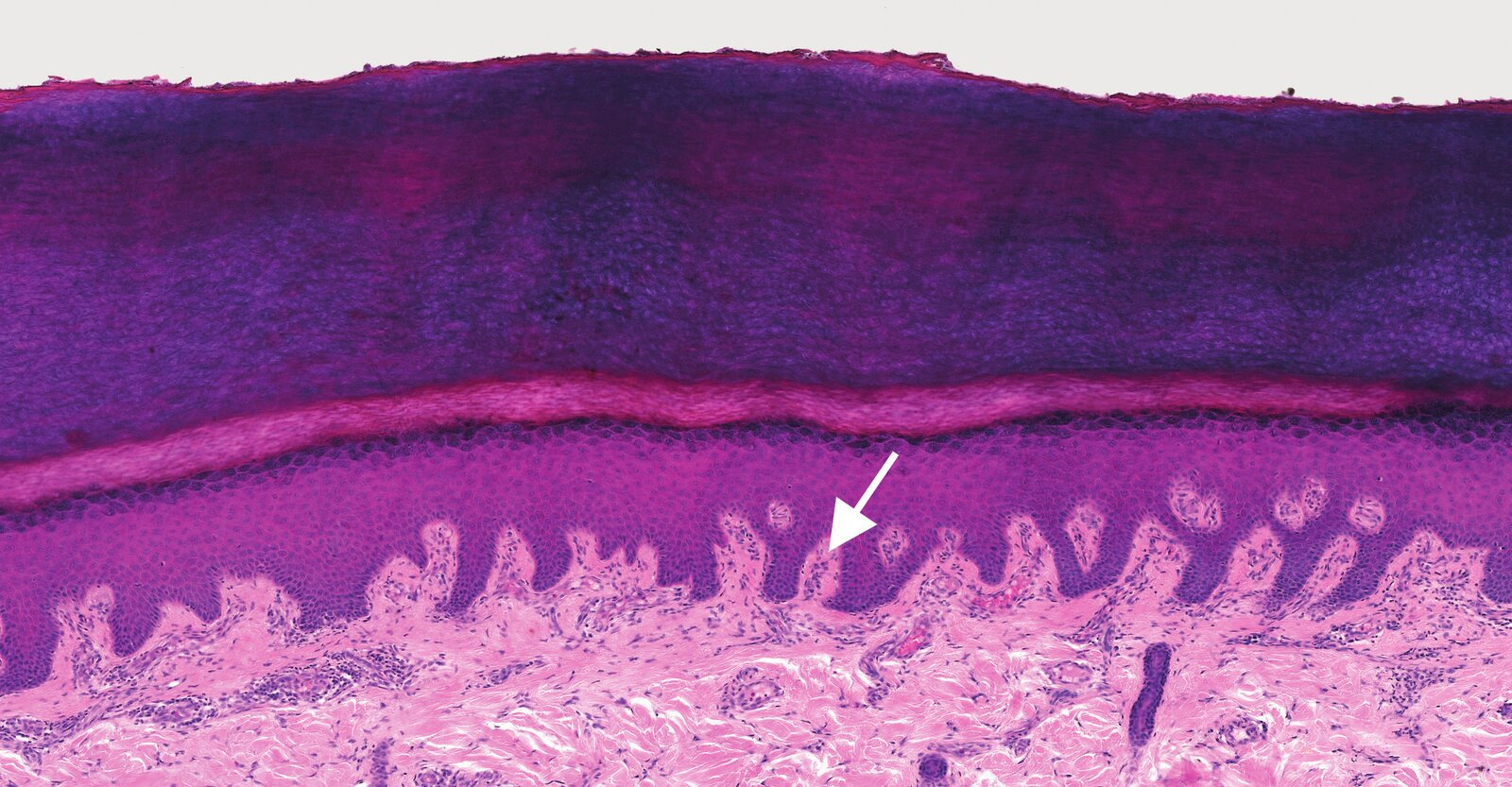
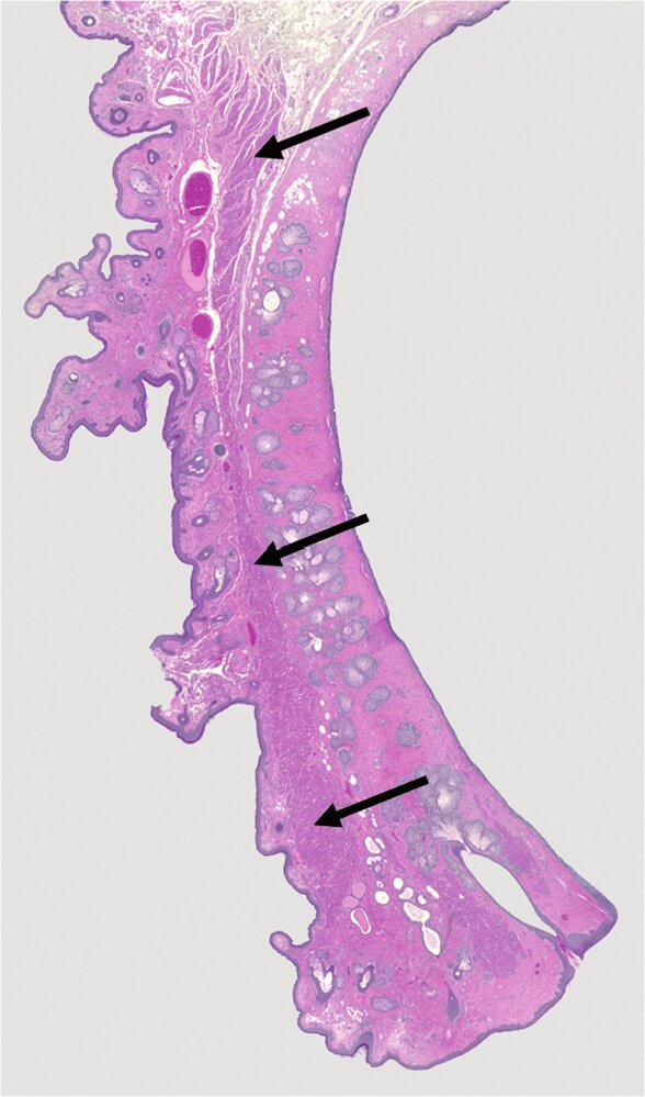
5. Which statement about the histological image is correct?"
A. The image shows a section through the fingertip.
B. The image shows a section through the lip.
C. Arrow 1 points to a transitional epithelium.
D. Arrow 2 points to lymph follicles.
E. Arrow 3 points to mucous glands.
B. The image shows a section through the lip.
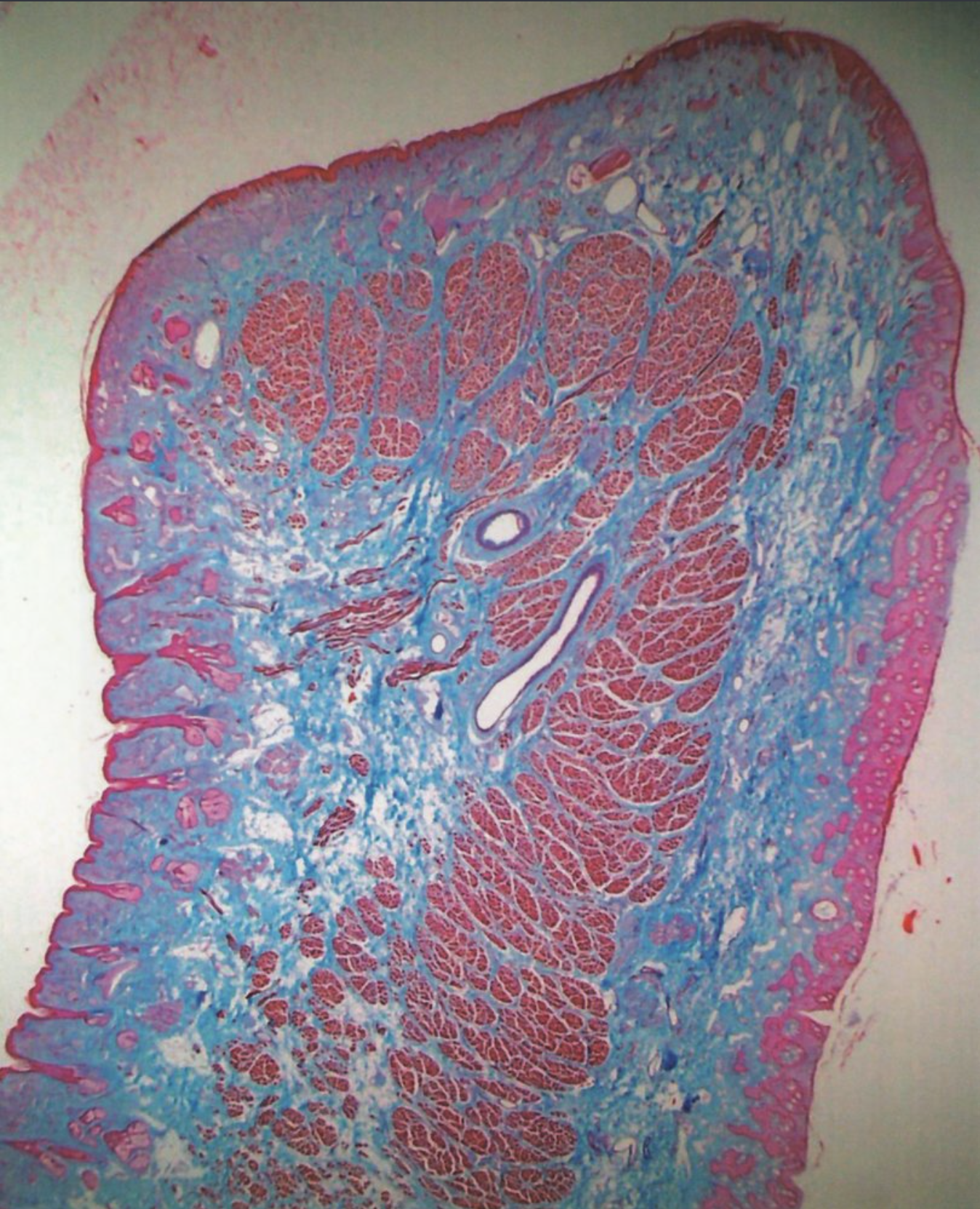
6. The structures shown in the image are:
A. Scent glands (apocrine sweat glands) in the armpit
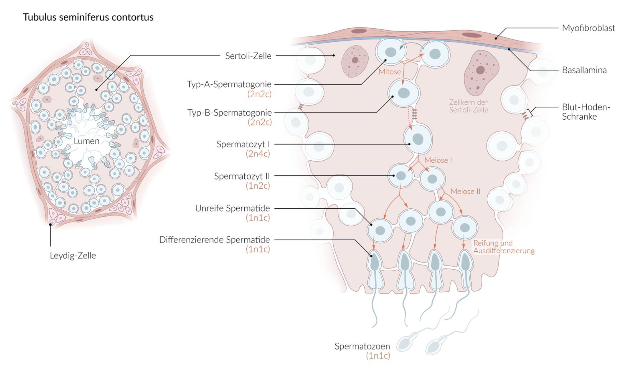
B. A section of the exocrine pancreas
C. A section of the parotid gland (Glandula parotidea)
D. A section of the submandibular gland (Glandula submandibularis)
E. Sweat glands in the deeper layer of the dermis
D. A section of the submandibular gland (Glandula submandibularis)
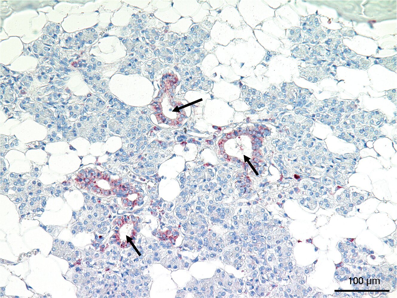
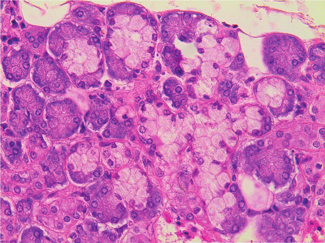
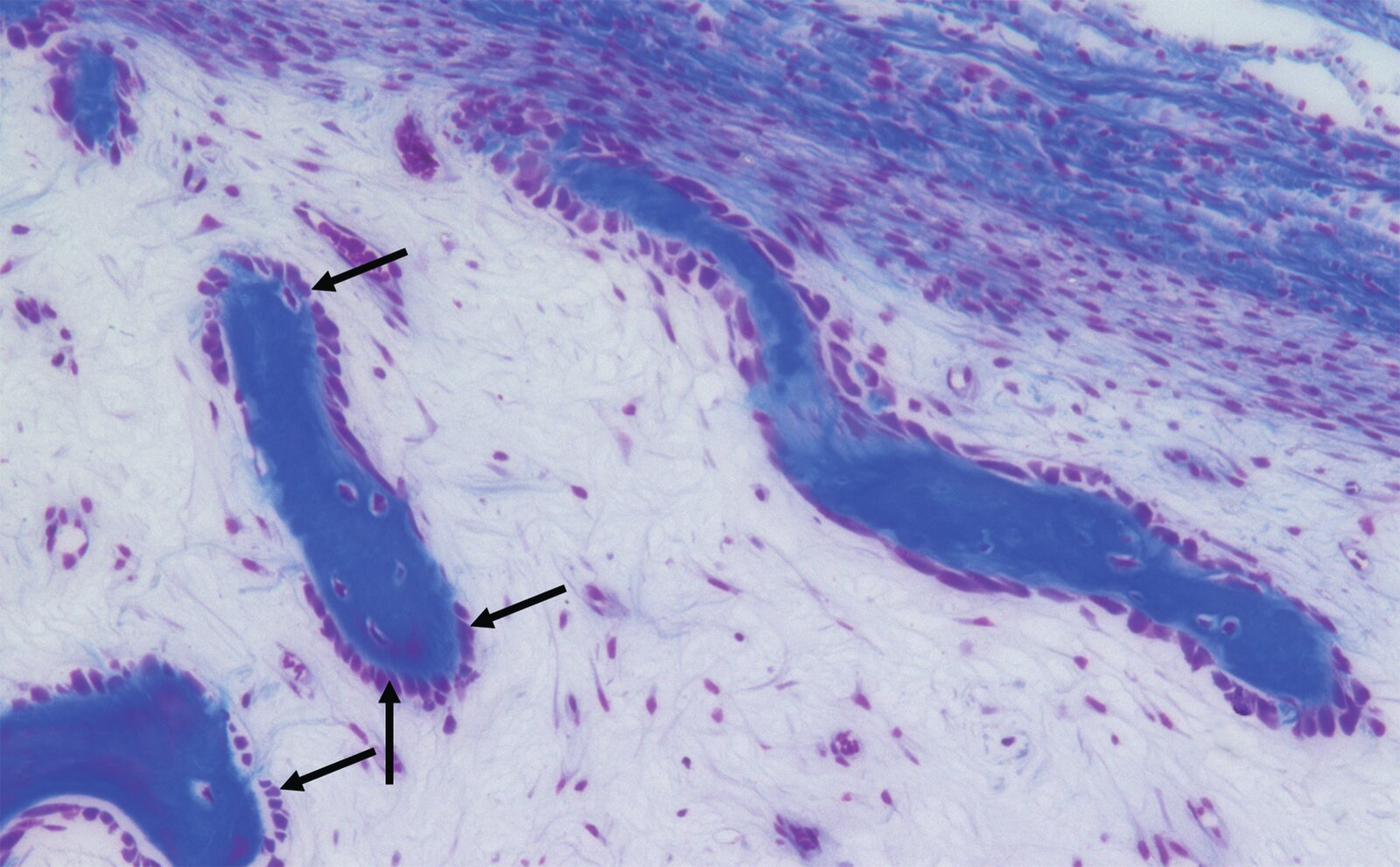
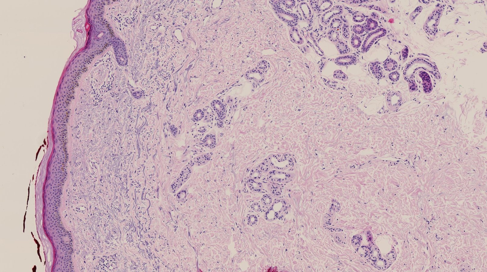
7. The epithelium of the structures marked with arrows in the image:
A. Has basal striations
B. Belongs to intercalated ducts
C. Lines interlobular excretory ducts
D. Is most commonly found in the sublingual gland
E. Secretes isotonic saliva
A. Has basal striations
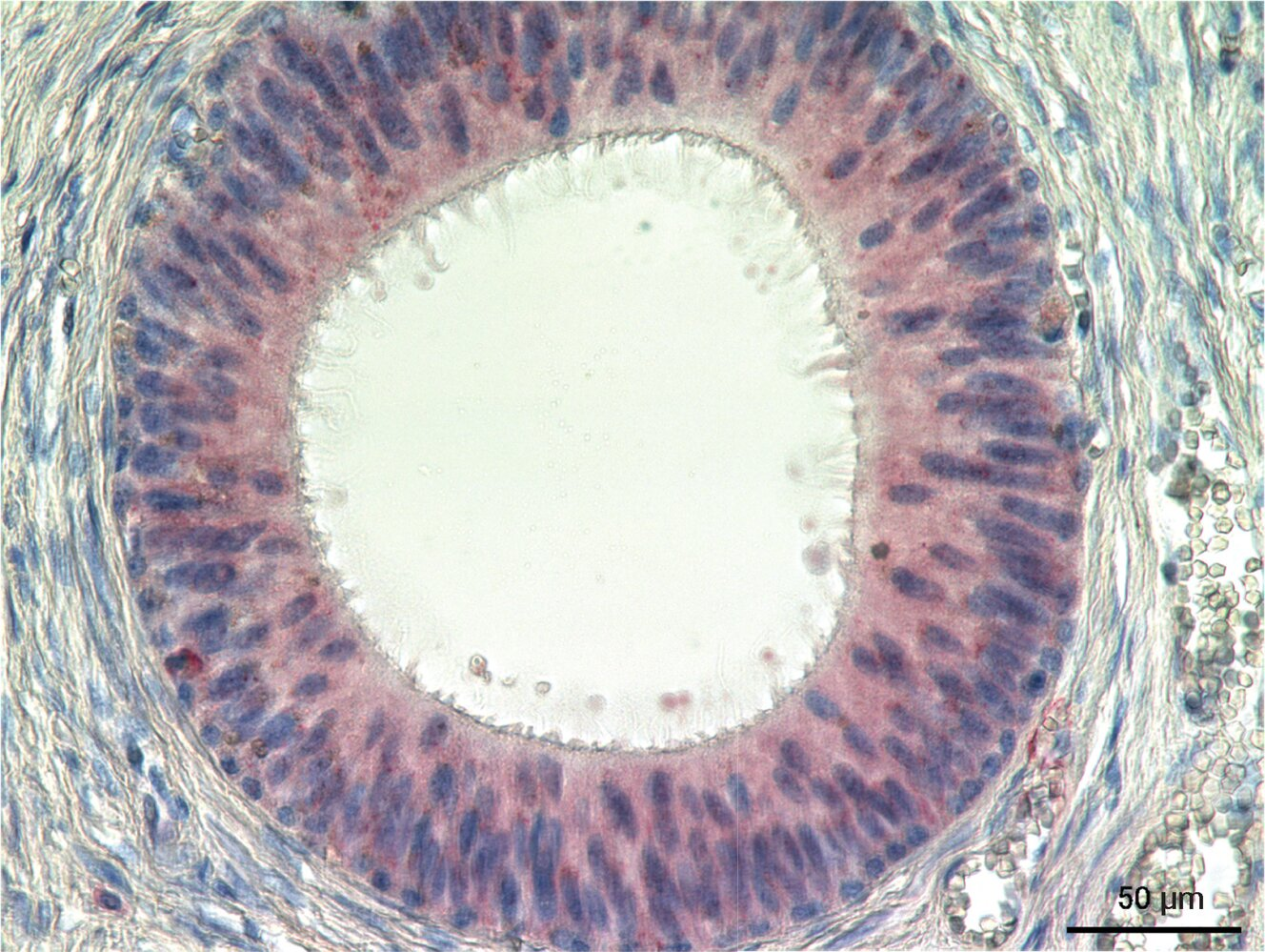
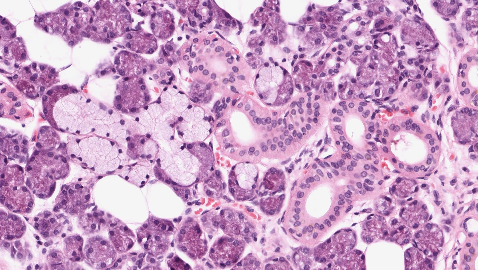
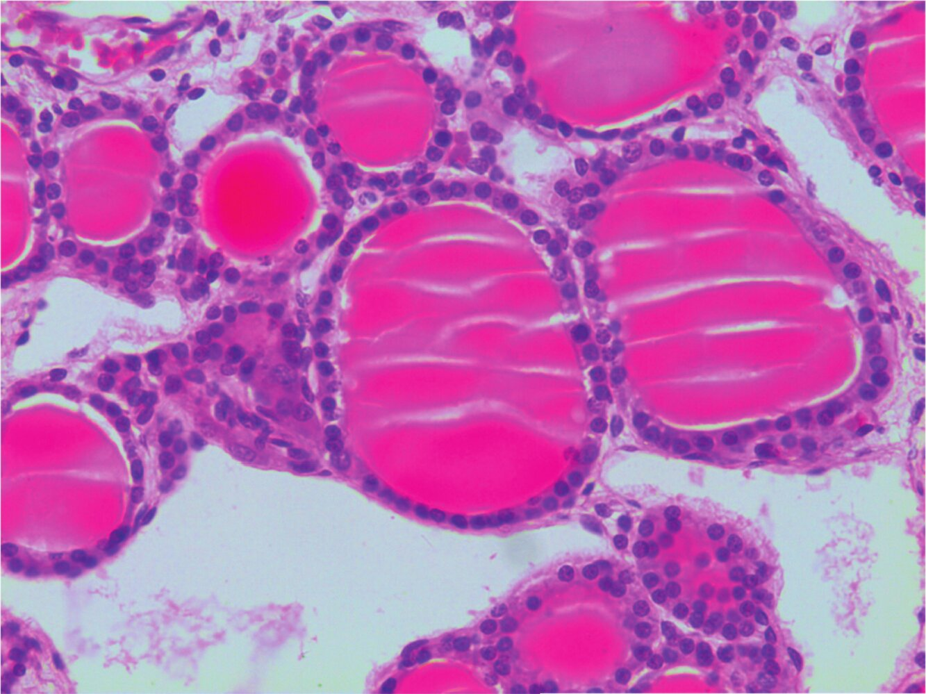
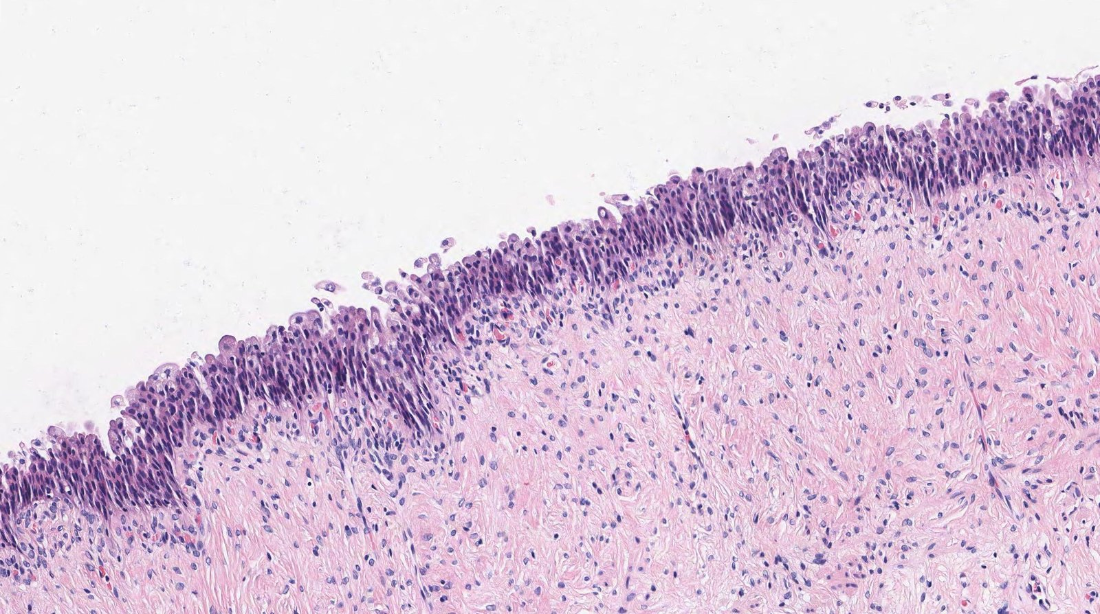
8. Which statement about the structure shown in the image is correct?
A. The epithelium is particularly characteristic of the periurethral zone.
B. The epithelial cells have long cilia (kinocilia).
C. It is a glandular end piece.
D. It is a transitional epithelium.
E. Below the basement membrane of the epithelium lie myofibroblasts or smooth muscle cells.
E. Below the basement membrane of the epithelium lie myofibroblasts or smooth muscle cells.
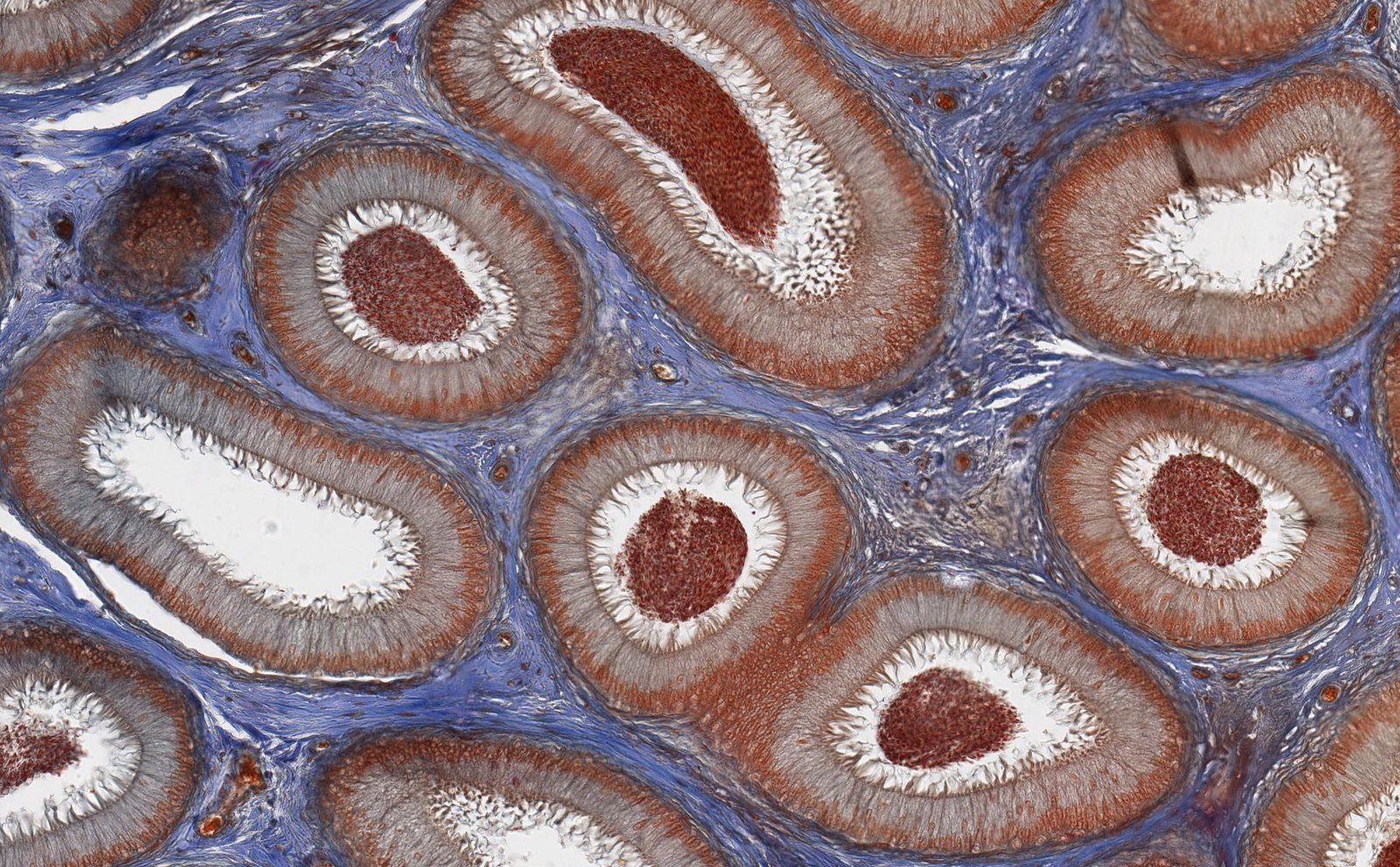
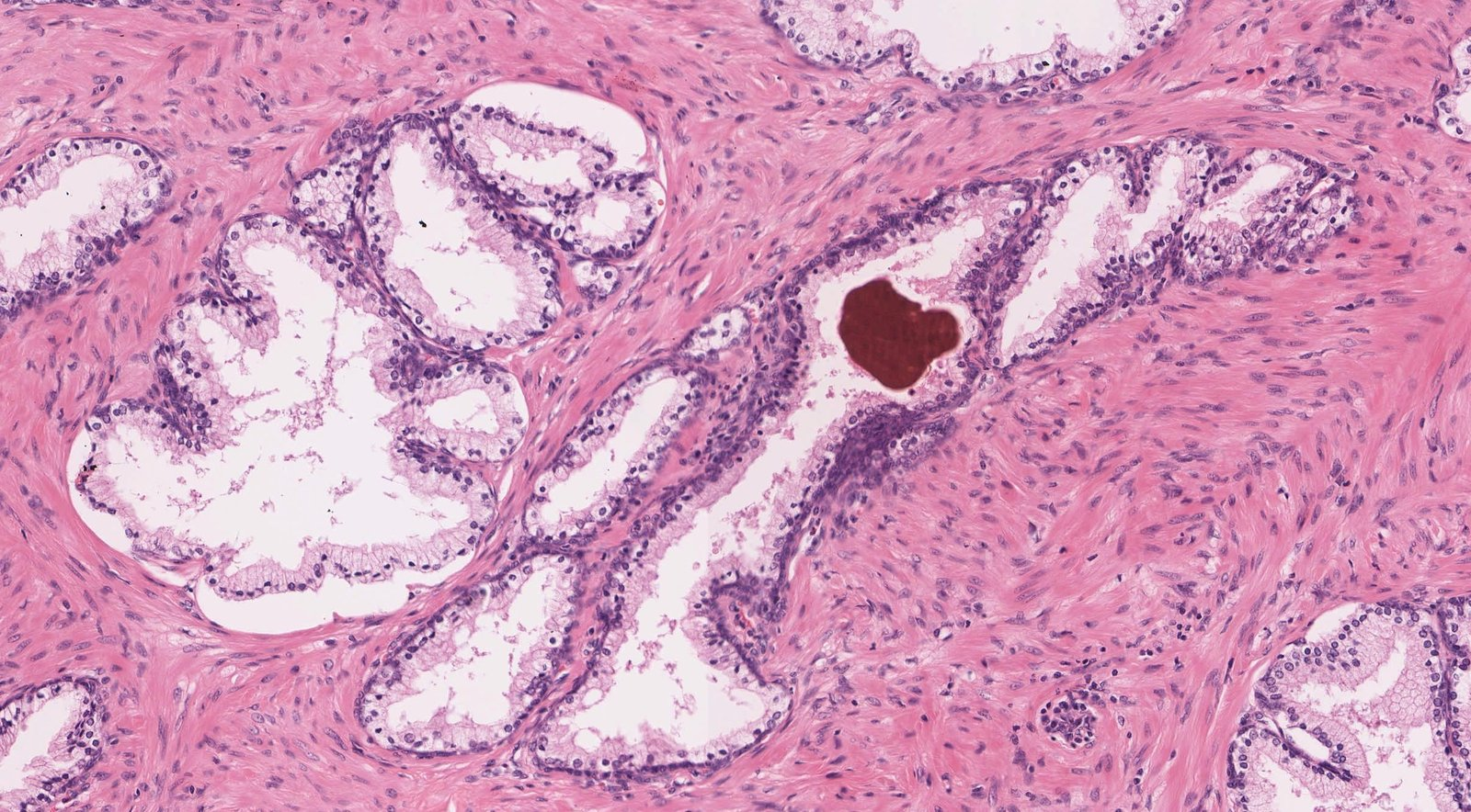
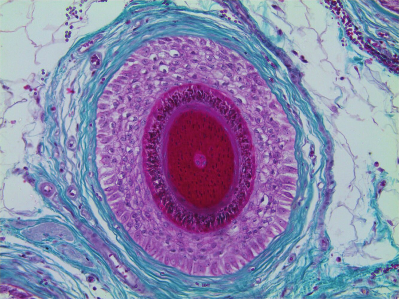
9. Which structure is shown in the image (van Gieson stain)?
A. Endometrium in the secretory phase
B. Lactating mammary gland (Mamma lactans)
C. Ovary
D. Prostate
E. Rete testis
D. Prostate
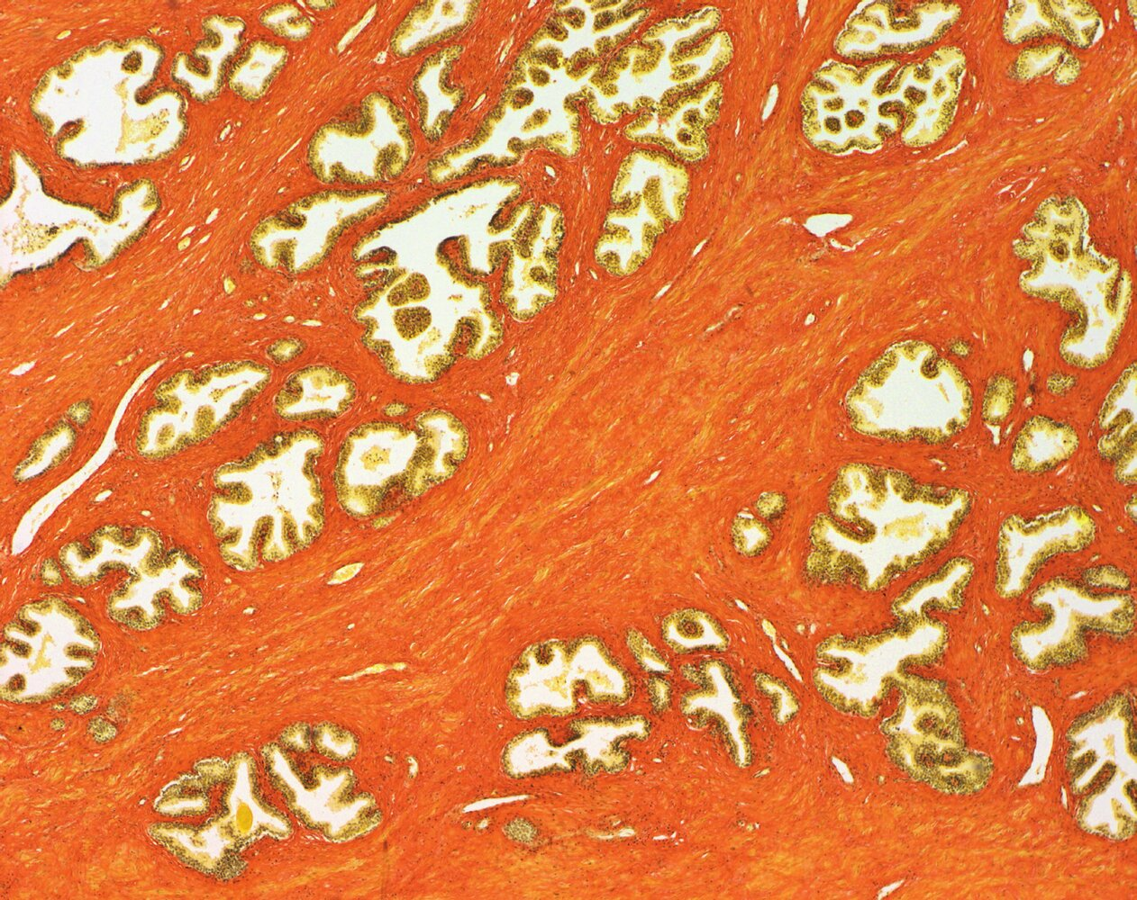
10. Which statement about the function of the organ shown in the image is NOT true?
A. It produces a hormone that influences calcium metabolism in cases of hypercalcemia.
B. It produces a hormone that regulates body temperature.
C. It produces a hormone that affects anterior pituitary cells via a feedback loop.
D. It produces a steroid hormone.
E. It produces an iodine-containing hormone.
D. It produces a steroid hormone.
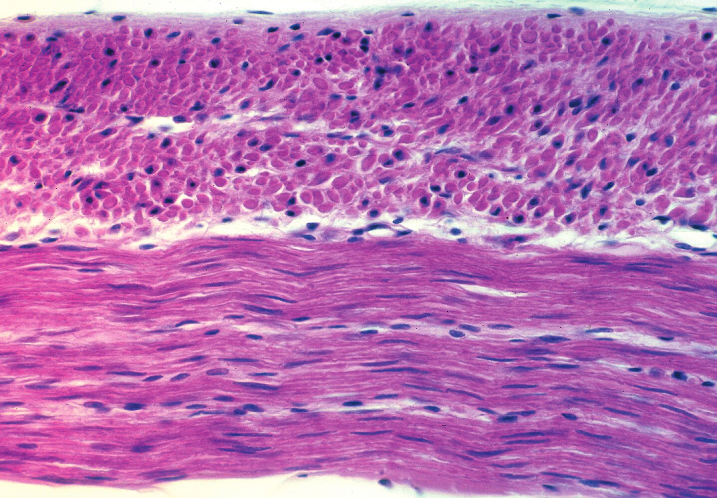
11. The interterritorial areas (regions between the chondrons) in fibrocartilage are characterized by a high content of:
A. Elastic fibers
B. Integrins
C. Type I collagen
D. Laminins
E. Reticular fibers
C. Type I collagen
12. The cells marked with arrows in the image:
A. Typically have multiple nuclei
B. Are inhibited by calcitriol
C. Partially differentiate into osteocytes
D. Are activated by the peptide hormone sclerostin
E. Are characterized by large lysosomes and vacuoles
C. Partially differentiate into osteocytes
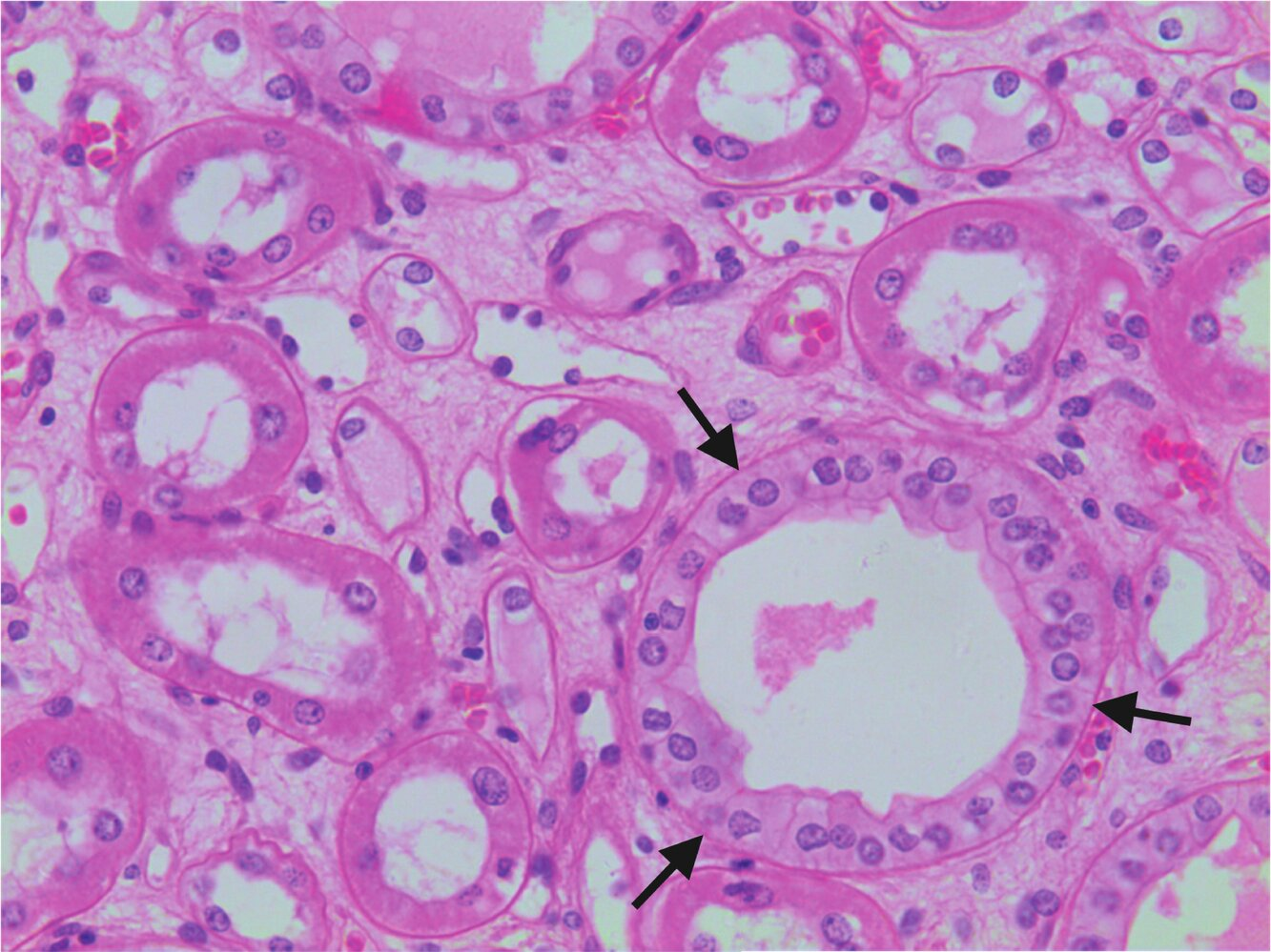
13. Which statement does not apply to the structure marked with arrows in the image?
A. It is located, among other places, in the renal medulla.
B. It opens into a papillary duct (Ductus papillaris).
C. Its epithelium is involved in water reabsorption.
D. It is influenced by a neuropeptide hormone released from the posterior pituitary.
E. It develops from the Müllerian ducts.
E. It develops from the Müllerian ducts.
14. Which statement about the histological image is correct?
A. The arrows point to the stratum granulosum.
B. The numbers 1 mark a zone with mucous glands.
C. The stars mark the adventitia of the organ shown.
D. The section shown contains plicae circulares.
E. The superficial cell layer of the epithelium contains uroplakins.
E. The superficial cell layer of the epithelium contains uroplakins.
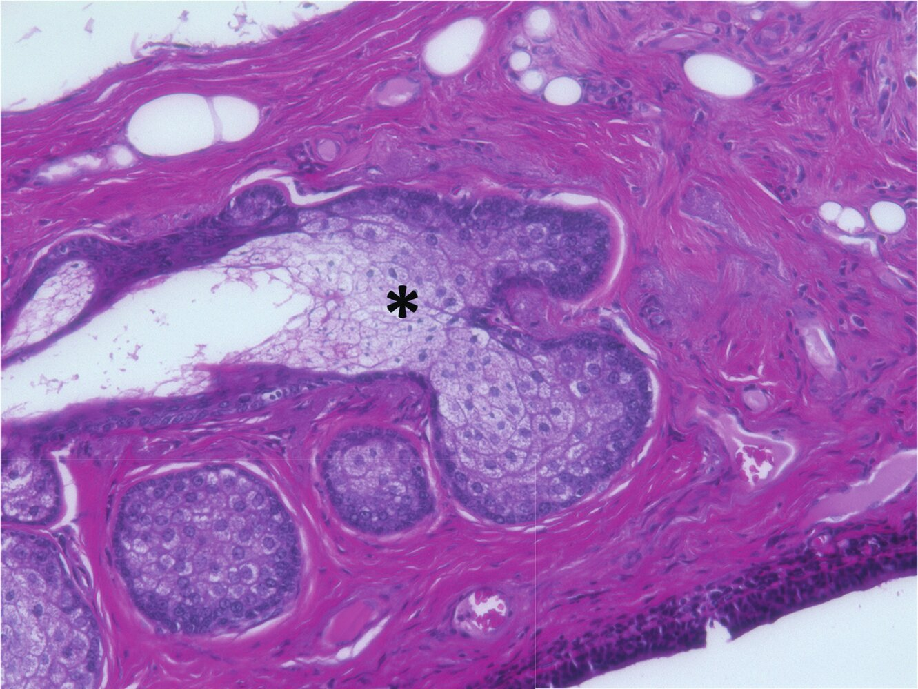
15. The cells of the gland marked with a star in the image:
A. Develop increasingly pyknotic nuclei during their differentiation
B. Typically die by necrosis
C. Accumulate large amounts of peptides in secretory granules during differentiation
D. Decrease in size during differentiation
E. Primarily serve in thermoregulation
A. Develop increasingly pyknotic nuclei during their differentiation
16. Which statement most likely applies to the structure shown in the image?
A. It carries sweat gland secretions.
B. It develops from a primordial follicle.
C. It is a typical component of thin (hairy) skin.
D. It plays a key role in the skin's elasticity.
E. It is moved by striated skeletal muscle.
C. It is a typical component of thin (hairy) skin.
17. Which statement about the histological image is correct?
A. Arrow 1 points to an oocyte.
B. Arrow 1 points to an intrafusal muscle fiber.
C. Arrow 1 points to a pseudounipolar ganglion cell.
D. Arrow 2 points to the urinary pole of the Bowman’s capsule.
E. Arrow 2 points to the perineurial sheath of a nerve.
B. Arrow 1 points to an intrafusal muscle fiber.
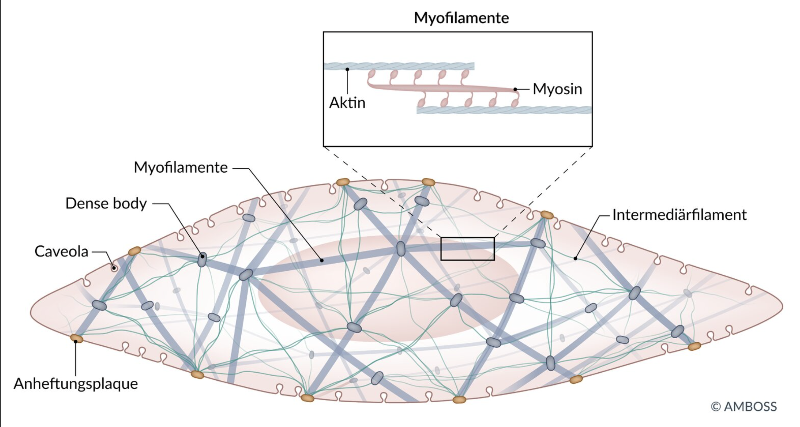
18. The image shows a histological preparation in which a specific type of muscle cells predominates.
What characterizes the muscle cells shown?
A. Their caveolae serve, among other things, to take up Ca²⁺ into the cell.
B. Their contraction is triggered by binding of Ca²⁺ to troponin C.
C. Their postsynaptic cell membrane contains clusters of nicotinic acetylcholine receptors.
D. Their abundant intermediate filament proteins are called cytokeratins.
E. Their cell membranes are invaginated as T-tubules at the junction of A and I bands.
A. Their caveolae serve, among other things, to take up Ca²⁺ into the cell.
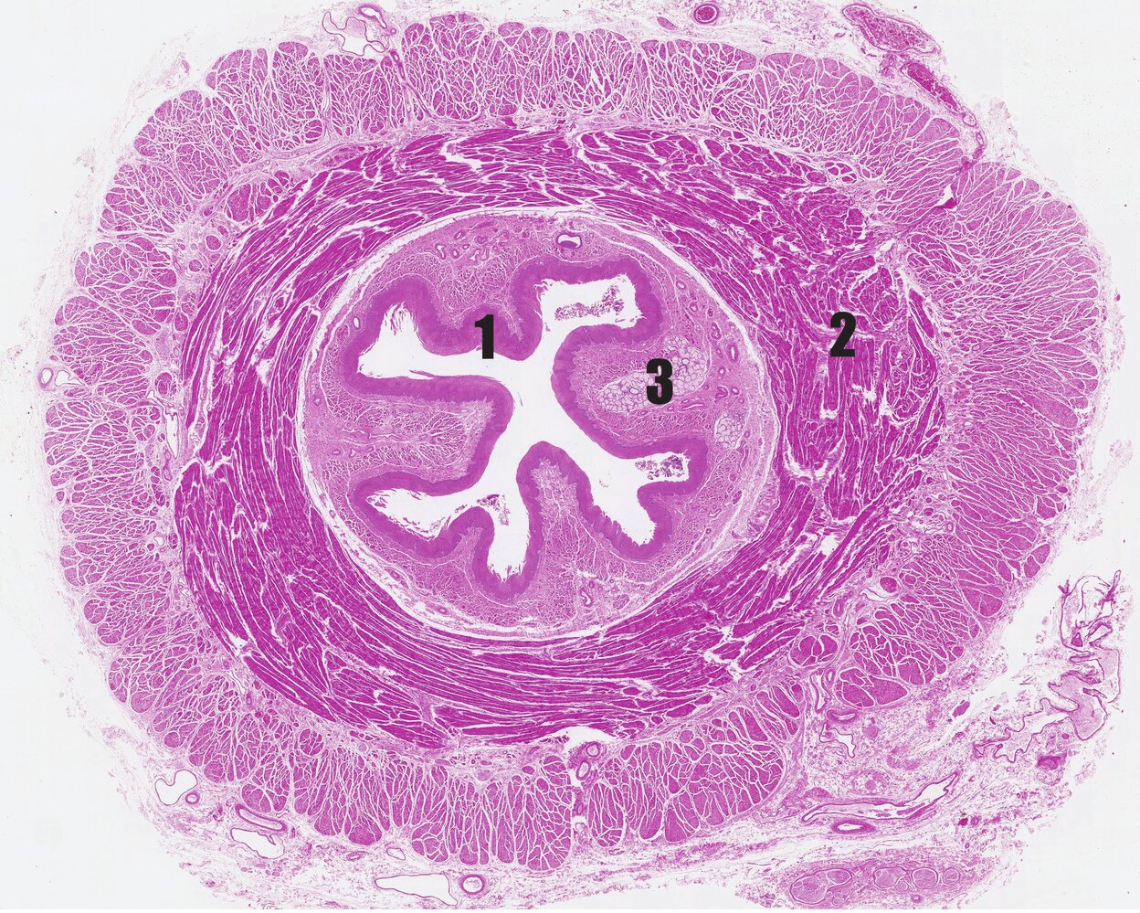
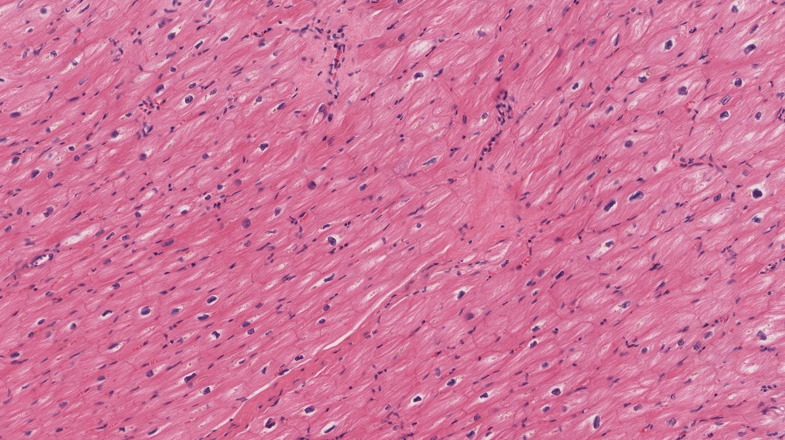
19. Which statement about the histological image is correct?
A. The organ shown contains skeletal muscle in its upper part.
B. The section of the organ shown is entirely covered by a tunica serosa.
C. Number 1 marks a pseudostratified ciliated epithelium.
D. Number 2 marks the lamina muscularis mucosae.
E. Number 3 marks the myenteric plexus (Plexus myentericus).
A. The organ shown contains skeletal muscle in its upper part.
20. The microscopic overview image in the illustration originates from:
A. The heart
B. The stratum reticulare of the skin
C. The organ capsule of the spleen
D. The tunica muscularis of the stomach
E. A skeletal muscle
A. The heart
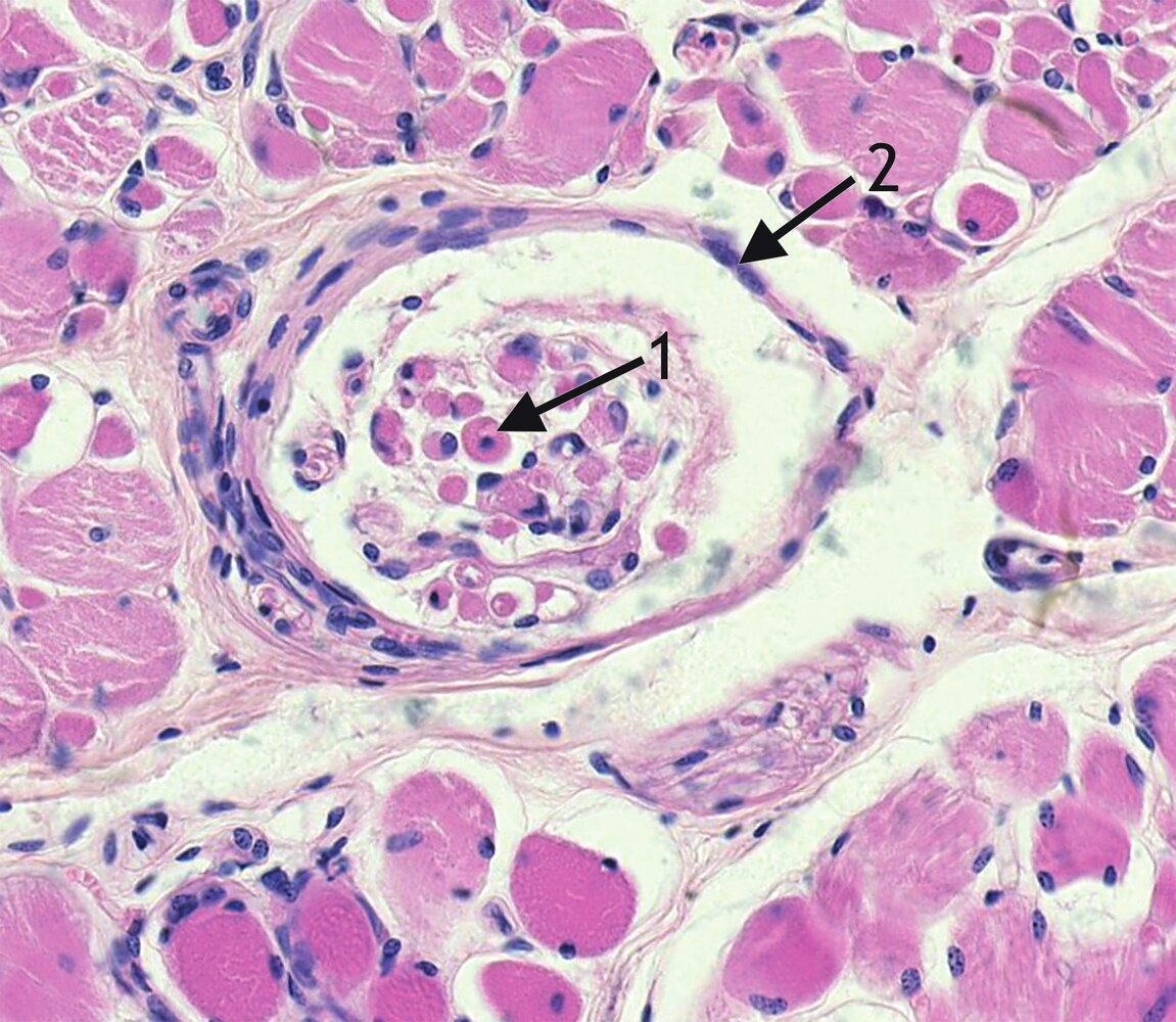
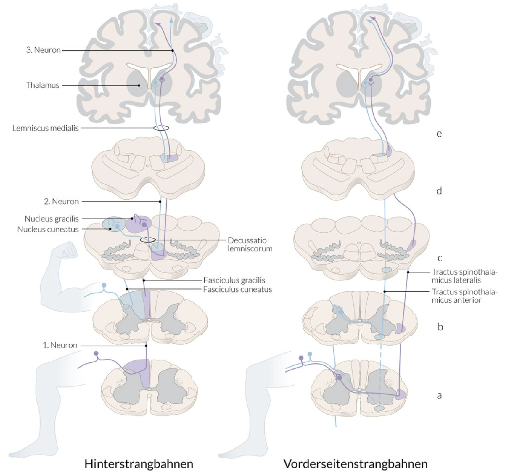
21. In the image, a location marked with an arrow is characteristic of the position of a sensory corpuscle.
The sensory stimuli perceived by this receptor are primarily transmitted through the:
A. Lateral funiculus (Funiculus lateralis)
B. Posterior funiculus (Funiculus posterior)
C. Anterior corticospinal tract (Tractus corticospinalis anterior)
D. Spinal tract of the trigeminal nerve (Tractus spinalis nervi trigemini)
E. Anterior spinothalamic tract (Tractus spinothalamicus anterior)
B. Posterior funiculus (Funiculus posterior)
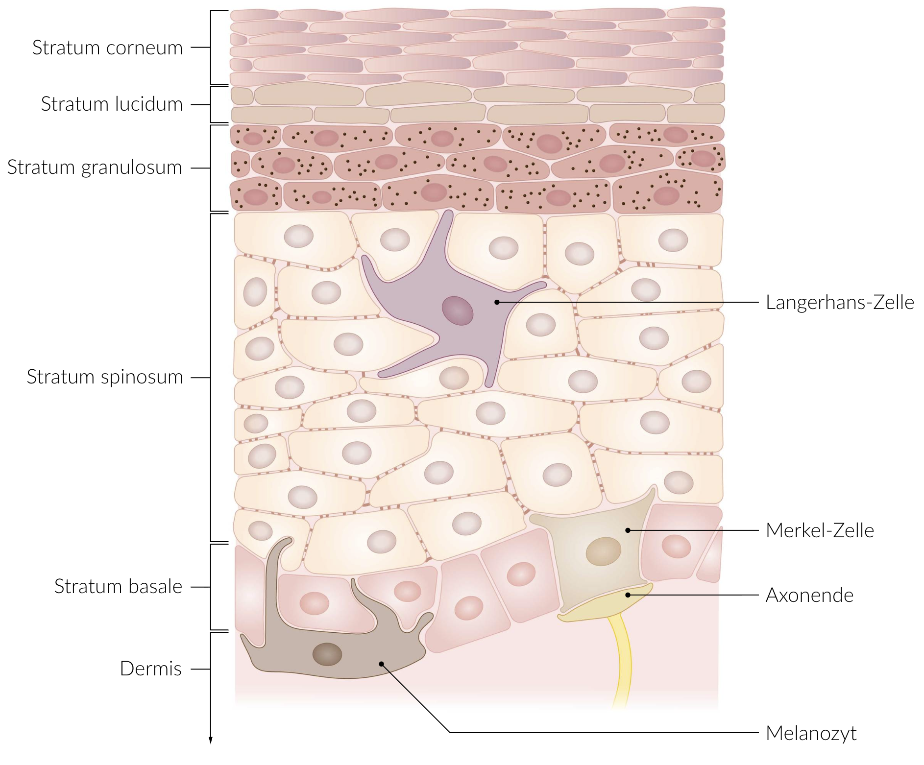
22. In blistering autoimmune skin diseases like pemphigus, the water barrier of the skin breaks down. As a result, patients are at risk of dying from dehydration due to uncontrolled fluid loss.
During normal maturation of the epidermis, the tight junctions (zonulae occludentes) that are essential for the skin’s barrier function are most likely found in which layer?
A. Stratum basale
B. Stratum spinosum
C. Stratum granulosum
D. Stratum lucidum
E. Stratum corneum
C. Stratum granulosum
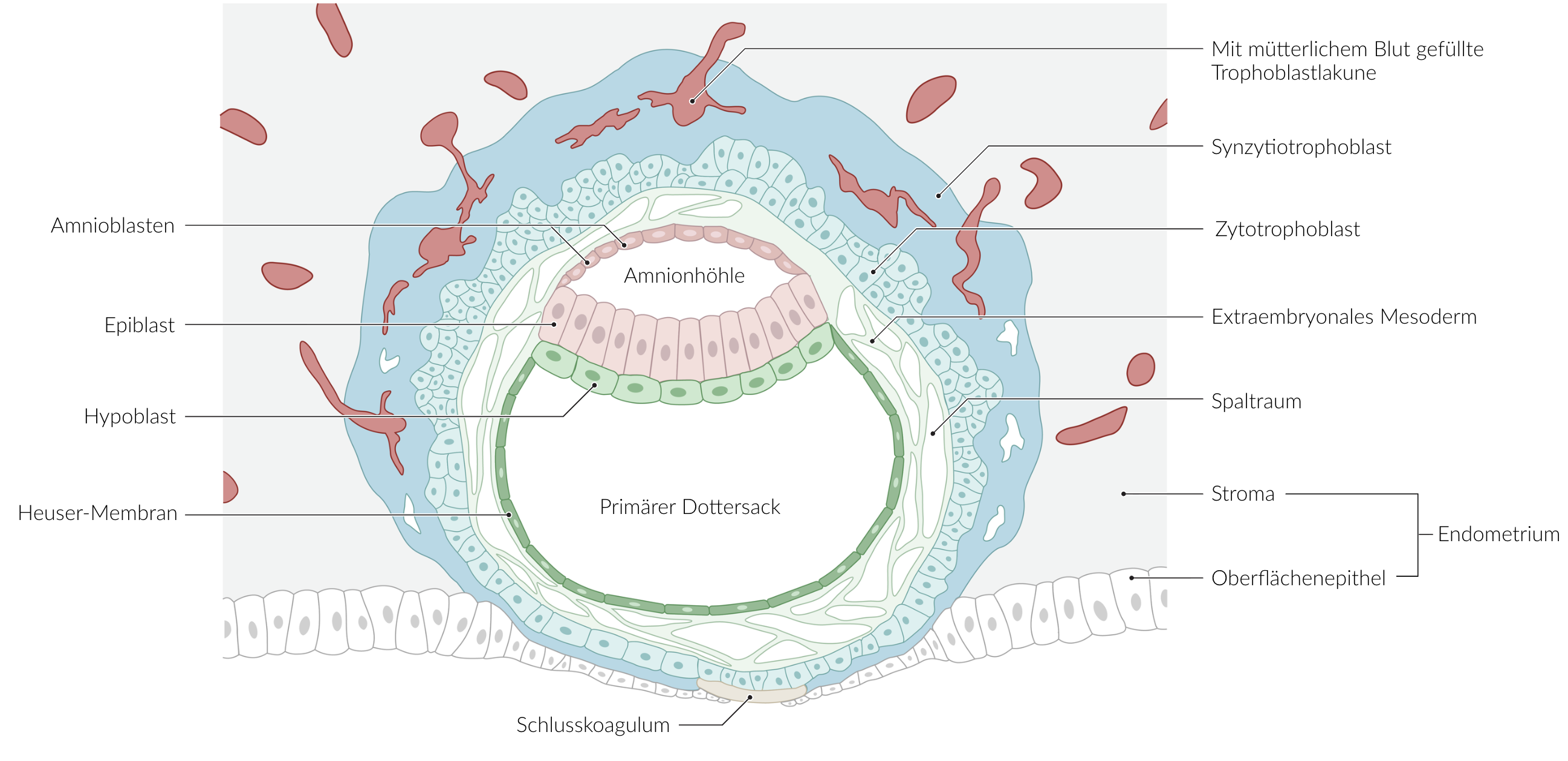
23. During embryonic development, hypoblast cells give rise to:
A. Endothelial cells of blood vessels
B. Alveolar epithelial cells
C. Epithelial cells of the yolk sac
D. Mesothelial cells of the visceral pleura
E. Mesothelial cells of the parietal peritoneum
C. Epithelial cells of the yolk sac
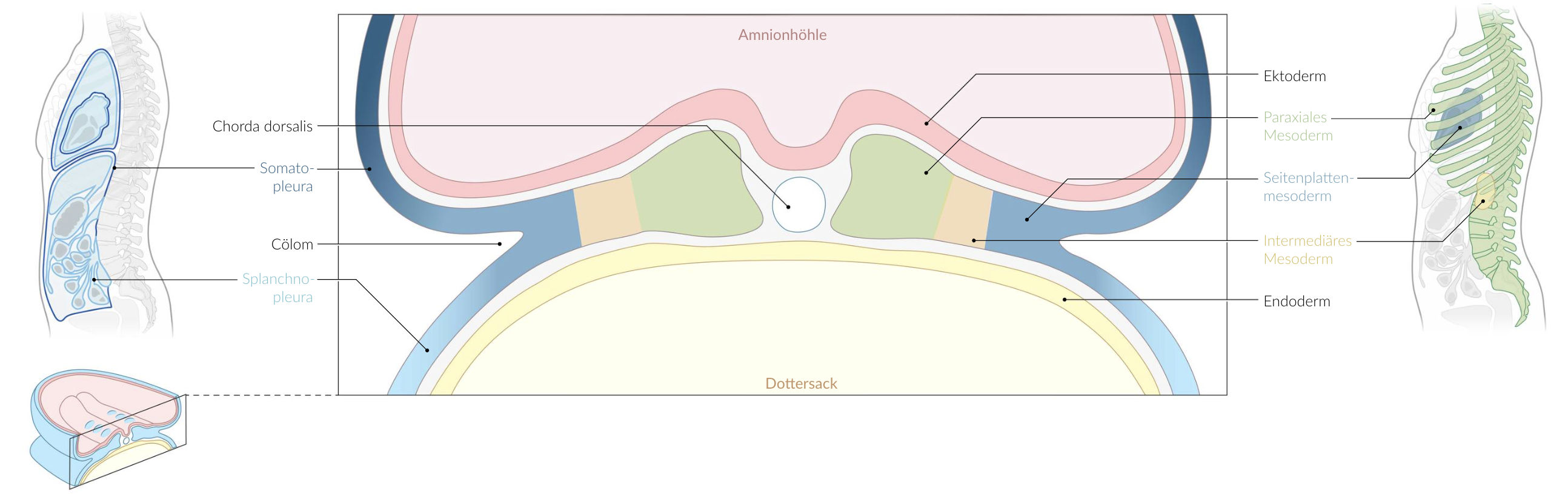
24. Which structure arises from the intermediate mesoderm?
A. Connective tissue of the lung
B. Distal part of the rectum
C. Egg cell of the ovary
D. Endothelium of the thoracic aorta
E. Proximal tubule of the kidney
E. Proximal tubule of the kidney
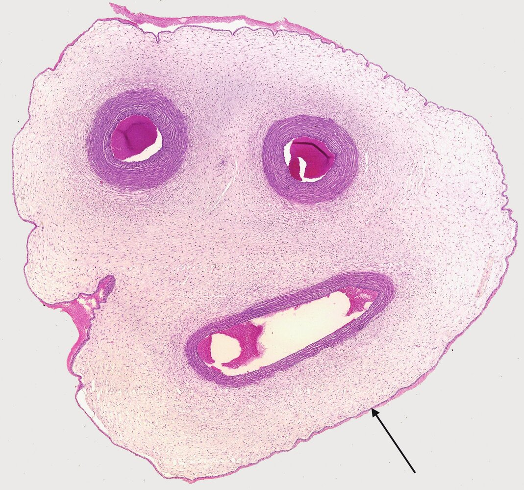
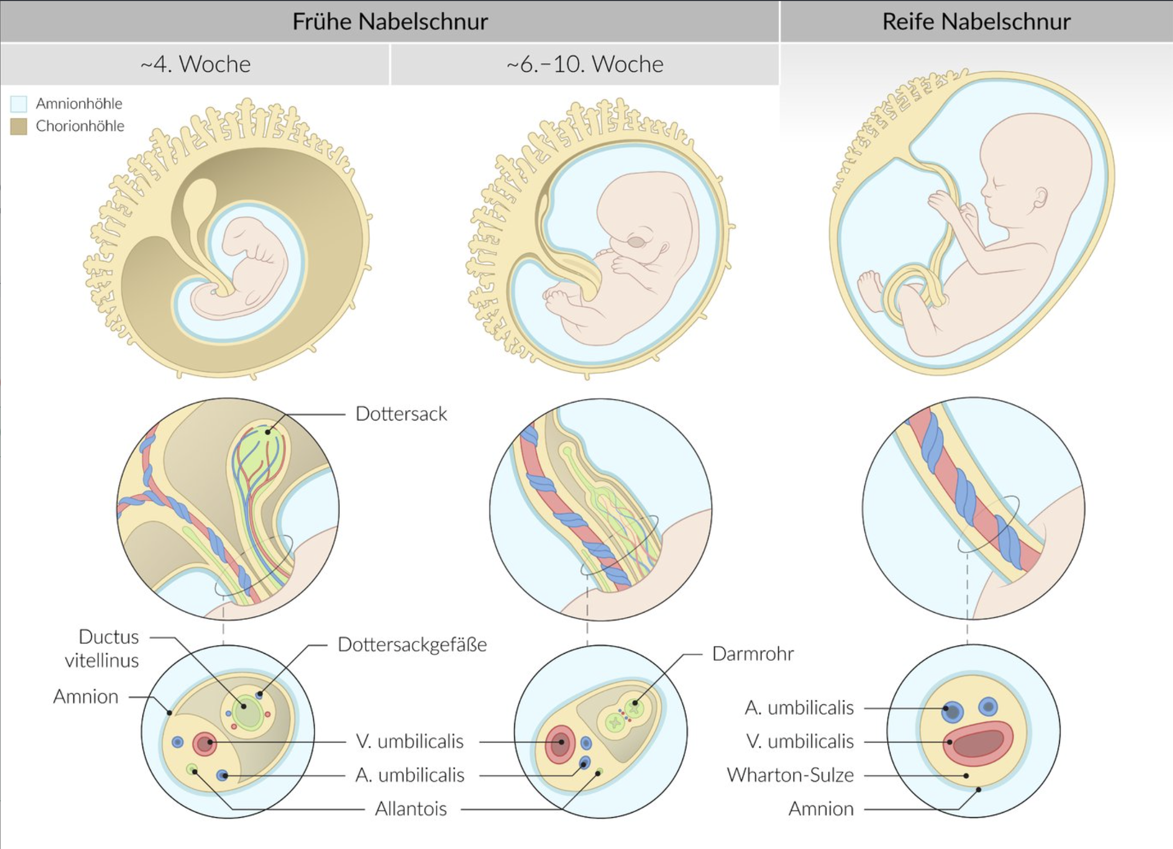
25. The image shows a cross-section of the umbilical cord.
What does the structure marked with an arrow correspond to?
A. Allantois
B. Amnion
C. Chorion
D. Yolk sac
E. Urachus
B. Amnion
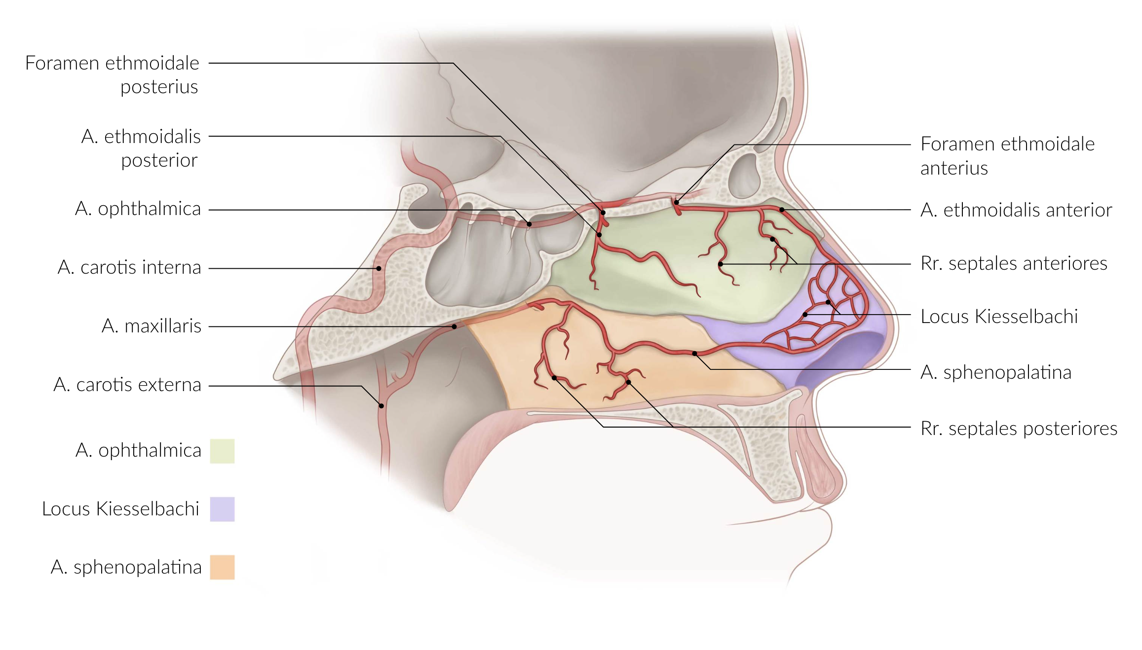
26. The Kiesselbach's plexus (Locus Kiesselbachi):
A. Is the site of herniation for a femoral hernia
B. Is a location where the sciatic nerve can be palpated
C. Is a group of norepinephrine-producing cells in the brainstem
D. Is one of the classic sites for the development of hemorrhoids
E. Is a common site of nosebleeds
E. Is a common site of nosebleeds
27. Which statement about the sympathetic nervous system is generally correct?
A. The axons of the sympathetic origin cells leave the spinal cord via the dorsal roots.
B. The postganglionic fibers are axons of pseudounipolar ganglion cells.
C. The postganglionic neurons are located in the lateral horns of the spinal cord’s gray matter.
D. The preganglionic fibers for the head synapse in the pterygopalatine ganglion.
E. The preganglionic fibers for the limbs and body wall form synapses in the sympathetic trunk (paravertebral ganglia).
E. The preganglionic fibers for the limbs and body wall form synapses in the sympathetic trunk (paravertebral ganglia).
28. Which of the following is a heteronymous (external) reflex?
A. Achilles tendon reflex
B. Biceps tendon reflex
C. Cremasteric reflex
D. Masseter reflex
E. Patellar tendon reflex
C. Cremasteric reflex
29. Which carpal bone is a sesamoid bone?
A. Capitate (Os capitatum)
B. Pisiform (Os pisiforme)
C. Scaphoid (Os scaphoideum)
D. Trapezium (Os trapezium)
E. Triquetrum (Os triquetrum)
B. Pisiform (Os pisiforme)
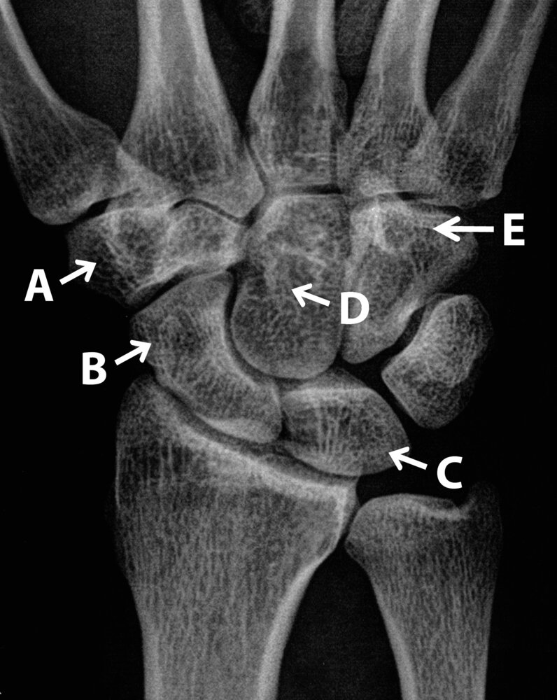
30. Carpal bones serve, among other things, as points of origin for short hand muscles.
The muscles flexor digiti minimi brevis and opponens digiti minimi originate directly from one of the bones labeled A to E in the X-ray image. (Any indirect origins via the flexor retinaculum from other bones are not considered.)
This typically refers to the bone labeled:
E
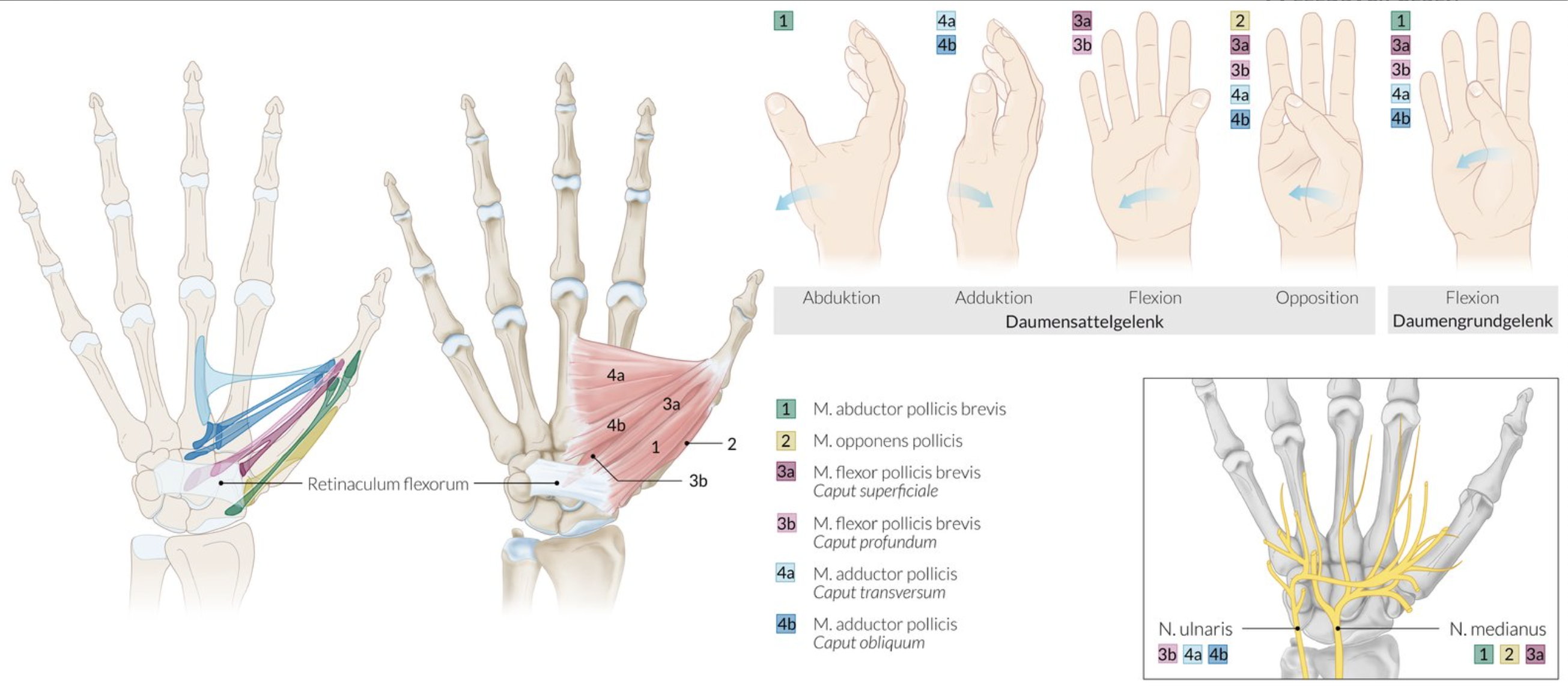
31. Which statement about the hand muscles is correct?
A. The adductor pollicis is a two-headed muscle.
B. The extensor pollicis brevis belongs to the hypothenar muscles.
C. The palmaris brevis flexes the fingers on the ulnar side.
D. Contraction of the lumbrical muscles leads to extension in the metacarpophalangeal joints.
E. The palmar interosseous muscles originate from the tendons of the flexor digitorum profundus.
A. The adductor pollicis is a two-headed muscle.
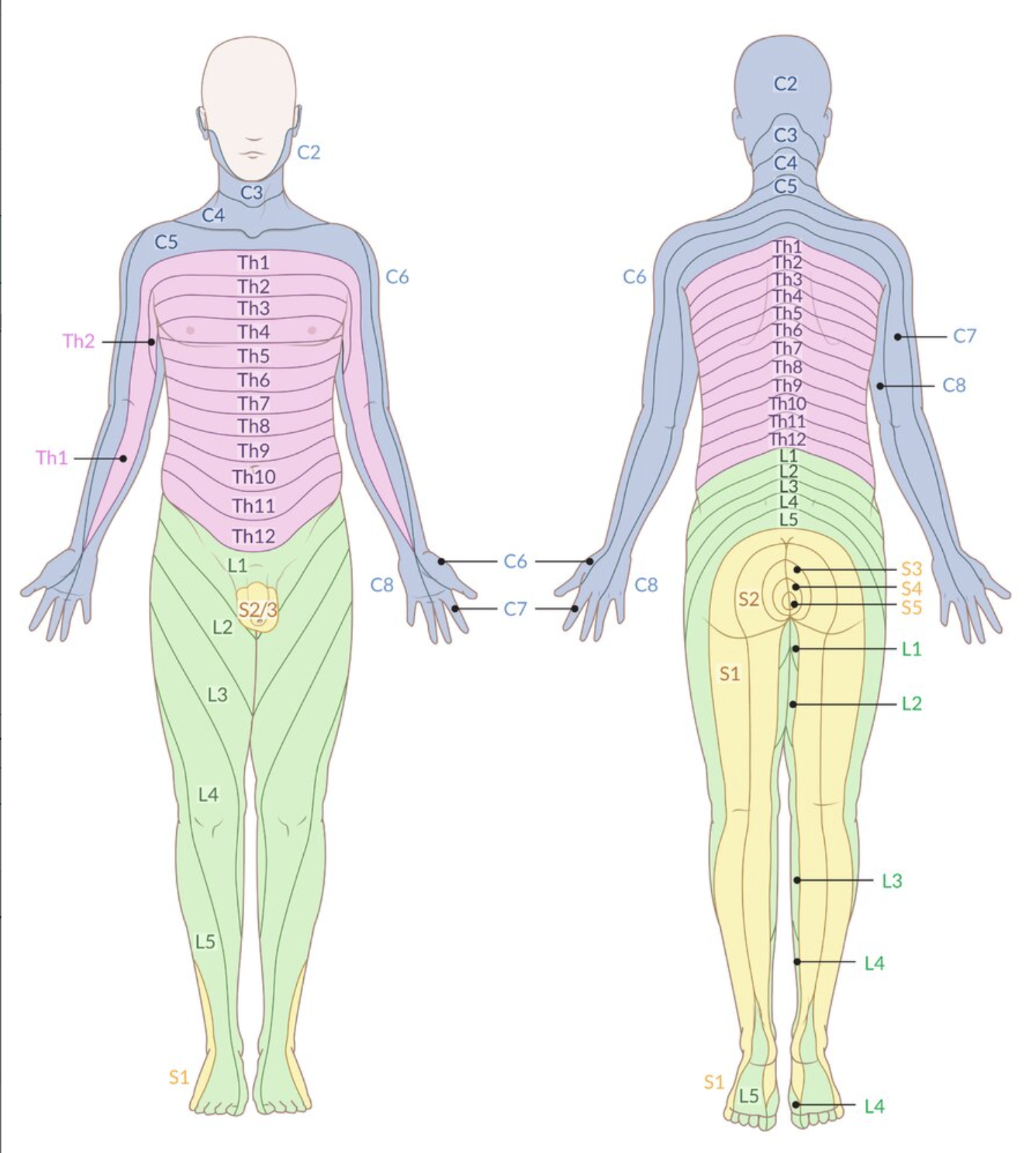
32. Herniated discs can cause mechanical irritation of dorsal root fibers, leading to pain in the skin areas innervated by the affected segment.
A patient with a herniated disc complains of pain in the right forearm, primarily felt on the palmar side of the middle finger.
Which of the following segments is most likely affected?
A. C4
B. C5
C. C6
D. C7
E. C8
D. C7
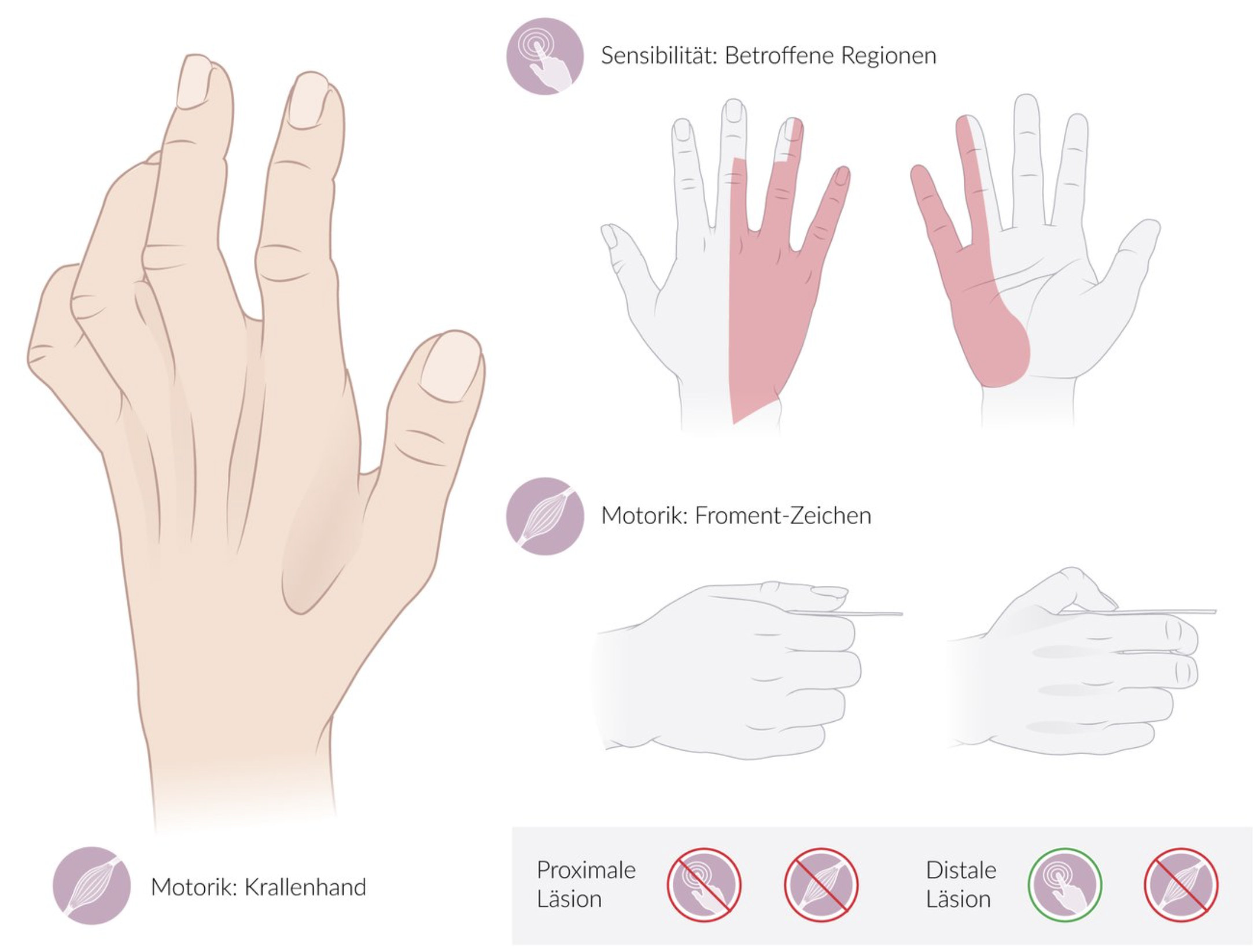
33. A patient sustained a stab injury to the flexor side of the wrist, without tendon damage. He presents to your clinic with limited finger movement.
Which symptom indicates an ulnar nerve lesion as a result of this injury?
A. Drooping of the hand toward the palm (= wrist drop)
B. Adducted position of the thumb (= ape hand)
C. Impaired active flexion of the thumb and adjacent two fingers (= pointing hand)
D. Limited abduction of the thumb’s saddle and metacarpophalangeal joints (= bottle sign)
E. Hyperextension of the finger base joints with flexion of the middle and end joints (= claw hand)
E. Hyperextension of the finger base joints with flexion of the middle and end joints (= claw hand)
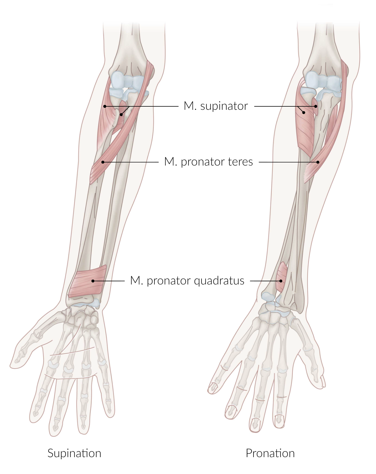
34. Which statement about the course of the median nerve (N. medianus) is generally correct?
A. The N. medianus crosses the brachial artery from medial to lateral along the upper arm.
B. It divides at the elbow into a superficial and a deep branch.
C. It passes between the heads of the pronator teres muscle.
D. It enters the palm through the Guyon’s canal.
E. Its terminal branches supply the hypothenar eminence both sensibly and motorically.
C. It passes between the heads of the pronator teres muscle.
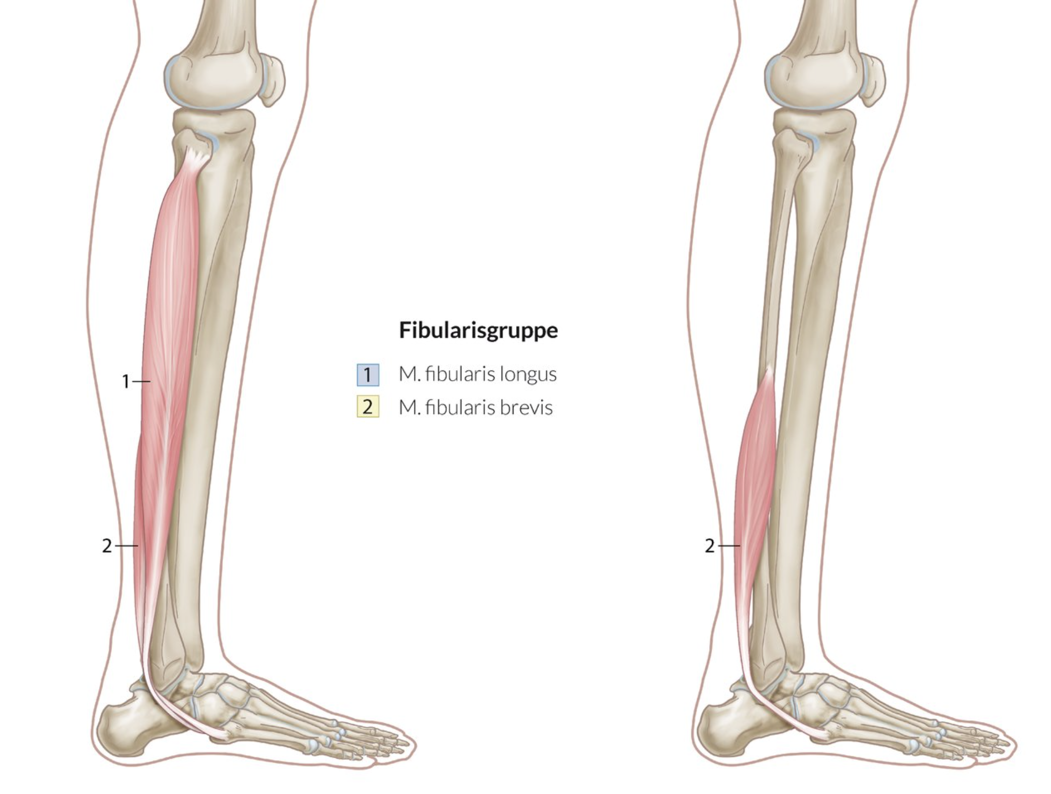
35. Which muscle primarily stabilizes the transverse arch of the foot actively?
A. Abductor hallucis
B. Fibularis (peroneus) longus
C. Flexor digitorum brevis
D. Flexor digitorum longus
E. Flexor hallucis longus
B. Fibularis (peroneus) longus
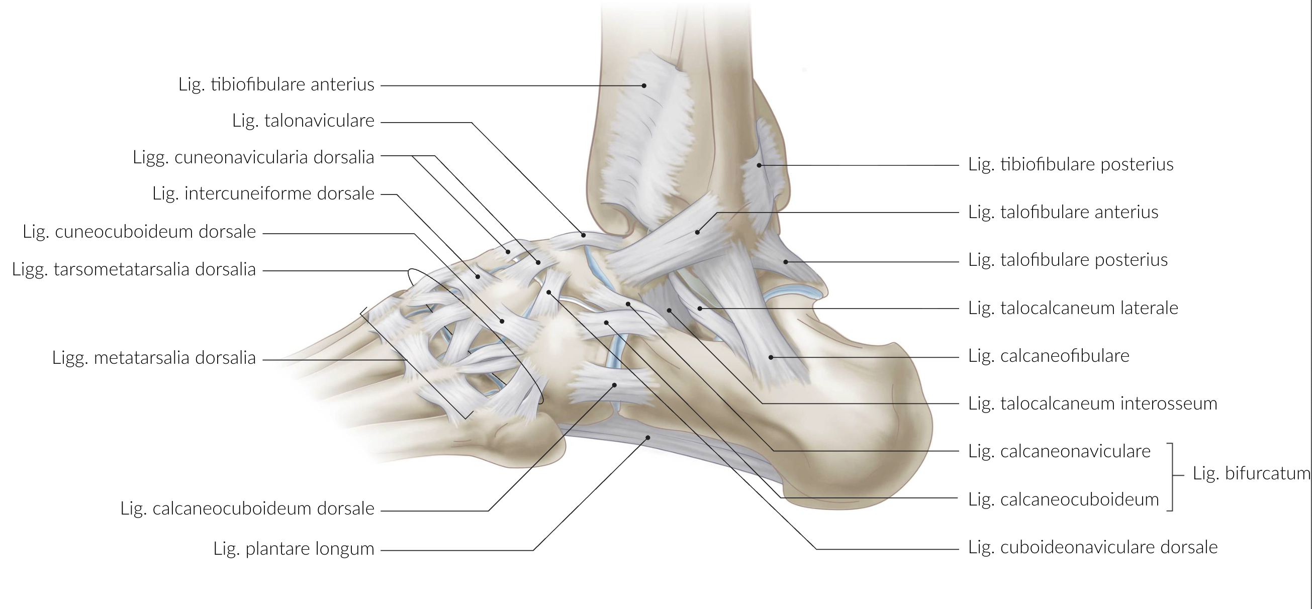
36. The upper and lower ankle joints are each stabilized by strong ligamentous structures.
Which ligament is part of the stabilization of the upper ankle joint?
A. Bifurcate ligament (Lig. bifurcatum)
B. Anterior talofibular ligament (Lig. talofibulare anterius)
C. Plantar calcaneonavicular ligament (Lig. calcaneonaviculare plantare)
D. Interosseous talocalcaneal ligament (Lig. talocalcaneum interosseum)
E. Dorsal talonavicular ligament (Lig. talonaviculare dorsale)
B. Anterior talofibular ligament (Lig. talofibulare anterius)
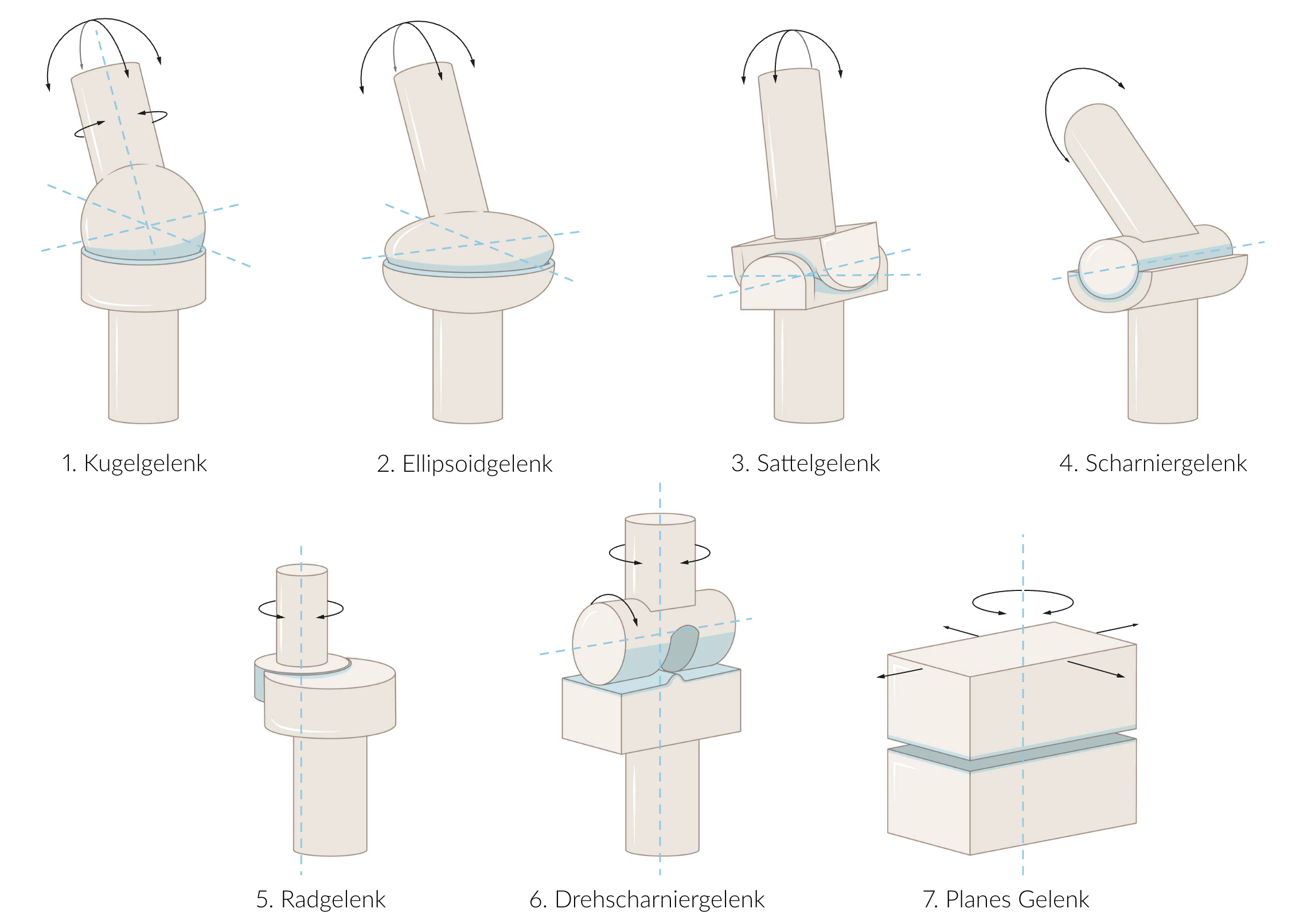
37. To describe the limited range of motion in osteoarthritis, knowledge of normal joint structures and their corresponding functions is essential.
Which statement correctly assigns joint types?
A. The hip joint (Articulatio coxae) is an ellipsoid joint.
B. The femorotibial joint is a hinge-pivot joint.
C. The upper ankle joint is a pivot joint.
D. The lower ankle joint is a hinge joint.
E. The tarsometatarsal joints (Articulationes tarsometatarsales) are ball-and-socket joints.
B. The femorotibial joint is a hinge-pivot joint.
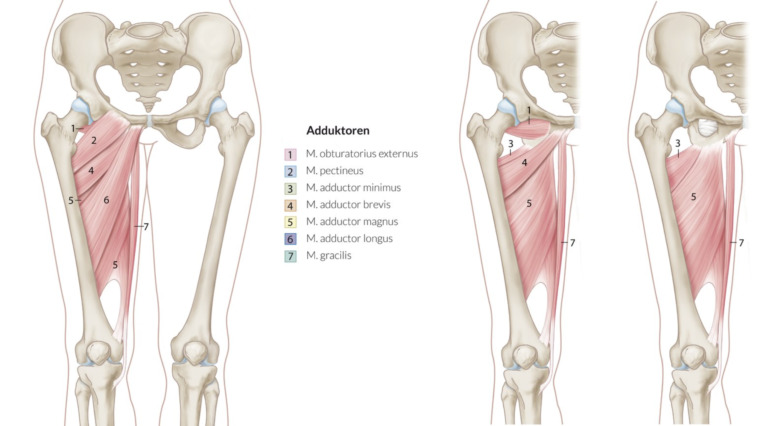
38. Which adductor muscle of the hip joint does not insert on the femur?
A. Adductor brevis
B. Adductor longus
C. Adductor magnus
D. Gracilis
E. Pectineus
D. Gracilis
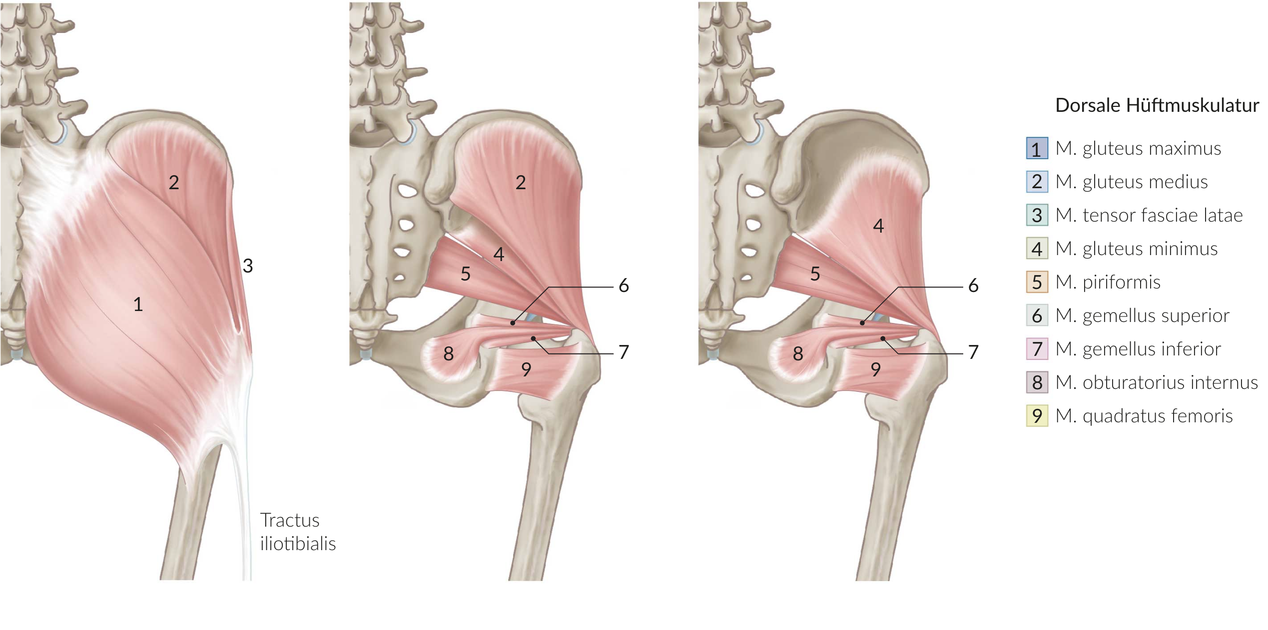
39. Which of the pelvitrochanteric muscles originates anteriorly from the sacrum (Os sacrum)?
A. Gemelli muscles
B. Obturator externus
C. Obturator internus
D. Piriformis
E. Quadratus femoris
D. Piriformis
40. Which statement about the obturator nerve (N. obturatorius) is correct?
A. It originates from the sacral plexus.
B. It innervates the obturator internus muscle.
C. It passes through the infrapiriform foramen to the inner thigh.
D. Its anterior branch (Ramus anterior) has both motor and sensory components.
E. Its posterior branch (Ramus posterior) provides motor innervation to the pectineus muscle.
D. Its anterior branch (Ramus anterior) has both motor and sensory components.
41. After surgery on the right hip, a patient comes to the clinic with movement impairments of the right leg.
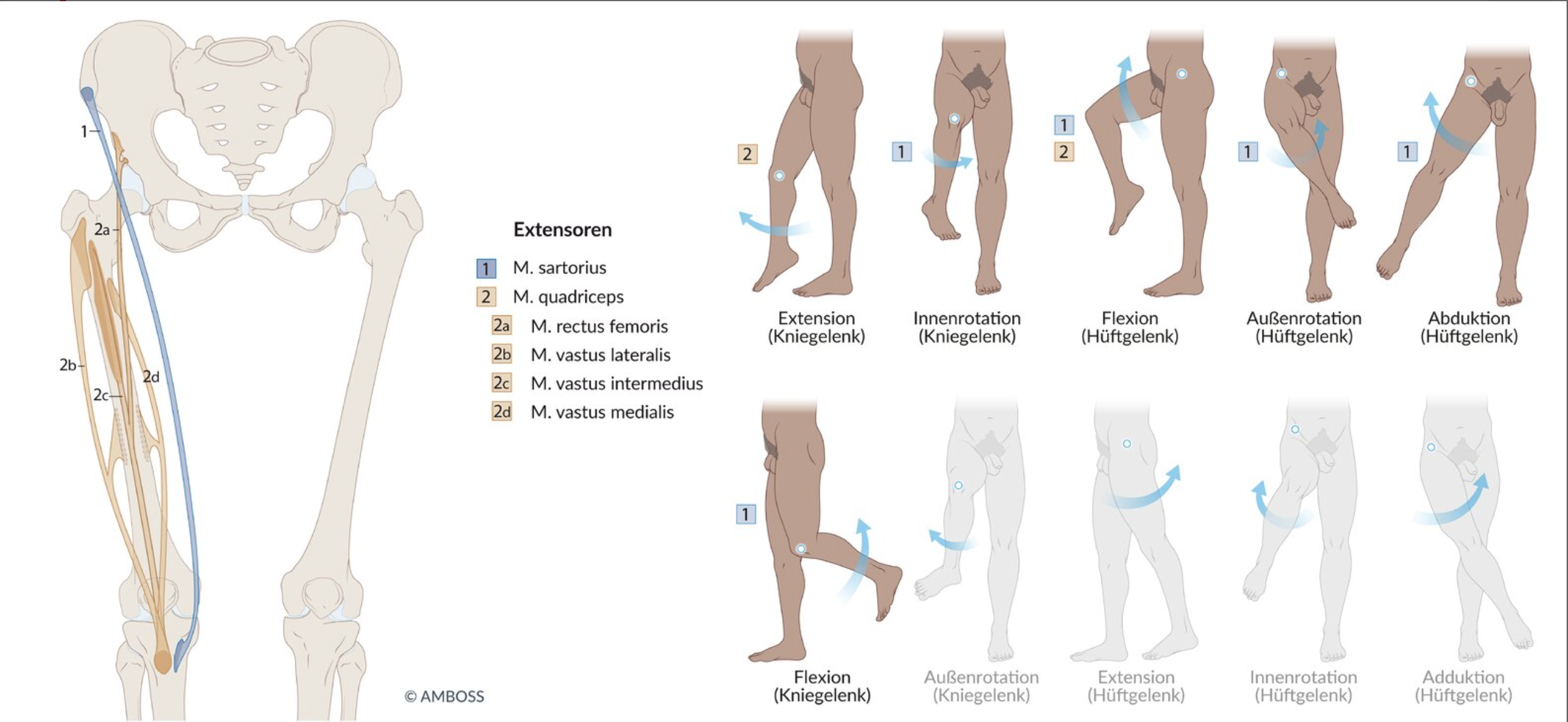
Which symptom combination suggests a lesion of the femoral nerve (N. femoralis) as a result of the procedure?
A. Impaired active abduction and internal rotation in the hip joint
B. Impaired adduction in the hip joint and internal rotation in the knee joint
C. Impaired external rotation in the hip joint and internal rotation in the knee joint
D. Impaired extension in the hip joint and external rotation in the knee joint
E. Impaired flexion in the hip joint and extension in the knee joint
E. Impaired flexion in the hip joint and extension in the knee joint
42. After receiving an injection into the gluteal muscles during a vaccination, a patient presents with movement difficulties in the right leg.
Which symptom indicates a lesion of the superior gluteal nerve (N. gluteus superior) as a result of this procedure?
A. Severe impairment of active extension in the hip joint
B. Flexion in the hip joint
C. External rotation in the hip joint
D. Abduction in the hip joint
E. Adduction in the hip joint
D. Abduction in the hip joint
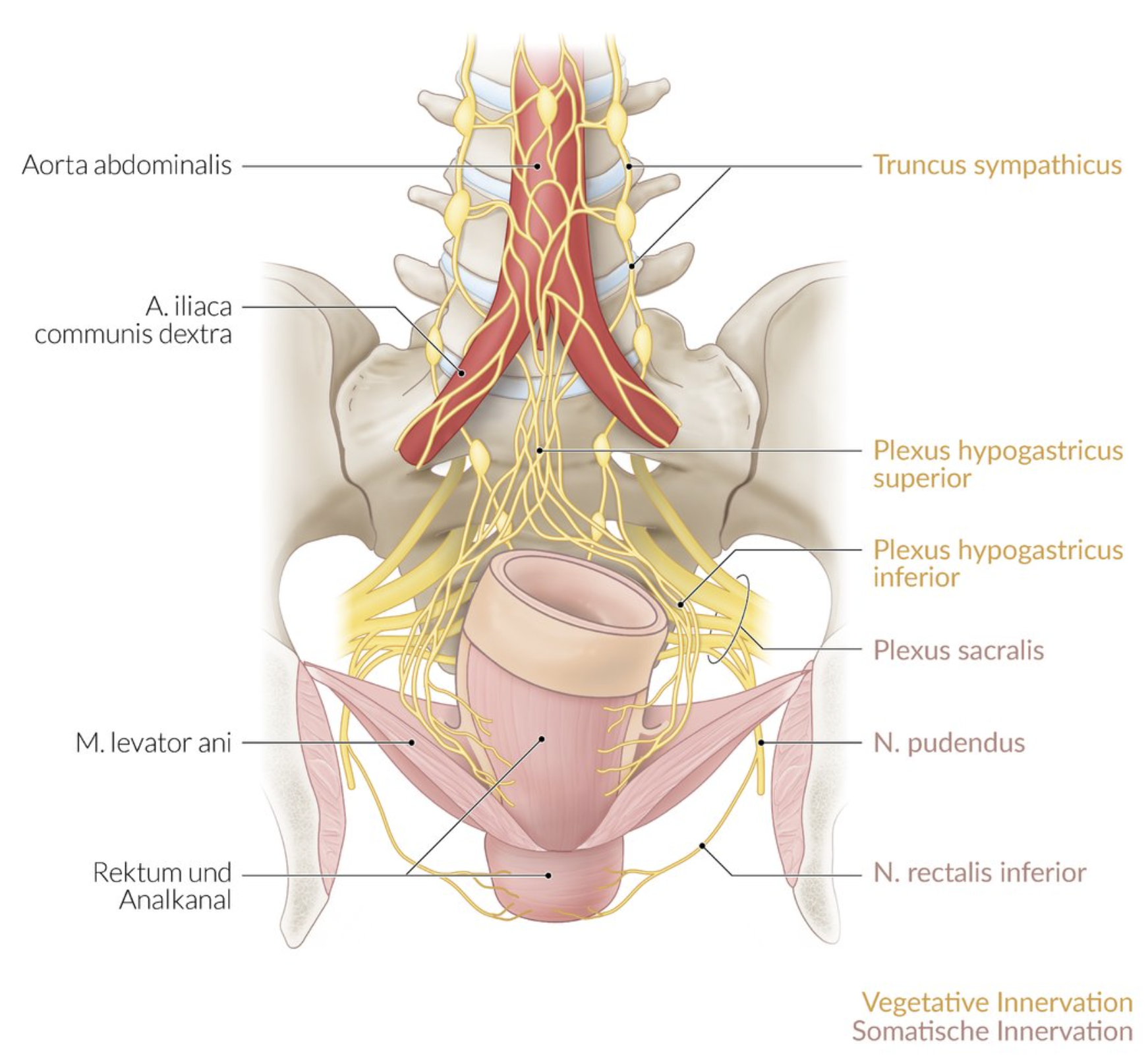
43. In the pelvis, the peritoneal, subperitoneal, and subfascial compartments can be topographically distinguished.
Which structure runs in the subperitoneal compartment?
A. Internal pudendal artery (A. pudenda interna) in the ischioanal fossa
B. Appendix vermiformis
C. Sigmoid colon (Colon sigmoideum)
D. Pudendal nerve (N. pudendus) in Alcock’s canal
E. Inferior hypogastric plexus (Plexus hypogastricus inferior)
E. Inferior hypogastric plexus (Plexus hypogastricus inferior)
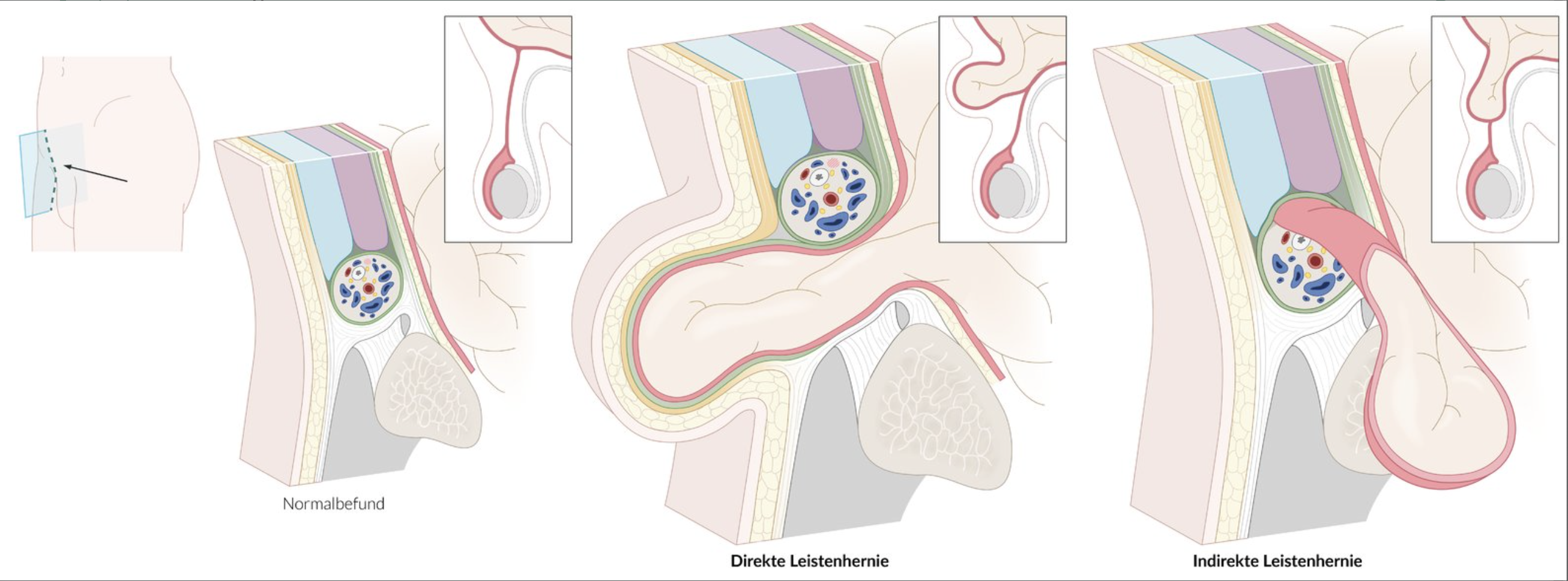
44. In which of the following regions is the internal hernia opening located in a direct inguinal hernia?
A. Acetabular fossa (Fossa acetabuli)
B. Iliac fossa (Fossa iliaca)
C. Lateral inguinal fossa (Fossa inguinalis lateralis)
D. Medial inguinal fossa (Fossa inguinalis medialis)
E. Supravesical fossa (Fossa supravesicalis)
D. Medial inguinal fossa (Fossa inguinalis medialis)
45. There is a large accumulation of fluid in a preformed cavity of the scrotum (hydrocele testis).
This fluid accumulation is most likely located between which two structures?
A. Between the external spermatic fascia and the cremasteric fascia
B. Between the cremasteric fascia and the internal spermatic fascia
C. Between the internal spermatic fascia and the parietal layer of the tunica vaginalis testis (periorchium)
D. Between the parietal layer and the visceral layer of the tunica vaginalis testis (epiorchium)
E. Between the visceral layer of the tunica vaginalis testis (epiorchium) and the tunica albuginea testis
D. Between the parietal layer and the visceral layer of the tunica vaginalis testis (epiorchium)
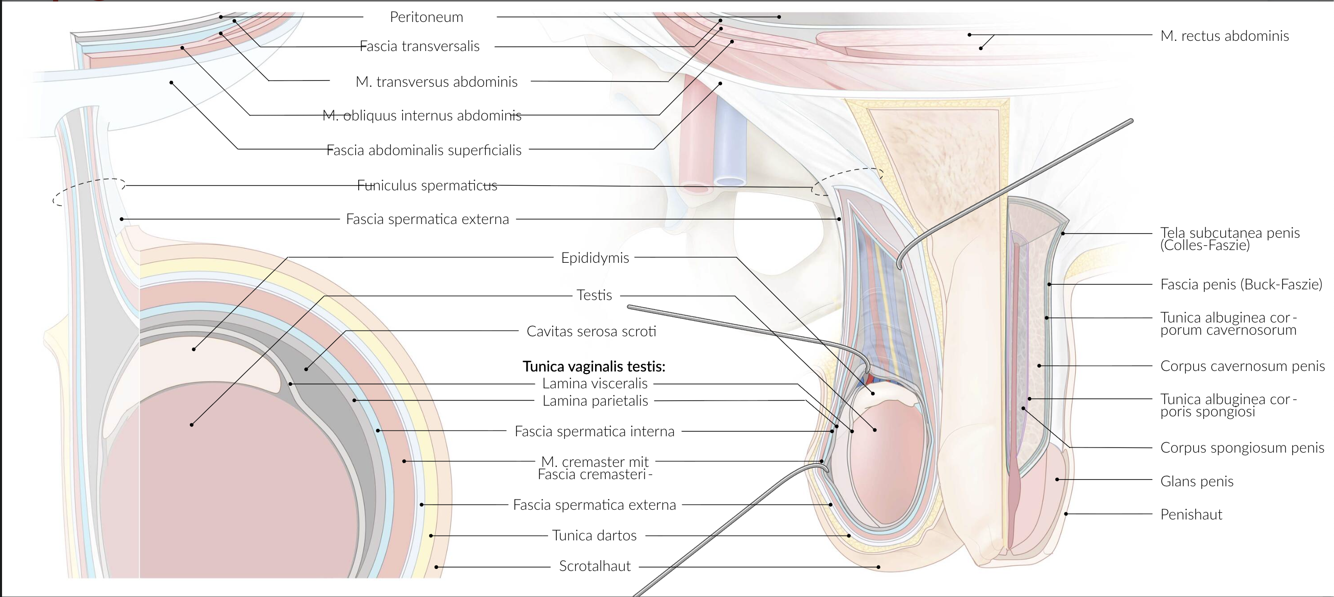
46. Which structure(s) form the main part of the blood-testis barrier?
A. Adherens junctions of the endothelial cells
B. Basement membrane of the capillaries
C. Desmosomes of the myofibroblasts
D. Gap junctions of the spermatogonia
E. Tight junctions of the Sertoli cells
E. Tight junctions of the Sertoli cells
47. In Germany, about 8–10 out of 100,000 men develop a malignant testicular tumor.
The regional lymph nodes of the testis and epididymis—and thus the typical primary site of metastasis—are the:
A. External iliac lymph nodes (Nodi lymphoidei iliaci externi)
B. Internal iliac lymph nodes (Nodi lymphoidei iliaci interni)
C. Superficial inguinal lymph nodes (Nodi lymphoidei inguinales superficiales)
D. Lumbar lymph nodes (Nodi lymphoidei lumbales)
E. Sacral lymph nodes (Nodi lymphoidei sacrales)
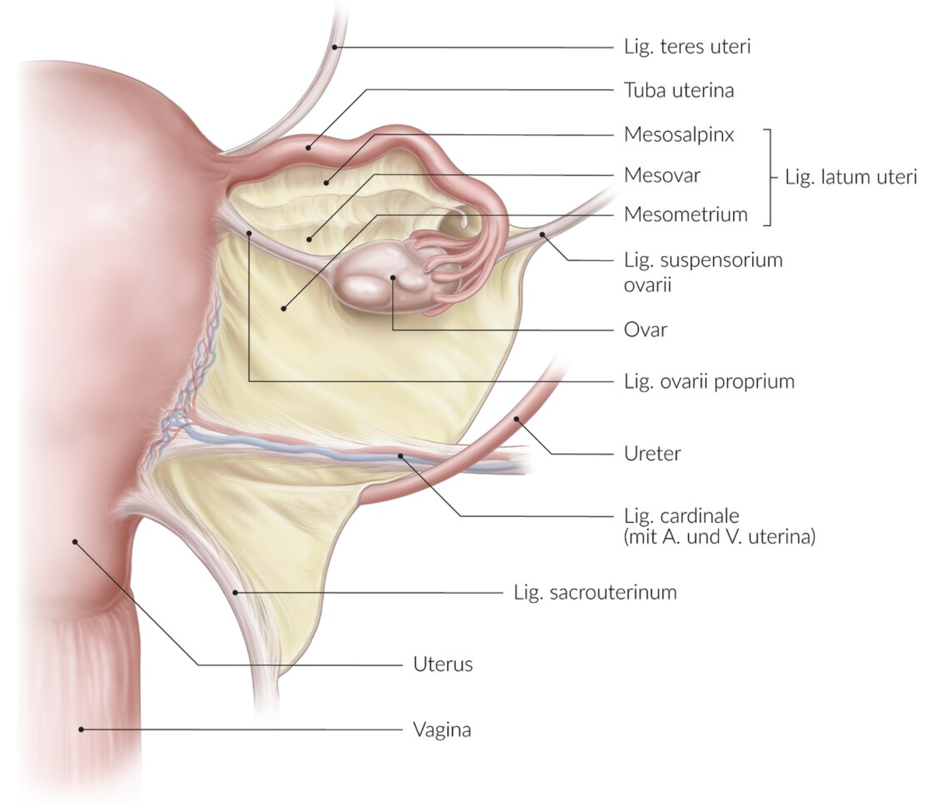
D. Lumbar lymph nodes (Nodi lymphoidei lumbales)
48. During surgery on a 50-year-old woman, the surgeon finds a large fibroid that has grown from the distal cervix uteri into the left broad ligament (Ligamentum latum). In addition to cycle-dependent bleeding, the patient suffers from other symptoms suggesting compression of a neighboring structure.
Which of the following left-sided structures is most likely compressed by the tumor due to its close anatomical location?
A. Obturator artery (A. obturatoria)
B. Ovarian artery (A. ovarica)
C. Inferior rectal artery (A. rectalis inferior)
D. Pudendal nerve (N. pudendus)
E. Ureter
E. Ureter
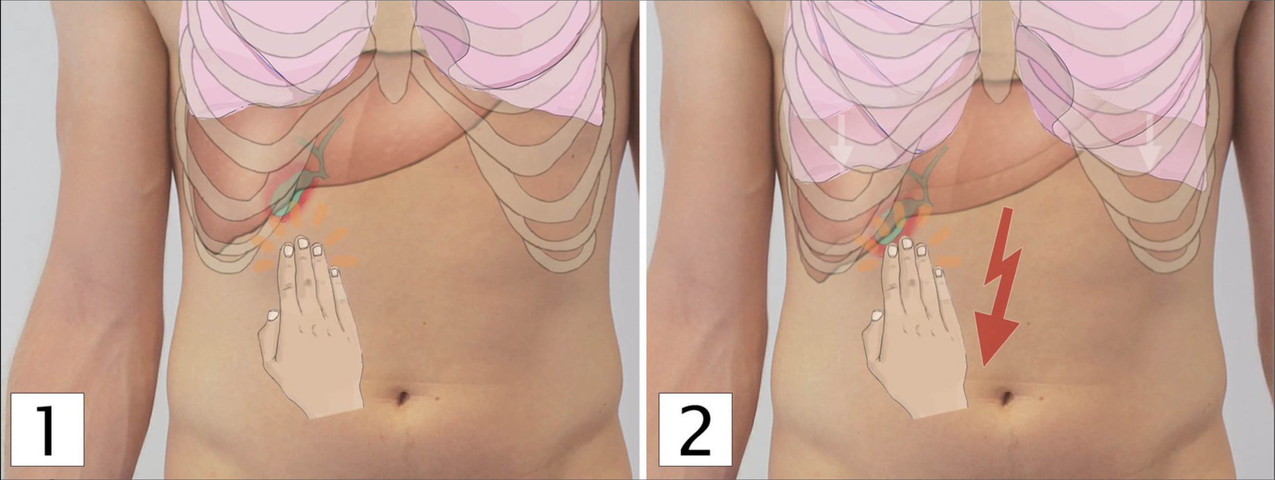
49. A 50-year-old female patient presents with abdominal pain radiating to the right shoulder. After ruling out a heart attack, you suspect acute inflammation of another internal organ.
Which of the following organs is most likely causing this pain if it is inflamed?
A. Gallbladder (Gallenblase)
B. Left kidney (linke Niere)
C. Stomach (Magen)
D. Spleen (Milz)
E. Pancreas (Pankreas)
A. Gallbladder (Gallenblase)
50. A patient presents with an acute aortic aneurysm (bulging of the aorta) in the region of the aortic arch.
Which of the following structures is most likely to be damaged by this space-occupying process?
A. Left recurrent laryngeal nerve (linker N. laryngeus recurrens)
B. Left phrenic nerve (linker N. phrenicus)
C. Left greater thoracic splanchnic nerve (linker N. splanchnicus thoracicus major)
D. Right vagus nerve (rechter N. vagus)
E. Right sympathetic trunk (rechter Truncus sympathicus)
A. Left recurrent laryngeal nerve (linker N. laryngeus recurrens)
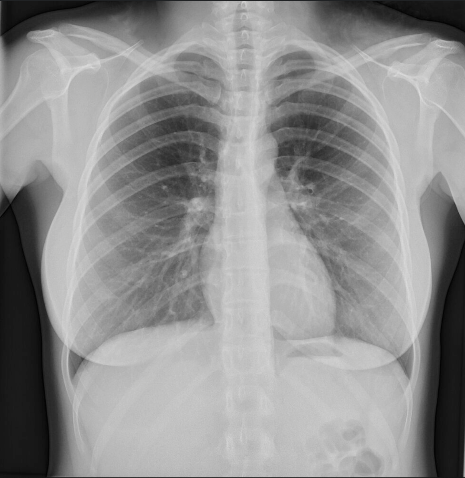
51. A chest X-ray overview image provides information about the size of the heart and can reveal additional findings.
Which of the following heart and vessel contours is typically well visible on the right side of the patient in a posteroanterior (p.a.) chest X-ray?
A. Apex of the heart (Apex cordis)
B. Aortic arch (Arcus aortae)
C. Pulmonary trunk (Truncus pulmonalis)
D. Superior vena cava (V. cava superior)
E. Right ventricle (Ventriculus dexter)
D. Superior vena cava (V. cava superior)
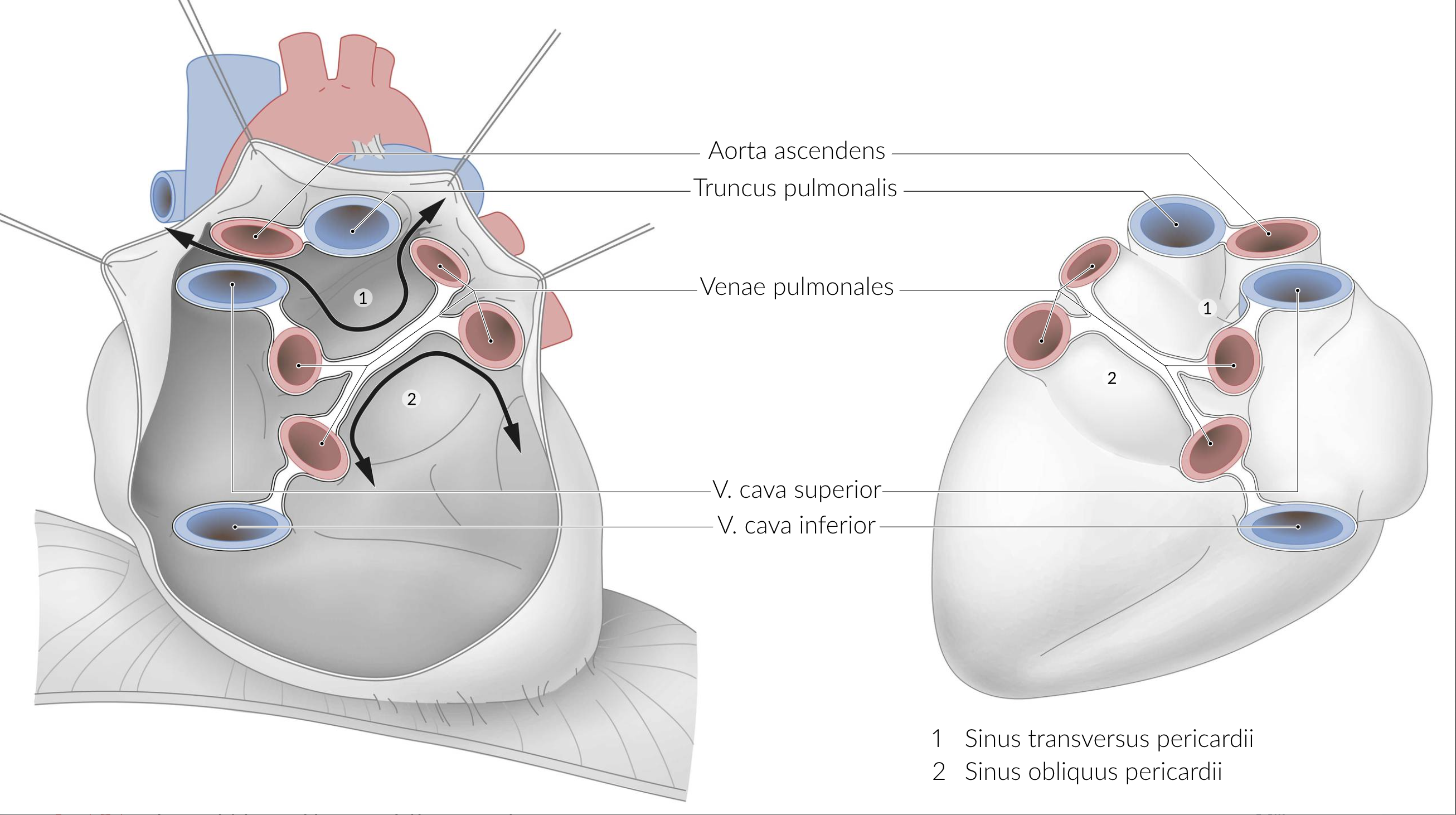
52. The pericardium allows unimpeded heart contractions while protecting the heart from overstretching.
Which statement about the pericardium is correct?
A. It forms the sinus transversus pericardii at the apex of the heart.
B. It forms the sinus obliquus pericardii dorsally as a fold.
C. It is fused with the diaphragm via the epicardium.
D. It extends cranially up to the aortic isthmus (Isthmus aortae).
E. It is completely overlapped by the pleural cavities.
B. It forms the sinus obliquus pericardii dorsally as a fold.
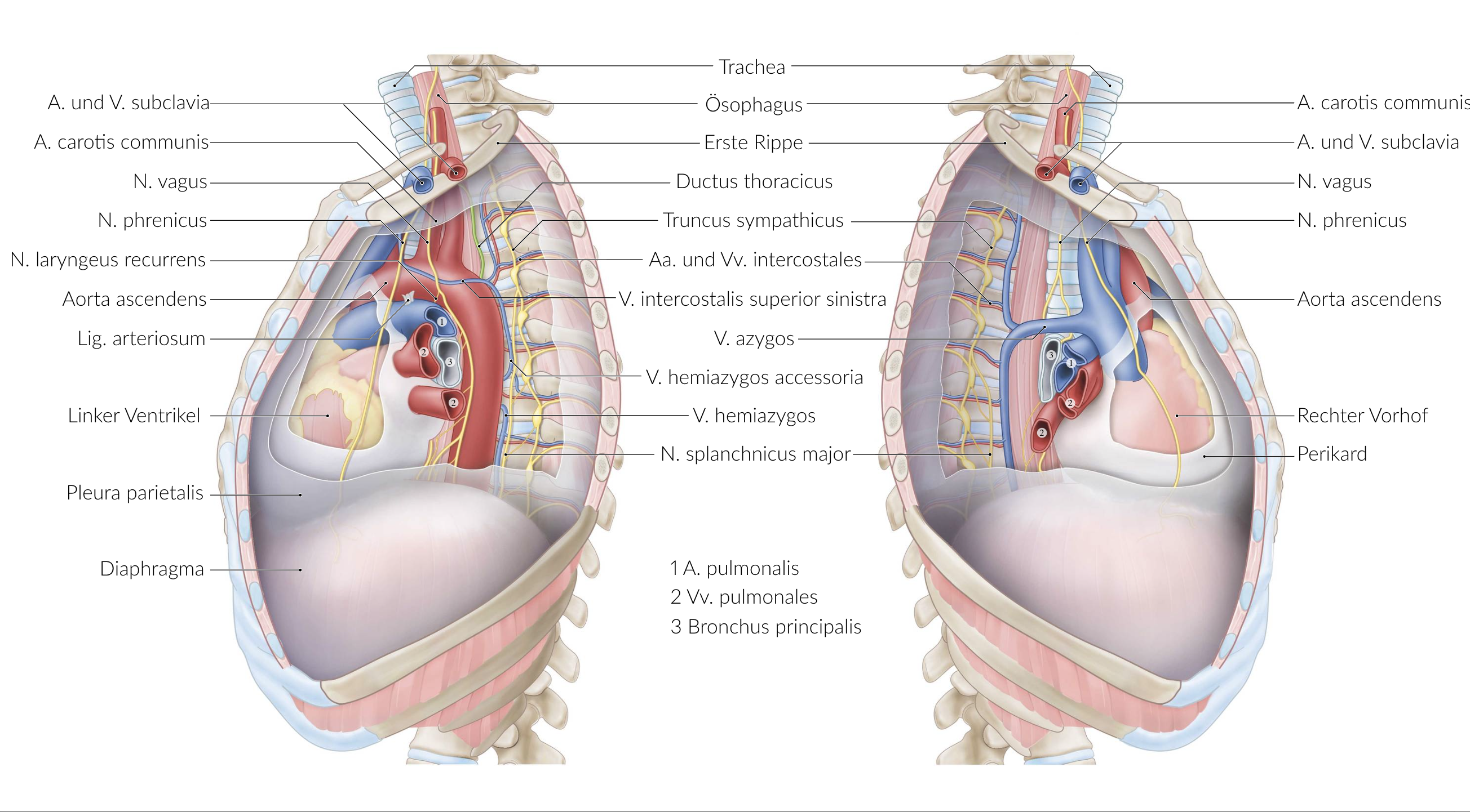
53. A 67-year-old patient is diagnosed with esophageal cancer, which is scheduled for surgery. As part of the preoperative counseling, the patient must be informed about the possible risk of injury to the thoracic duct (Ductus thoracicus).
Which statement about its location and course is correct?
A. It drains into the right internal jugular vein (V. jugularis).
B. It passes through the centrum tendineum of the diaphragm along with the inferior vena cava (V. cava inferior).
C. It runs lateral to the sympathetic trunk (Truncus sympathicus).
D. It runs ventral to the esophagus.
E. It passes through the superior and inferior mediastinum.
E. It passes through the superior and inferior mediastinum.
54. Which of the following statements about the development of the gastrointestinal tract is correct?
A. The greater omentum (Omentum majus) develops from the dorsal mesogastrium.
B. The lesser omentum (Omentum minus) connects the liver to the anterior abdominal wall.
C. The gallbladder develops in the dorsal mesogastrium.
D. The spleen develops in the ventral mesogastrium.
E. The omental bursa (Bursa omentalis) develops in the ventral mesogastrium.
A. The greater omentum (Omentum majus) develops from the dorsal mesogastrium.
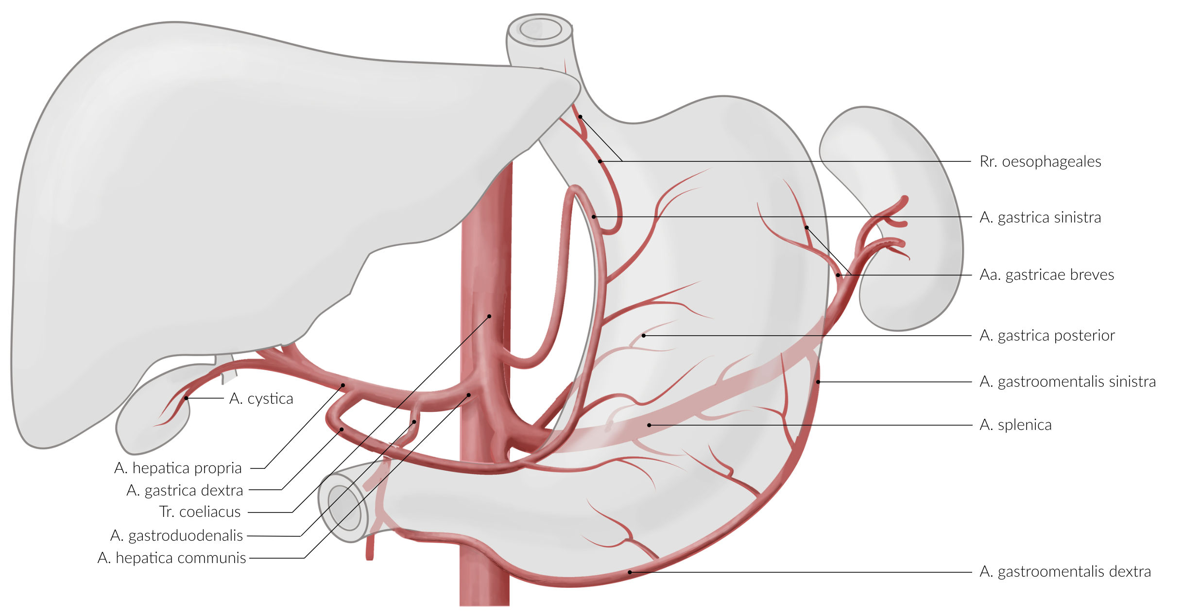
55. The stomach is supplied by several arteries. Knowing their locations is important for surgical procedures involving the stomach.
Which arteries commonly anastomose in the ligamentum gastrocolicum?
A. Right gastro-omental artery (A. gastroomentalis dextra) and right gastric artery (A. gastrica dextra)
B. Left gastro-omental artery (A. gastroomentalis sinistra) and left gastric artery (A. gastrica sinistra)
C. Proper hepatic artery (A. hepatica propria) and gastroduodenal artery (A. gastroduodenalis)
D. Right and left gastric arteries (Aa. gastricae dextra et sinistra)
E. Right and left gastro-omental arteries (Aa. gastroomentales dextra et sinistra)
E. Right and left gastro-omental arteries (Aa. gastroomentales dextra et sinistra)
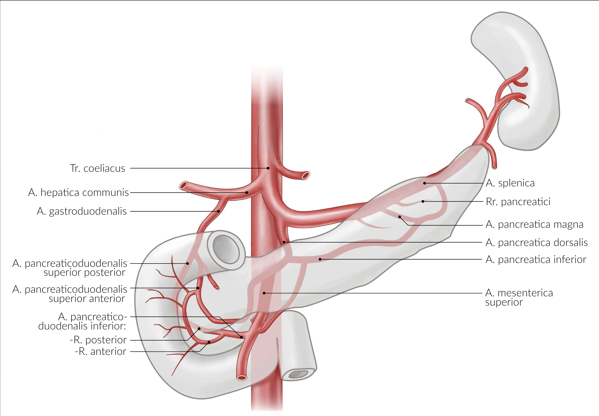
56. In a CT scan, you detect a malignant tumor in the area of the uncinate process of the pancreas, located near the horizontal part of the duodenum (Pars horizontalis duodeni).
Which vessel is the tumor most likely to invade due to its anatomical location?
A. Left gastric artery (A. gastrica sinistra)
B. Gastroduodenal artery (A. gastroduodenalis)
C. Superior mesenteric artery (A. mesenterica superior)
D. Inferior pancreatic artery (A. pancreatica inferior)
E. Splenic artery (A. splenica)
C. Superior mesenteric artery (A. mesenterica superior)
57. Inflammation of the appendix (appendicitis) is a common condition, especially in the second and third decades of life.
Therefore, knowledge of its position and projection is very important.
Which position of the appendix vermiformis is most common?
A. Hanging downward (herabhängend)
B. Pre-ileal (präileal)
C. Retro-ileal (retroileal)
D. Pre-cecal (präzäkal)
E. Retrocecal (retrozäkal)
E. Retrocecal (retrozäkal)
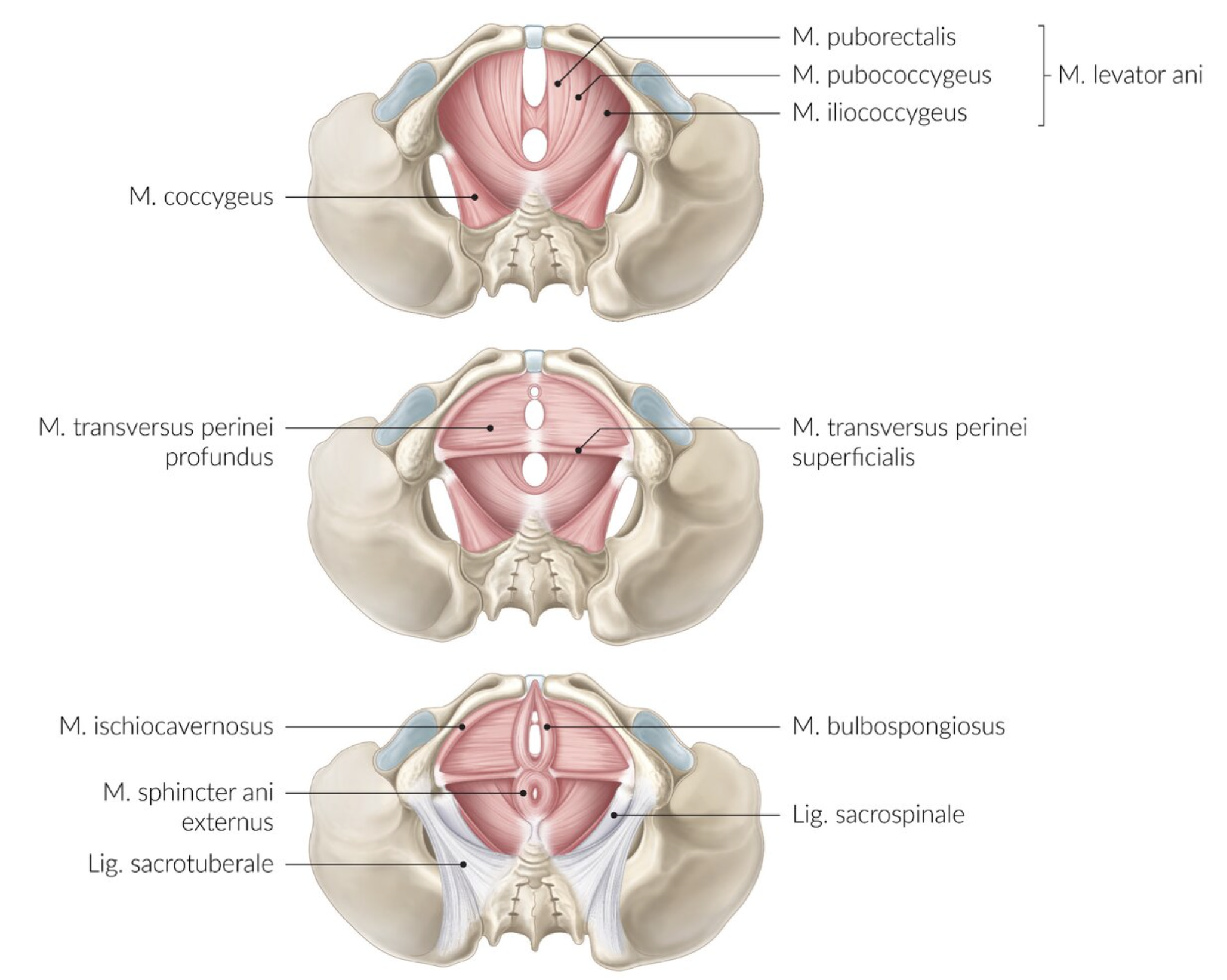
58. Defecation disorders are a common medical issue.
Which of the listed structures is not involved in maintaining fecal continence?
A. Corpus cavernosum recti
B. Puborectalis muscle (M. puborectalis)
C. External anal sphincter (M. sphincter ani externus)
D. Internal anal sphincter (M. sphincter ani internus)
E. Deep transverse perineal muscle (M. transversus perinei profundus)
E. Deep transverse perineal muscle (M. transversus perinei profundus)
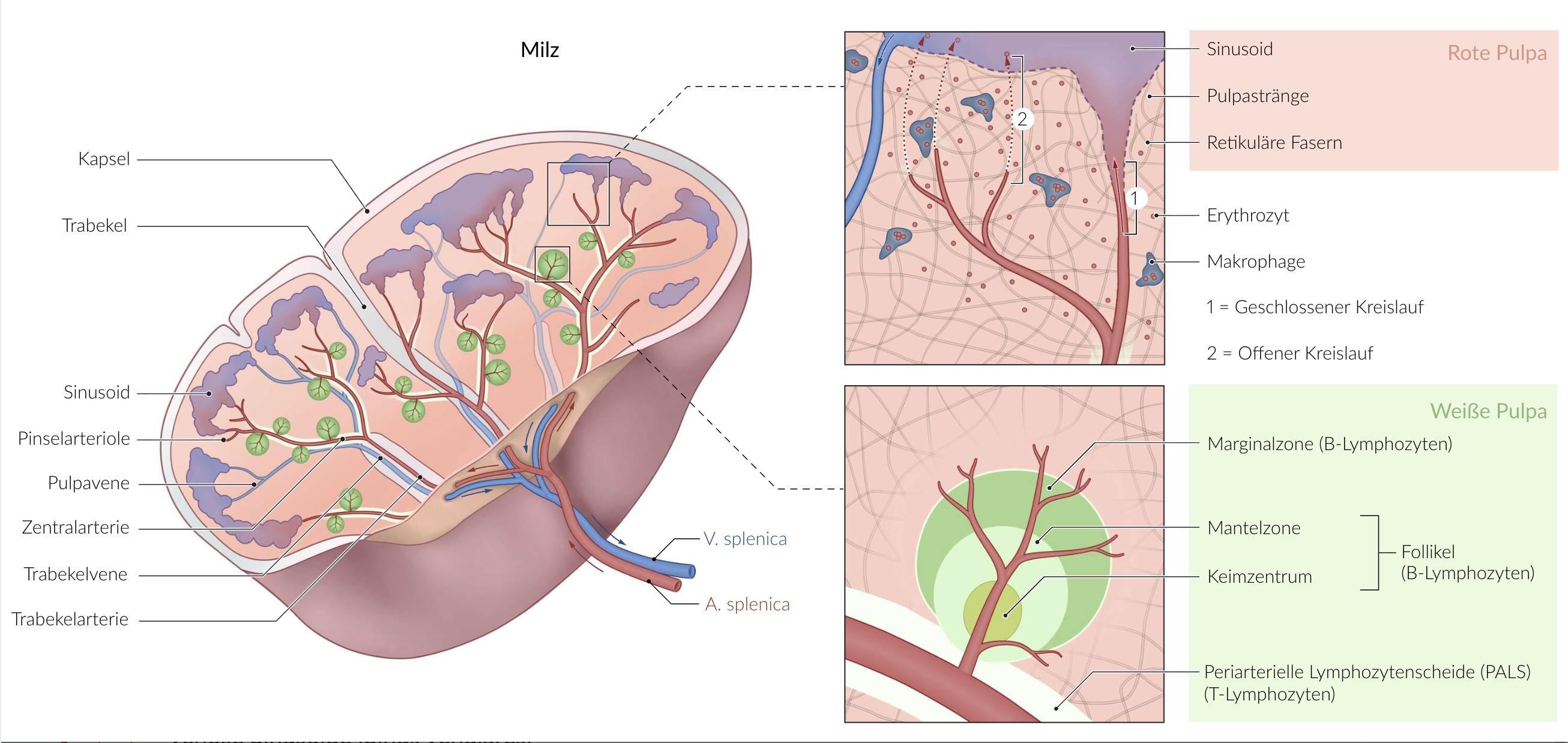
59. Which statement is not correct?
Histological characteristics of the red pulp of the spleen include:
A. Blood vessels with discontinuous endothelium
B. Marginal zones
C. Pulp cords
D. Reticular cells
E. Venous sinusoids (Sinus venulares)
B. Marginal zones
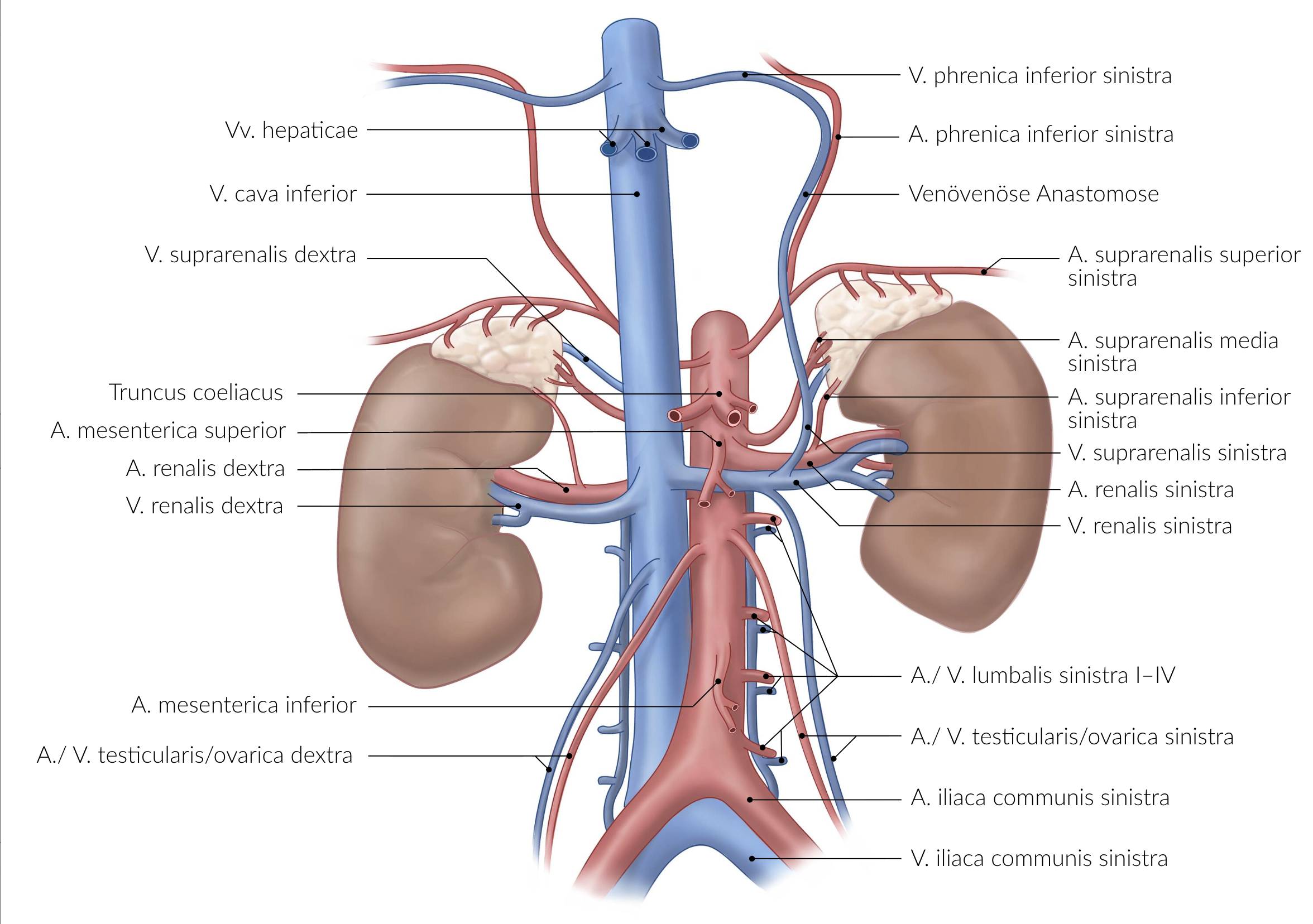
60. Which of the following statements about the adrenal gland or its supplying structures is most accurate?
A. The adrenal gland has no connective tissue capsule.
B. The superior suprarenal artery (A. suprarenalis superior) is a branch of the inferior phrenic artery (A. phrenica inferior).
C. Lymphatic drainage occurs via the common iliac lymph nodes (Nodi lymphoidei iliaci communes).
D. The adrenal medulla develops embryonically from the mesoderm.
E. Sympathetic innervation of the adrenal medulla is provided by postganglionic fibers from the celiac plexus (Plexus coeliacus).
B. The superior suprarenal artery (A. suprarenalis superior) is a branch of the inferior phrenic artery (A. phrenica inferior).
61. The transverse cervical nerve (N. transversus colli / cervicalis):
A. Arises from the Ansa cervicalis (profunda)
B. Innervates the platysma muscle motorically
C. Crosses the path of the sternocleidomastoid muscle (M. sternocleidomastoideus)
D. Runs beneath the pretracheal layer of the cervical fascia
E. Is accompanied by the transverse cervical artery (A. transversa colli)
C. Crosses the path of the sternocleidomastoid muscle (M. sternocleidomastoideus)
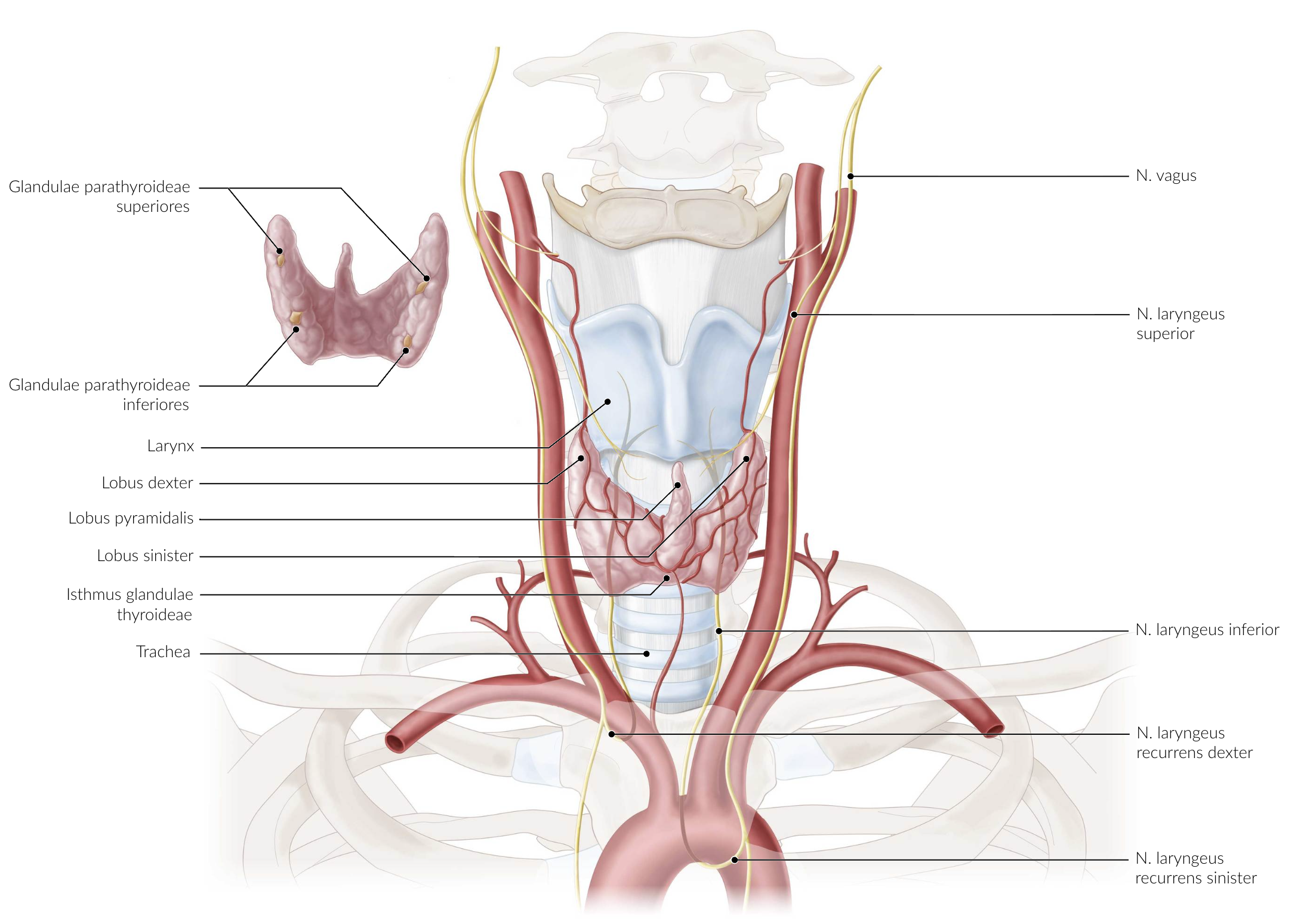
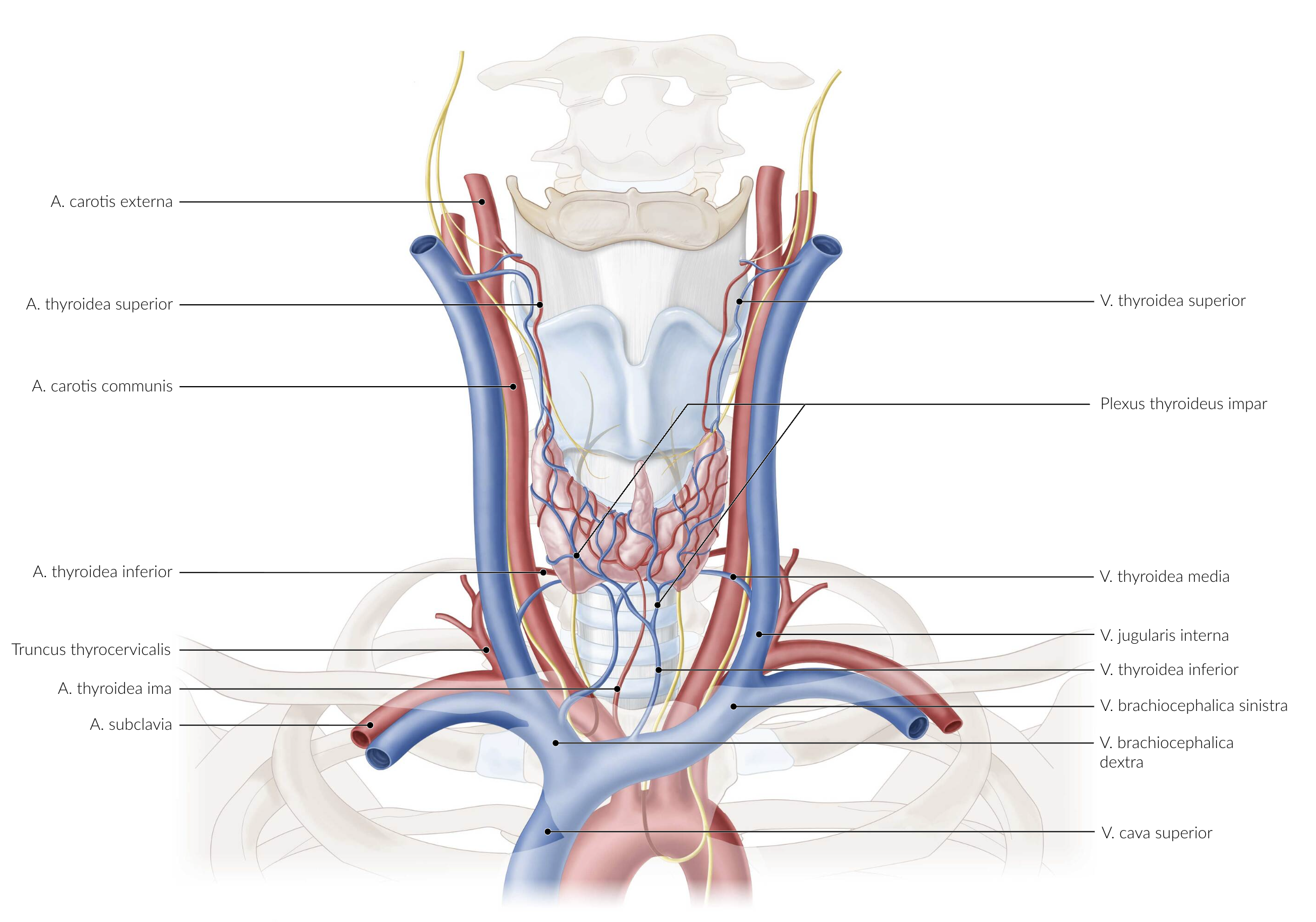
62. A patient is scheduled to undergo a lower tracheotomy (Tracheotomia inferior), in which the trachea is incised from the front just below the thyroid gland.
Which of the following blood vessels is most likely to be injured during this procedure?
A. Inferior thyroid artery (A. thyroidea inferior)
B. Superior thyroid artery (A. thyroidea superior)
C. Costocervical trunk (Truncus costocervicalis)
D. Right brachiocephalic vein (V. brachiocephalica dextra)
E. Inferior thyroid vein (V. thyroidea inferior)
E. Inferior thyroid vein (V. thyroidea inferior)
63. Which muscle is enclosed by the middle layer (Lamina pretrachealis) of the cervical fascia?
A. Omohyoid muscle (M. omohyoideus)
B. Rectus capitis anterior muscle (M. rectus capitis anterior)
C. Middle scalene muscle (M. scalenus medius)
D. Sternocleidomastoid muscle (M. sternocleidomastoideus)
E. Platysma
A. Omohyoid muscle (M. omohyoideus)

64. After a surgical procedure on the floor of the mouth, tongue movement is impaired. When the patient sticks out their tongue, it deviates to the right.
Which nerve is most likely damaged?
A. Left glossopharyngeal nerve (N. glossopharyngeus links)
B. Right glossopharyngeal nerve (N. glossopharyngeus rechts)
C. Left hypoglossal nerve (N. hypoglossus links)
D. Right hypoglossal nerve (N. hypoglossus rechts)
E. Right lingual nerve (N. lingualis rechts)
D. Right hypoglossal nerve (N. hypoglossus rechts)
65. Parasympathetic fibers that supply the head organs travel with cranial nerves III, VII, and IX to one of the four parasympathetic ganglia in the head.
Postganglionic parasympathetic fibers from the pterygopalatine ganglion go to the:
A. Lacrimal gland (Gl. lacrimalis)
B. Parotid gland (Gl. parotidea)
C. Sublingual gland (Gl. sublingualis)
D. Ciliary muscle (M. ciliaris)
E. Dilator pupillae muscle (M. dilatator pupillae)
A. Lacrimal gland (Gl. lacrimalis)
66. Where is the geniculate ganglion (Ganglion geniculi) of the facial nerve (N. facialis) located?
A. In the subarachnoid space
B. Within the petrous part (Pars petrosa) of the temporal bone (Os temporale)
C. Between the periosteum and the petrous part of the temporal bone
D. Between the arachnoid mater and the dura mater
E. Between the dura mater and the periosteum
B. Within the petrous part (Pars petrosa) of the temporal bone (Os temporale)
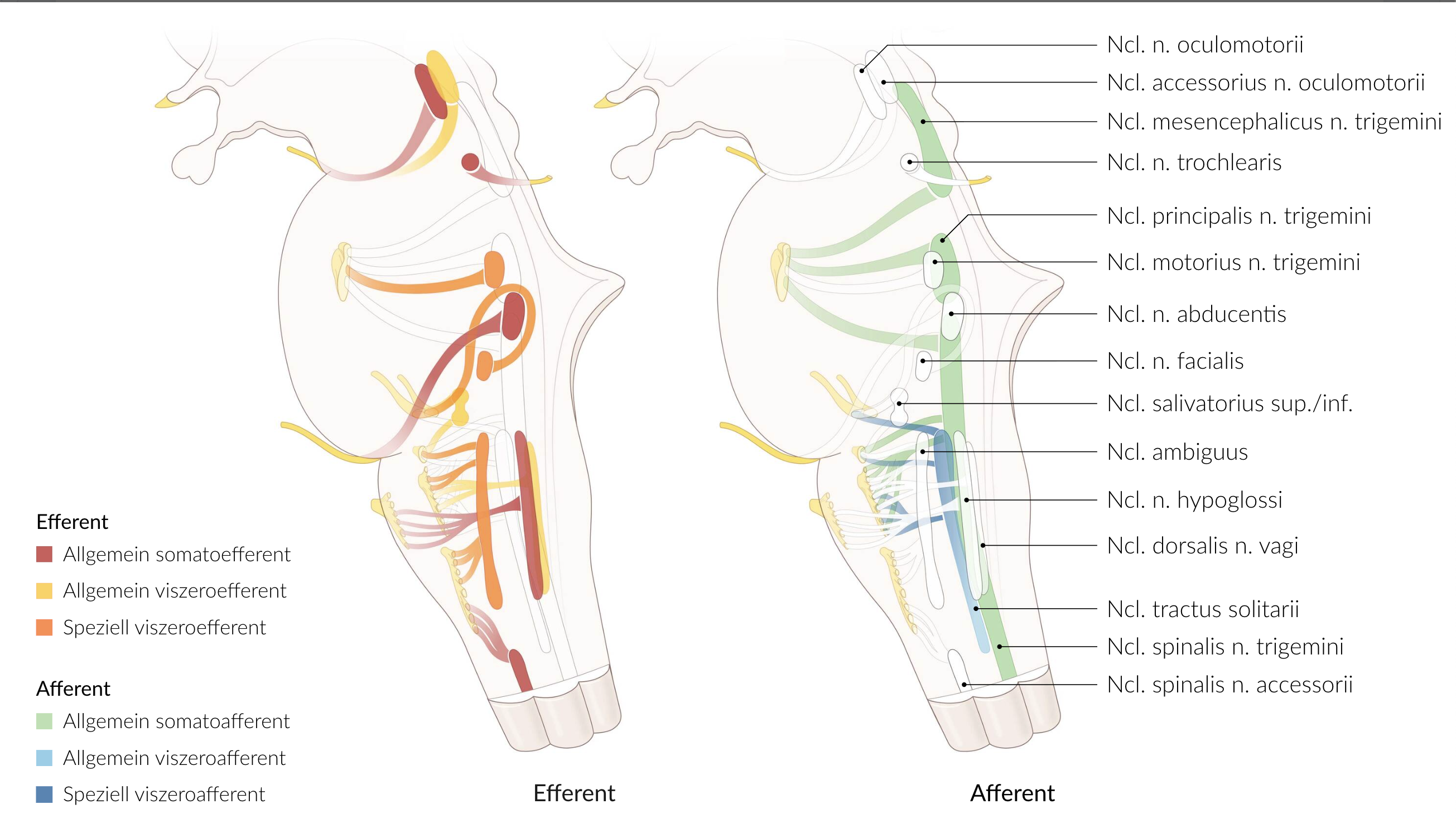
67. A patient has neurological deficits due to a brain tumor in the midbrain (Mesencephalon).
Which structure does not belong to the midbrain and is therefore least likely to be affected?
A. Accessory nucleus of the oculomotor nerve (Ncl. accessorius n. oculomotorii)
B. Motor nucleus of the trigeminal nerve (Ncl. motorius n. trigemini)
C. Nucleus of the oculomotor nerve (Ncl. n. oculomotorii)
D. Nucleus of the trochlear nerve (Ncl. n. trochlearis)
E. Red nucleus (Ncl. ruber)
B. Motor nucleus of the trigeminal nerve (Ncl. motorius n. trigemini)
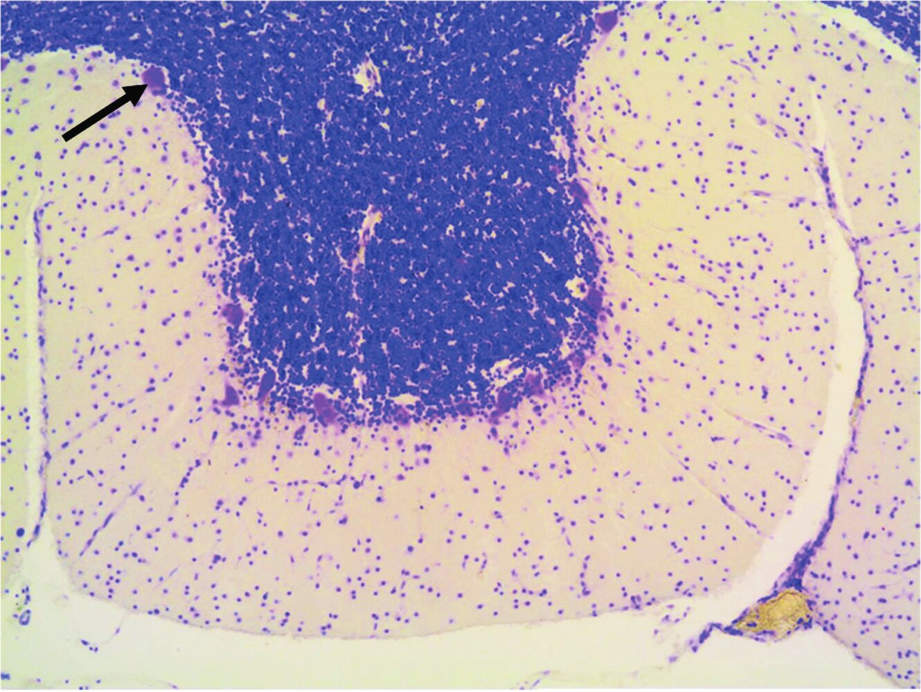
68. Which statement about the nerve cell marked with an arrow in the image is correct?
A. Its axon projects to neurons of the cerebellar nuclei.
B. It is an interneuron.
C. It receives afferents from the superior olivary complex.
D. It is a Golgi type II neuron.
E. It forms parallel fibers in the molecular layer (Stratum moleculare).
A. Its axon projects to neurons of the cerebellar nuclei.

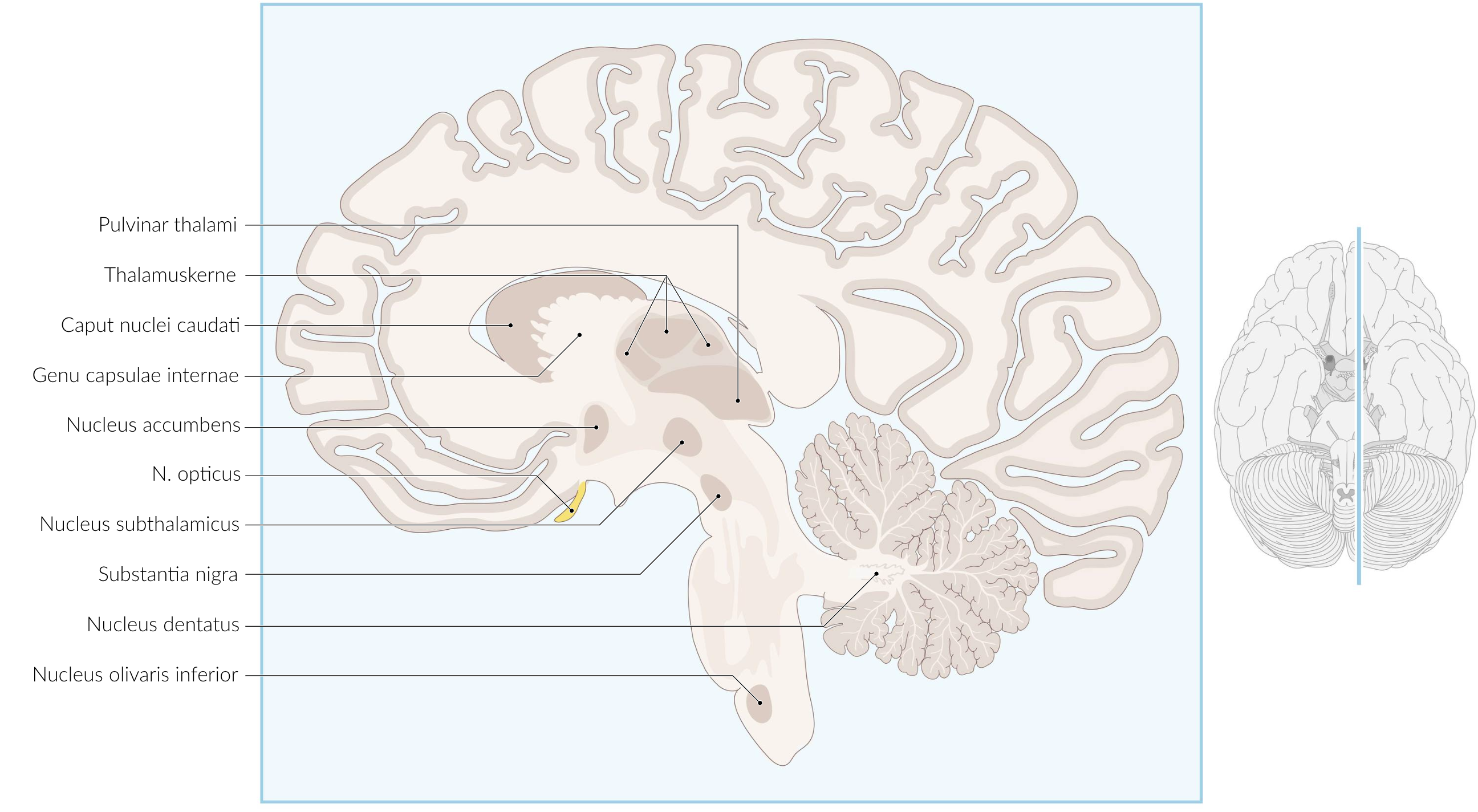
69. The hippocampal formation is associated with learning, memory, and spatial representation functions.
Which brain region is not neuroscientifically considered part of the hippocampal formation or its associated cortical areas of the limbic system?
A. Entorhinal cortex (Area/Cortex entorhinalis)
B. Cornu ammonis
C. Dentate gyrus (Gyrus dentatus)
D. Subiculum
E. Substantia nigra
E. Substantia nigra
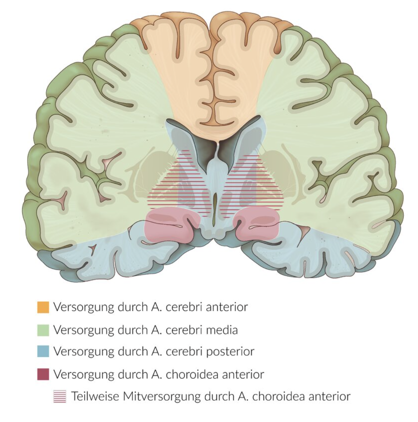
70. Which of the following disorders is most likely caused by a cortical lesion due to an ischemic event in the left anterior cerebral artery (A. cerebri anterior)?
A. Loss of the right visual field
B. Bilateral hearing loss
C. Sensory disturbance in the right hand
D. Paresis (weakness) of the right leg
E. Impaired language comprehension
D. Paresis (weakness) of the right leg
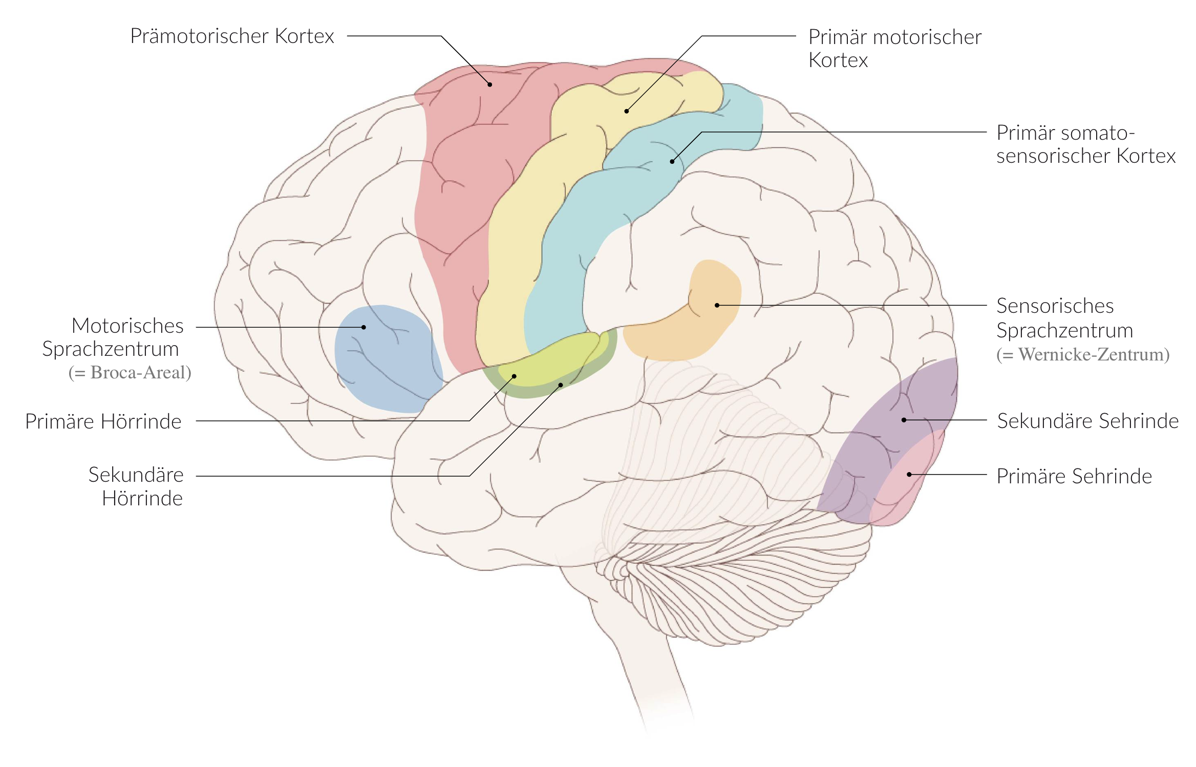
71. In a 58-year-old female patient, you observe the following findings: She speaks significantly faster than healthy individuals, her speech lacks content, sentences are often interrupted, and words are either mixed up or newly invented. You suspect sensory aphasia.
In which of the following structures on the language-dominant side is a lesion most likely?
A. Inferior frontal gyrus (Gyrus frontalis inferior)
B. Middle frontal gyrus (Gyrus frontalis medius)
C. Superior frontal gyrus (Gyrus frontalis superior)
D. Postcentral gyrus (Gyrus postcentralis)
E. Superior temporal gyrus (Gyrus temporalis superior)
E. Superior temporal gyrus (Gyrus temporalis superior)
72. An adult female patient has feet more than twice the size of her healthy twin sister (despite having the same height). She also has a noticeably large tongue and undefined visual field impairments. You suspect an intracranial mass lesion and plan to determine its location through imaging.
Based on the symptoms described, the lesion is most likely located in which structure(s)?
A. Anterior pituitary gland (Adenohypophysis)
B. Mammillary body (Corpus mammillare)
C. Paraventricular nucleus of the hypothalamus (Nucleus paraventricularis hypothalami)
D. Supraoptic nucleus (Nucleus supraopticus)
E. Tegmental nuclei (Nuclei tegmentales)
A. Anterior pituitary gland (Adenohypophysis)
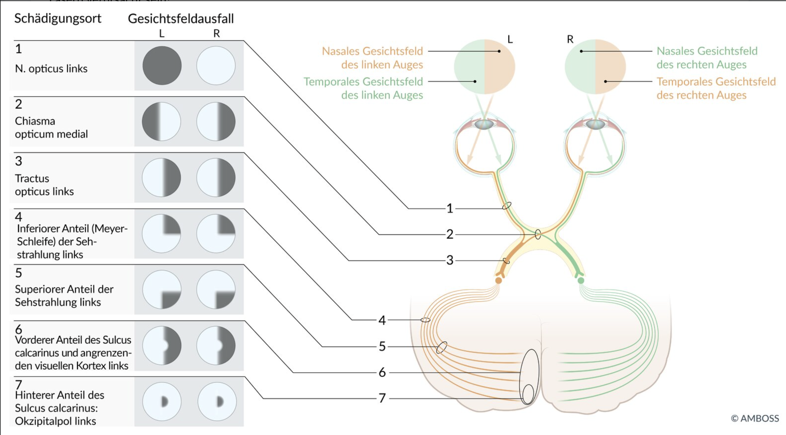
73. Which of the following visual field defects is most likely caused by a complete lesion of the fibers crossing in the optic chiasm (Chiasma opticum)?"
A. Loss of the nasal visual fields in both eyes
B. Loss of the temporal visual fields in both eyes
C. Loss of the entire visual field of the left eye
D. Loss of the left visual field in both eyes
E. Loss of the right visual field in both eyes
B. Loss of the temporal visual fields in both eyes
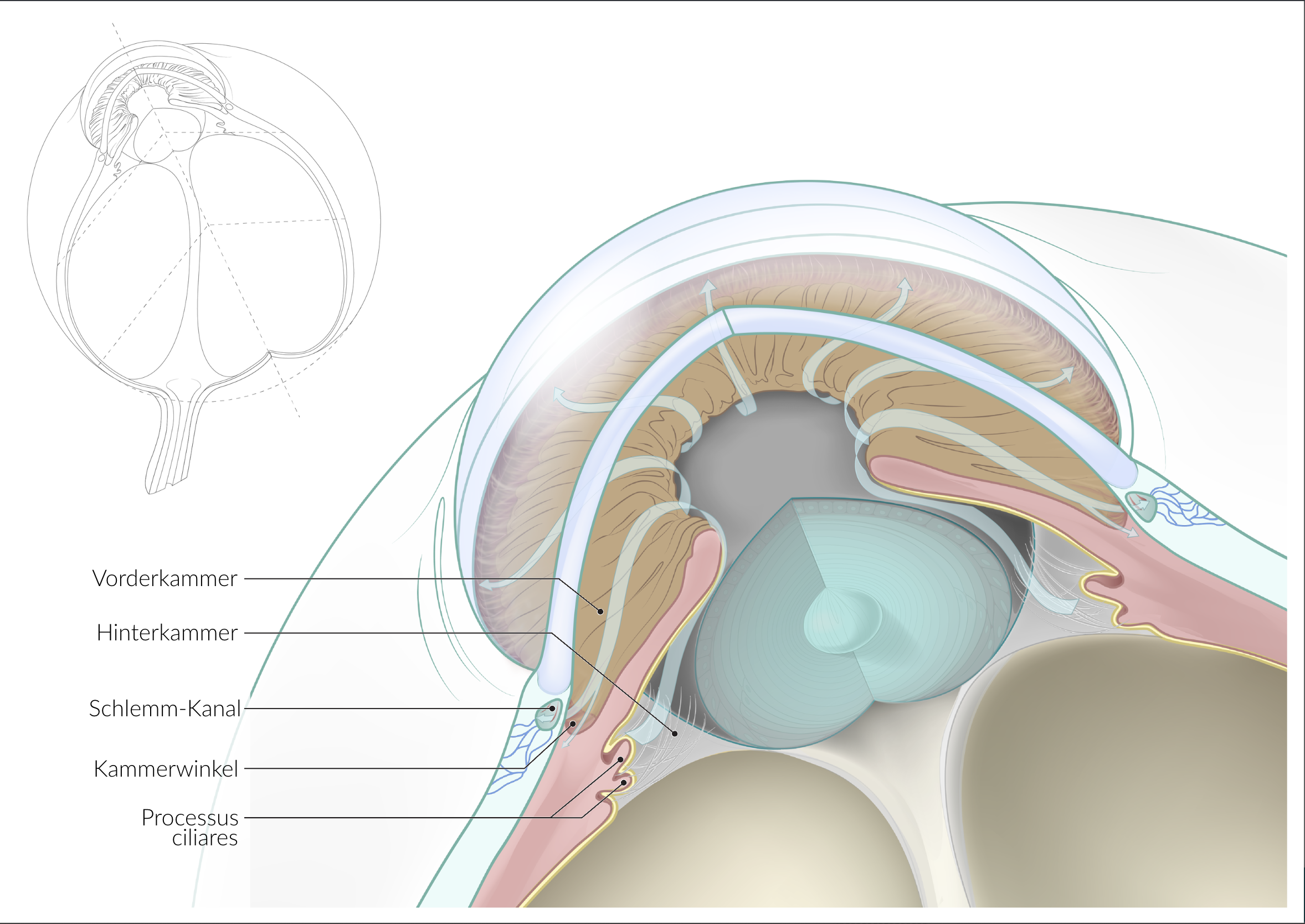
74. Through which part of the eye does the majority of the aqueous humor drain?
A. Iridocorneal angle (Angulus iridocornealis)
B. Bruch’s membrane (Bruch-Membran)
C. Choroid (Choroidea)
D. Vitreous body (Corpus vitreum)
E. Ciliary processes (Processus ciliares)
A. Iridocorneal angle (Angulus iridocornealis)
75. Which of the listed nerves typically does not pass through the common tendinous ring (Anulus tendineus communis) into the orbit?
A. Abducens nerve (N. abducens)
B. Oculomotor nerve (N. oculomotorius)
C. Nasociliary nerve (N. nasociliaris)
D. Optic nerve (N. opticus)
E. Trochlear nerve (N. trochlearis)
E. Trochlear nerve (N. trochlearis)
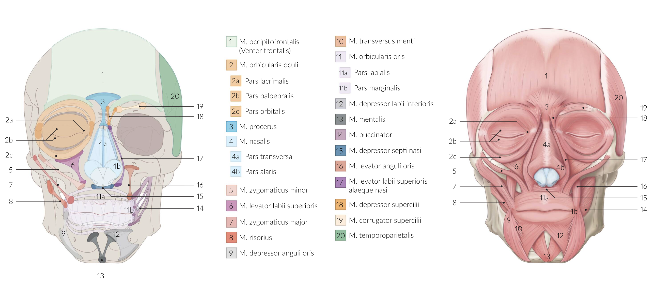
76. The muscle marked with arrows in the image:
A. Has an oblique part (Pars obliqua)
B. Consists of smooth muscle fibers
C. Elevates the eyelid
D. Is enclosed by a fascia
E. Is innervated by the facial nerve (N. facialis)
E. Is innervated by the facial nerve (N. facialis)
77. Which of the following statements about the cells of the stria vascularis of the cochlear duct (Ductus cochlearis) is correct?
A. The basal cells form a loose cellular network that is highly permeable to water and ions.
B. The basal cells are located on the wall of the cochlear duct facing the modiolus.
C. The intermediate cells show mitochondria-rich folds on their basolateral surface.
D. The marginal cells receive K⁺ ions via recirculation through gap junctions.
E. The marginal cells produce perilymph.
D. The marginal cells receive K⁺ ions via recirculation through gap junctions.
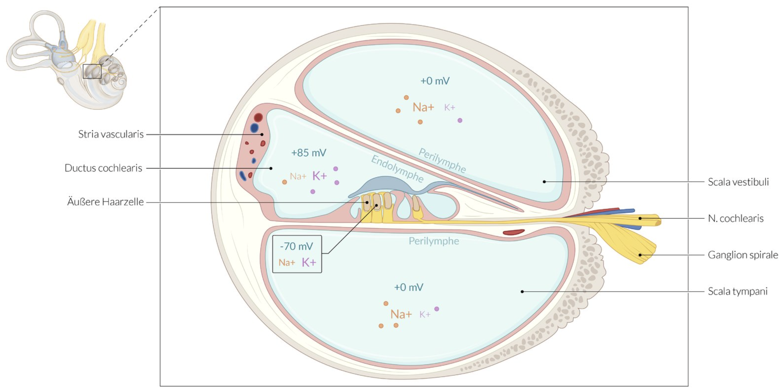
78. Which of the following cells in the inner ear detect head movements and transmit this information via the vestibulocochlear nerve (N. vestibulocochlearis) to central nuclei?
A. Outer phalangeal cells (äußere Phalangenzellen)
B. Hair cells of the maculae (Haarzellen der Maculae)
C. Hensen cells (Hensen-Zellen)
D. Inner phalangeal cells (innere Phalangenzellen)
E. Pillar cells (Pfeilerzellen)
B. Hair cells of the maculae (Haarzellen der Maculae)