BI1014 - enzymology and proteins
1/47
There's no tags or description
Looks like no tags are added yet.
Name | Mastery | Learn | Test | Matching | Spaced | Call with Kai |
|---|
No analytics yet
Send a link to your students to track their progress
48 Terms
enzymes
- Catalysts for reactions under mild conditions
- High specificity for substrates and reactions
- Reversible binding to substrate → enzyme-substrate complex
- Reaction occurs → product released
low substrate concentration
few enzymes bound to substrate
high substrate concentration
saturation, most enzymes have substrate bound
initial velocity
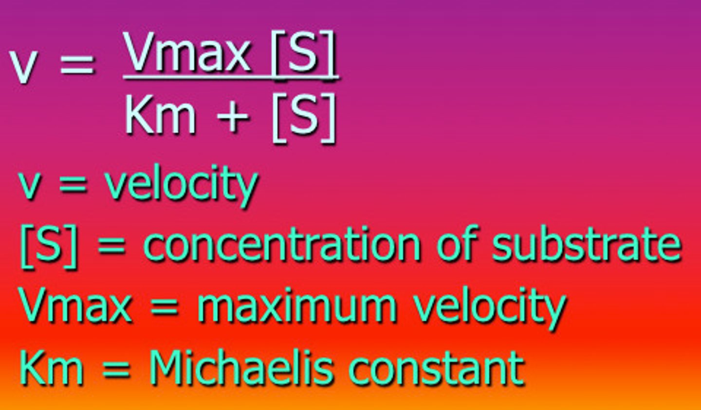
V₀ (initial velocity) vs. substrate concentration = hyperbolic curve
Vmax
Vmax: max velocity at enzyme saturation
Km
Km: substrate concentration at ½ Vmax (approximates affinity)
- Michaelis constant
Kcat
Kcat: catalytic constant (turnover number)
Vmax equation
Vmax = Kcat × [Etot]
![<p>Vmax = Kcat × [Etot]</p>](https://knowt-user-attachments.s3.amazonaws.com/d2c10e23-28c6-4647-a476-fcca44d79b45.png)
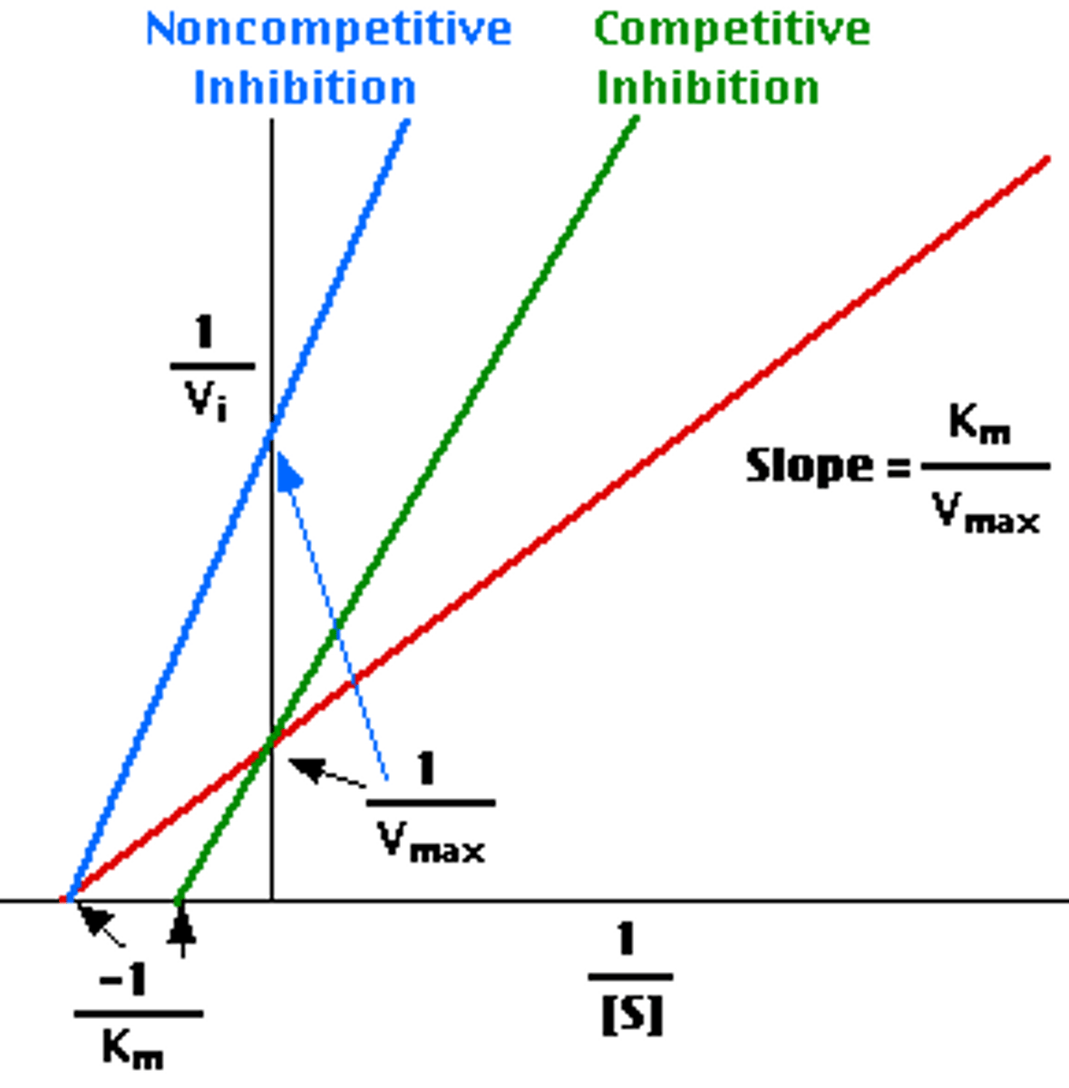
Lineweaver - burk plot
(1/v vs. 1/[S]):
x-intercept = -1/Km
y-intercept = 1/Vmax
![<p>(1/v vs. 1/[S]):</p><p>x-intercept = -1/Km</p><p>y-intercept = 1/Vmax</p>](https://knowt-user-attachments.s3.amazonaws.com/b53d96fa-9662-4320-b19c-66584f4be8fd.png)
Michaelis - Menten equation
kinetic parameters
V0
Vmax
Km
Kcat
enzyme inhibition
reversible and irreversible
reversible inhibition
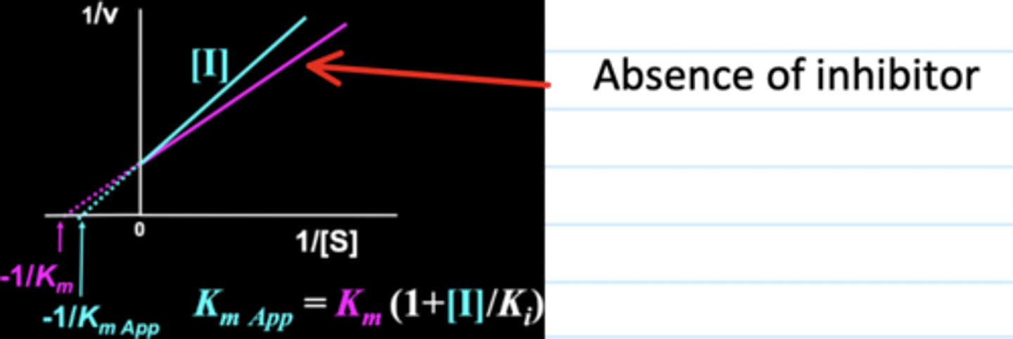
Competitive: inhibitor binds active site
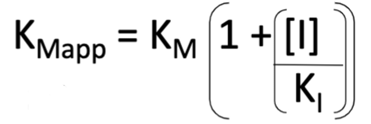
- Vmax unchanged, Km appears increased (KmApp)
- Overcome by increasing substrate
Ki: inhibitor constant
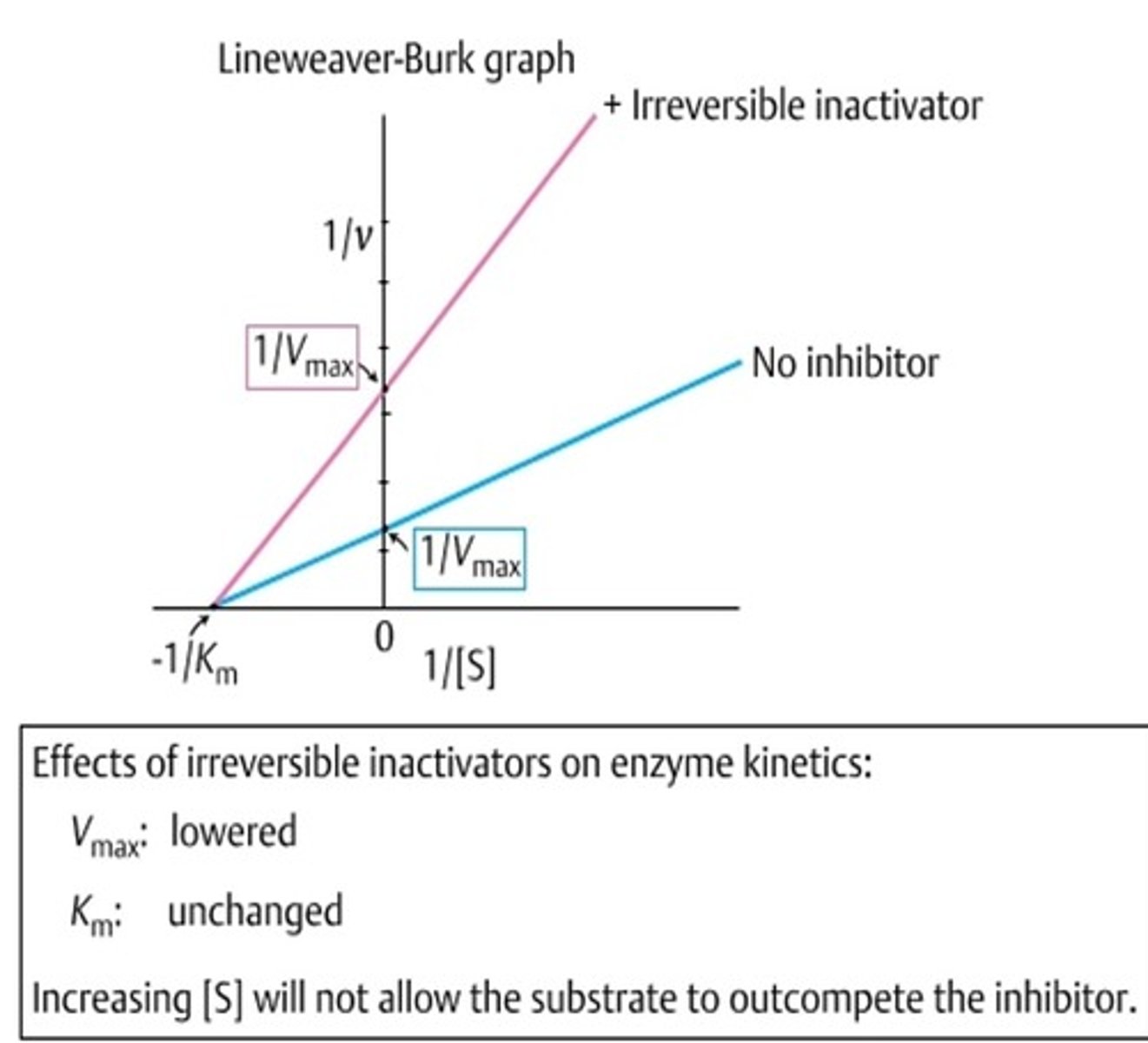
irreversible inhibition
- Covalent bond to enzyme
- Vmax decreases, Km unchanged
clinical relevance
Myasthenia gravis → treat by inhibiting acetylcholinesterase
enzymes
- Facilitate reactions
- Make/break bonds
- Facilitated electron distribution
KmApp
- With a competitive inhibitor, Km stays the same but seems to have changed when measured because more substrate is needed to reach Km/2 (known as KmApp)
determining Ki
ki is the inhibitor constant
functions of proteins
- Defence (keratin, antibodies)
- Structure (muscles, skin)
- Catalysis (enzymes)
- Transport (haemoglobin)
structures of proteins
- Polymers of L-amino acids
- Side chains (R groups) define identity
- Peptide bonds (partial double-bond character, no rotation)
- Written N- to C-terminal
amino acids
- d-amino acids exist but not in proteins encoded by DNA
- Amino acids in proteins all have L-configuration
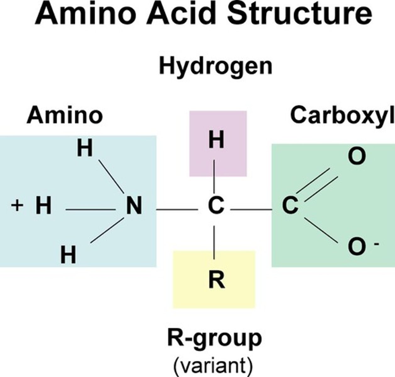
side chains
- Side chains are made up of the R group
- The R groups contain different groups
- The name of the amino acid is determined by its side chain -> We shorten the name to the first 3 letters to make it easier
peptide bonds
- amino acids are joined by peptide bonds
- A delocalization of electrons across this region gives double bond like characteristics and stops the bond from rotating
- A polypeptide is multiple proteins bonded together
- Always write from the N-terminal end to the C-terminal end (carboxyl group)
secondary structures
- alpha helix
- beta pleated sheets
alpha helix
- Right-handed, 3.6 residues/turn
- H-bonds: N to N+4
- Side chains project outward
Proline breaks helix (no H-bonding, rigid)
beta sheets
- Antiparallel: strong H-bonds, alternating directions
- Parallel: weaker H-bonds, same direction
- R groups alternate up/down
Example: spider silk (Gly-Ala)n → flexibility
folding in tertiary structure
- Driven by hydrophobic effect (entropic gain)
- Side chain interactions (hydrophobic, hydrophilic)
stabilisation in tertiary structure
- Van der Waals, H-bonds, disulphide bonds (oxidized cysteines)
- Salt bridges (Asp-Lys, pH-sensitive)
domains in tertiary structure
- Independently folding regions
- Often linked to specific functions
quaternary structure
- Assembly of multiple polypeptide chains
- Examples: haemoglobin, RNA polymerase, HIV protease
- Critical for function, drug targeting, protein arrangement
sidechain interactions in tertiary structure
- Some are hydrophobic (valine, leucine, phenylalanine)
- some are hydrophilic (aspartate, lysine, serine)
hydrophobic effect
- Oil and water do not mix
- Hydrophobic effect: most important determinant of tertiary structure
- Hydrophobic collapse -> hydrophobic chains interact with other hydrophobic chains, hydrophilic with hydrophilic - entropic effect
entropic effect
- Hydrophobic molecules make water molecules form a "cage"
- Water molecules thus are more ordered than if the hydrophobic molecule was not there
- More order = less entropy
disulphide bonds
- Covalent bonds between two cysteine side chains
- Lose a proton and form a disulphide bond
- Oxidization reaction to form the bond, usually in secreted or Golgi apparatus proteins because of oxidising environments
salt bridge
- Important but pH sensitive
- Aspartate residues can form salt bridges with lysine residues
summary
Primary structure – sequence of amino acids
Secondary structure – helices and sheets
Tertiary structure – 3d structure – folded polypeptide
Quaternary structure – assembly of more than one folded polypeptide
protein purification
- recognition and assay
- criteria of purity
recognition and assay
identify and quantify target protein
criteria of purity
high-resolution separation methods
techniques of protein purification
SDS page, ammonium sulphate precipitation, isoelectric focusing
SDS page
- Denatures proteins with SDS
- Separates by mass
- Visualized with Coomassie/silver stain
ammonium sulphate precipitation
"Salting out" → reduced solubility separates proteins
isoelectric focussing
pH gradient separates proteins at their isoelectric point (pI)
chromatography
size exclusion, ion exchange, affinity chromatography
size exclusion
(SEC):
Separates by size; larger elute first
ion exchange
Separates by charge (cation or anion exchange)
affinity chromatography
High specificity (e.g., enzyme-substrate, antibody-antigen)
determining structure
Combine SEC + SDS-PAGE to deduce quaternary structure (e.g., dimers)