ch 2 - the heart
1/50
There's no tags or description
Looks like no tags are added yet.
Name | Mastery | Learn | Test | Matching | Spaced | Call with Kai |
|---|
No analytics yet
Send a link to your students to track their progress
51 Terms
found in the ____ cavity in a space called the ____. covered by the _____ (tissue) and sits in the ______ cavity.
[thoracic cavity] [mediastinum]
[pericardium] [pericardial cavity]
pericardial cavity
serous fluid filled cavity - located btwn visceral and parietal pericardium
most BVs emerge and enter through the ____. the most inferior tip of the heart is the _____
[base] [apex]
name the 4 chambers of the heart
atria (upper receiving chambers)
right atrium
left atrium
ventricles (lower pumping chambers)
right ventricle
left ventricle
auricles
external muscular pouches increasing atria volume
list the 2 circulation system types in the human body
pulmonary
systemic
pulmonary circulation
transports deoxygenated blood from the heart to the lungs for O2 absorption, CO2 release
systemic circulation
carries oxygenated blood from heart to all body tissues to deliver nutrients, collect waste
why does pulmonary circulation send deoxygenated blood to the lungs if it’s deoxygenated? don’t the lungs breathe in oxygen?
we are talking about blood flow, not the actual gas of O2 inhaled into the lungs. deoxygenated blood undergoes gas exchange when it is sent to the lungs (aka CO2 is dropped off and fresh O2 is absorbed).
the fresh O2 travels back to the heart through the pulmonary veins (systemic circulation) and is then sent out to body tissues
what part of the heart pumps blood into the following:
pulmonary circulation circuit
systemic circulation circuit
pulmonary = right ventricle (pumping chamber)
systemic: left ventricle (pumping chamber)
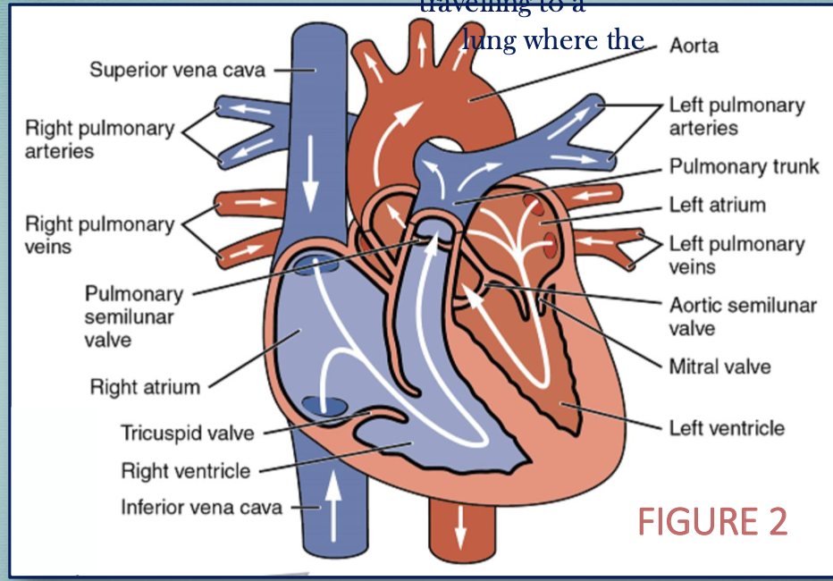
describe the flow of blood intro, through, and out of the heart during the pulmonary circulation circuit (specific structure names)
deoxygenated blood enters heart through superior and inferior venae cavae
blood drains into the right atrium
blood pumps into the right ventricle
from there, blood travels through the pulmonary semilunar valve to the pulmonary trunk (superior structure)
from pulmonary trunk, blood travels through L+R pulmonary arteries
blood travels to lungs where pulmonary capillaries exhibit gas exchange (CO2 → O2)
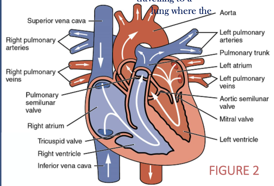
describe the flow of blood intro, through, and out of the heart during the systemic circulation circuit (specific structure names)
after travelling to the lungs to pick up O2, oxygenated blood travels back to the heart
blood returns into the heart through the L+R pulmonary veins
blood drains into the left atrium
blood is pumped into the left ventricle
blood is pumped through the aortic semilunar valve into the aorta
aorta branches out allowing blood to travel to body tissues
systemic capillaries exhibit gas exchange (O2 → CO2)
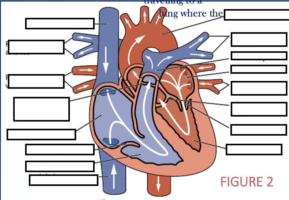
label all structures within the figure
membrane surrounding heart and the pericardial cavity
pericardium
pericardium (where + 2 layers + sublayers)
also define pericardial cavity
membrane surrounding heart and its pericardial cavity
2 layers
outer: fibrous pericardium
inner: serous pericardium
parietal layer: fused to fibrous pericardium
visceral layer: physically part of heart wall
pericardial cavity
serous-fluid filled space inbtwn visceral and parietal pericardium

identify the membrane layers of the heart
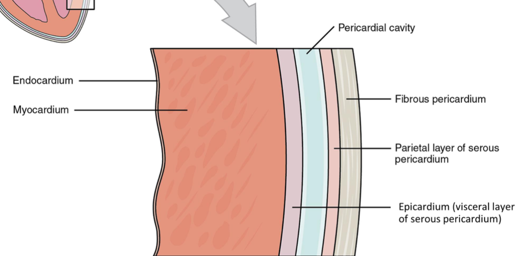
identify the 3 layers of the heart (NOT the serous/fibrous membranes)
epicardium
myocardium
endocardium
epicardium
(1/3 heart layers)
visceral pericardium
myocardium (thick/thin layer, cell type, unique function, septa)
(2/3 heart layers)
thickest layer of hearty
cardiac muscle cells
cardiac muscle tissue contracts to pump blood through the heart
endocardium (thick/thin layer, cell type, septa)
(3/3 heart layers)
innermost layer, thinnest layer
simple squamous epithelium - endothelium
the myocardium surrounding the ______ ventricle is thicker so it can generate large pressure amounts, overcoming the […]
[left ventricle] - [greater resistance in the systemic circuit]
![<p>[left ventricle] - [greater resistance in the systemic circuit]</p>](https://assets.knowt.com/user-attachments/86ec3f87-574c-44c6-91d3-1993033b29ad.png)
septa (3) (other internal structures of heart)
extension of myocardium that divides heart into its 4 chambers
separating atria: interatrial septum
separating ventricles: interventricular septum
separating atria from ventricles: atrioventricular septum
thicker
reinforced with cardiac skeleton (dense connective tissue)
fossa ovalis (other internal structures of heart)
oval depression in the interatrial septum (septum separating the 2 atria) - leftover from foramen ovale (opening in fetal heart)
valves (2) (other internal structures of heart)
openings btwn atria and ventricles: atrioventricular valves
openings btwn ventricles and arteries (leading to aorta and pulmonary arteries): semilunar valves (pulmonary, aortic)
purpose of having valves
ensures unidirectional flow of blood; no backflow
coronary sinus (other internal structures of heart)
vein draining blood from the heart myocardium
autorhythmicity
cardiac muscle initiating action potentials that spread rapidly from cell to cell
contractile cells
conduct impulses and perform contractions to pump blood
conducting cells
initiate and propagate action potential to trigger contractions
sinoatrial (SA) node
pacemaker setting normal heart rhythm
generates nerve impulse initiating sinus rhythm → sends impulse to atrioventricar node
atrioventricular (AV) node
electrocardiogram (ECG)
tracing the electrical signal of the heart using electrodes (3, 5, 12)
5 points of the ECG
P Wave = atrial depolarization, contraction begins
QRS Complex = ventricular depolarization, contraction begins at R wave
T Wave = repolarization of the ventricles (atria repolarize during QRS wave)
systole
period where blood is pumping out of the heart
diastole
relaxation period where heart chambers fill with blood
S1 vs S2
S1 = atrioventricular valves