Unit 1.2: Normal Wound Healing
1/34
There's no tags or description
Looks like no tags are added yet.
Name | Mastery | Learn | Test | Matching | Spaced | Call with Kai |
|---|
No analytics yet
Send a link to your students to track their progress
35 Terms
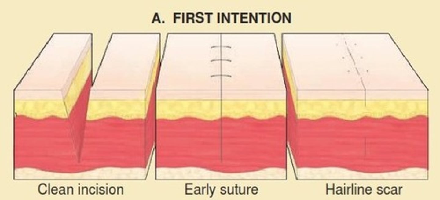
What is the primary intention of wound healing/closure?
This is the simplest and fastest type of closure, occurring when wound edges are physically approximated using sutures, staples, adhesive tapes, or biologic glues
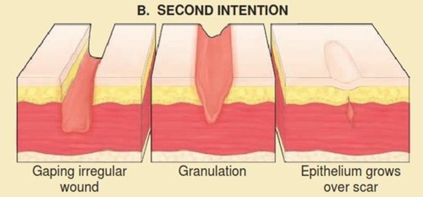
What is the secondary intention of wound healing/closure?
This method is used when wound edges cannot be approximated (e.g., a large crater or "pothole"). The body must build a matrix of granulation tissue to fill the wound defect. Healing occurs through a combination of granulation, wound contraction, and epithelialization from the wound periphery. Where PTs spend most of wound care.
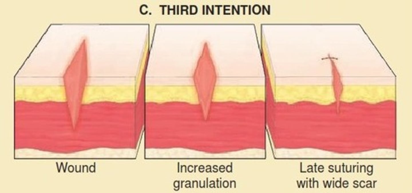
What is the delayed closure (tertiary) wound healing/closure?
This is a combination of primary and secondary closure. The wound is initially left open to be cleansed and observed for signs of infection or contamination (common in traumatic injuries or if the wound has been untreated for more than 4 hours). Once the wound bed appears clean and free of debris, it is surgically closed. Ex. SKIN GRAFT
What is the window in which an open wound is considered to have definite potential for infection?
4 hours
What are the phases of wound healing?
1. Inflammation
2. Proliferation
3. Re-epithelization in partial-thickness wounds
4. Remodeling
What is the duration of inflammation?
Days 1-4
What occurs during the inflammation stage?
Involves a vascular response (initial vasoconstriction to control bleeding, followed by vasodilation) and a cellular response (PMNs/neutrophils arrive first to kill bacteria, followed by macrophages, which direct the repair process)
What are the characteristics of the inflammation stage?
cardinal signs: Redness (rubor), swelling (tumor), warmth (calor), and pain (dolor). The wound bed may appear glossy due to transudate or exudate.
What do polymorphonuclear leukocytes (PNMs) do during inflammation?
- Secretes pro-inflammatory cytokines
- destroy bacteria
- Release proteases, collagenases, and elastases to break down dead tissue
- Produce Matrix Metalloproteases (MMPs)
What do Matrix Metalloproteases (MMPs) do during inflammation?
They help with tissue breakdown
- Normal levels should peak a couple of days after injury and steadily decrease
What is the goal of the later response of inflammation?
Increase circulation to the area via vasodilation and angiogenesis
What is chemotaxis?
The process of active movement toward the area of the highest concentration of a chemical signal
- How inflammatory cells find the injury
What does the autolytic debridement by macrophages originate from?
mononuclear leukocytes
What occurs once the macrophages die?
The formation of fibroblasts occurs
What is apoptosis?
programmed cell death; achieved by cells such as NK cells contacting the host and causing it to die
What is phagocytosis?
Cell eating: cells are engulfed or broken up by cells such as macrophages
What is the duration of proliferation?
Days 4-24
What occurs during the proliferation stage?
Focuses on building new tissue through angiogenesis (vessel formation), granulation (fibroblasts laying a collagen scaffolding), wound contraction (myofibroblasts pulling edges together), and epithelialization
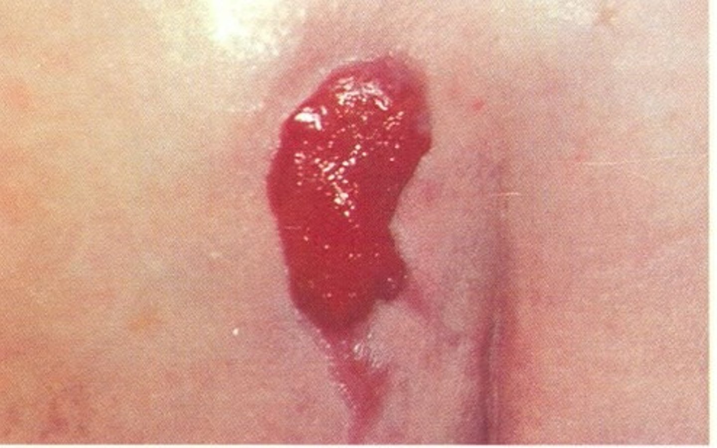
What are the characteristics of the proliferation stage?
The wound bed develops "beefy red" granulation tissue. You may see small endothelial buds (angiogenesis) and a pale pink border at the wound edges (epithelialization)
What is angiogenesis?
The formation of small vessels that bring nutrients and remove waste products
What is granulation?
Highly vascularized tissue scaffolding that fills the wound void
What is re-epithelialization?
Fibroblasts lay down collagen in the wound bed that strengthens the granulation tissue
What is wound contraction?
Myofibroblasts cause the wound to contract toward the center
What is the duration of re-epithelialization in partial-thickness wounds?
Begins within 24 hours
What occurs during the re-epithelialization in partial-thickness wounds?
keratinocytes migrate not just from the wound edges, but also from epidermal appendages (hair follicles and sweat glands) that remain in the dermis.
What are the characteristics of the re-epithelialization in partial-thickness wounds?
Appears as pale pink or silvery skin resurfacing the wound. It may appear as "islands" of new skin forming around hair follicles within the wound bed
What is the duration of remodeling in partial-thickness wounds?
21 days to 2 years
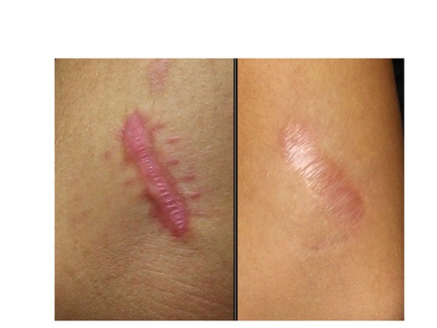
What occurs during the remodeling stage?
Collagen fibers reorganize along lines of stress (Davis's Law). Fibroblasts convert to myofibroblasts for continued contraction, and tensile strength increases to a maximum of 80% of the original tissue.
What are the characteristics of the remodeling stage?
The scar undergoes blanching (losing its red/pink color) and matures from a thickened state to a thinner, smoother appearance. Melanocyte aggregation begins to restore skin pigmentation.
What is blanching?
temporary whitening of the skin due to pressure over an area of skin, causing restricted blood flow
What is the Extracellular Matrix (ECM)? What types of cells/tissues are contained in the ECM?
It is a structural scaffolding that consists of cells, fibers, and ground substance, organized into the layers of the skin to provide strength and flexibility. During the proliferative phase of healing, the body must produce a new ECM to fill the wound defect left by damaged or debrided tissues.
What is ground substance?
A gelatinous matrix in which fibers are embedded, primarily found in the papillary dermis.
What are the fibers found in EMC?
The matrix is composed mainly of collagen (for structural strength) and elastin (for pliability and flexibility).
What are fibroblast cells found in EMC?
These are the primary cells within the dermis responsible for producing the collagen and elastin fibers and the growth factors necessary for the ECM.
What are the regulatory enzymes found in EMC?
The ECM is constantly balanced by Matrix Metalloproteases (MMPs), which break down tissue components, and Tissue Inhibitors of Matrix Metalloproteases (TIMPs), which bind to MMPs to maintain a balance between repair and degradation