Lab Machines
1/128
There's no tags or description
Looks like no tags are added yet.
Name | Mastery | Learn | Test | Matching | Spaced | Call with Kai |
|---|
No analytics yet
Send a link to your students to track their progress
129 Terms
what is it
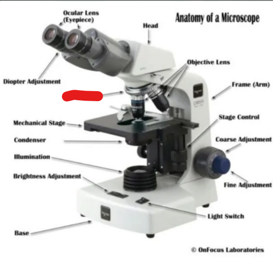
diopter adjustment
what is it
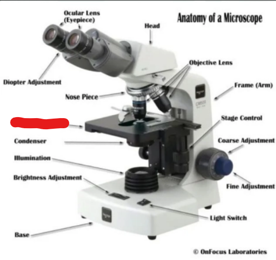
nose piece
what is it
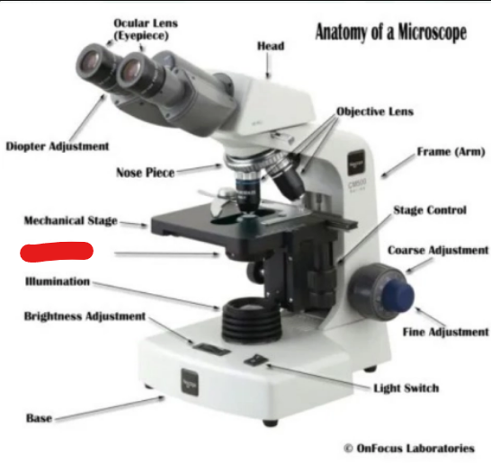
mechanical stage
what is it
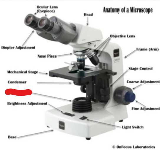
condenser
what is it
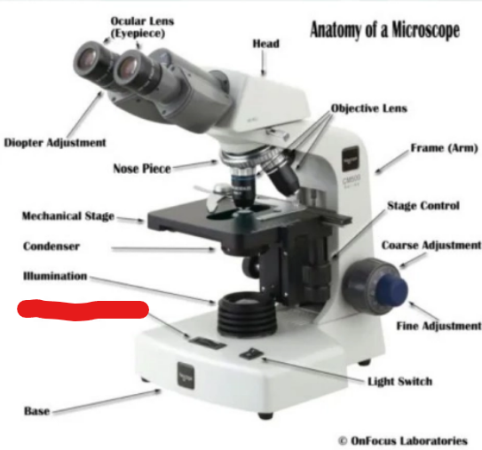
illumination
what is it
brightness adjustment
what is it
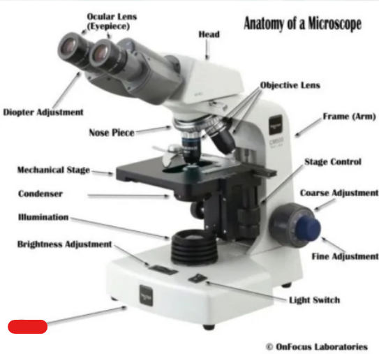
base
what is it
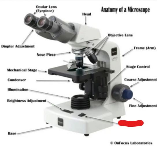
light switch
what is it
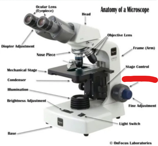
fine adjustment
what is it
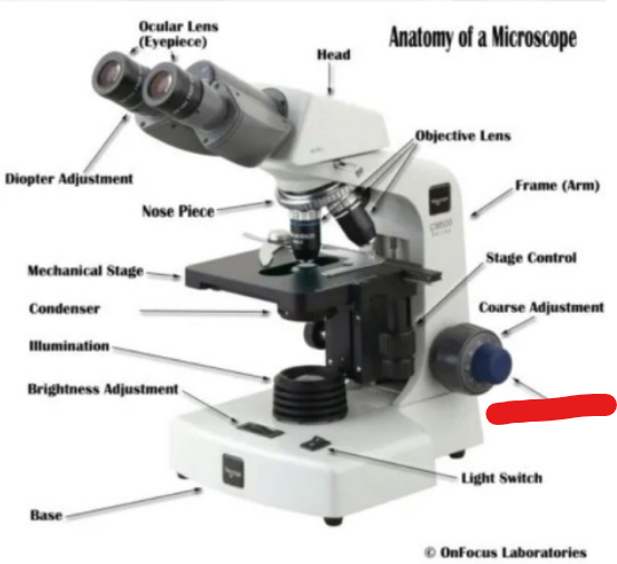
coarse adjustment
what is it
stage control
what is it
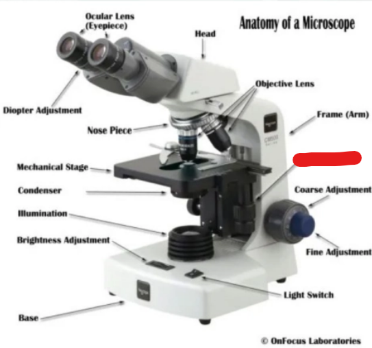
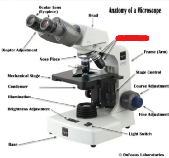
frame (arm)
what is it
objective lens
what is it
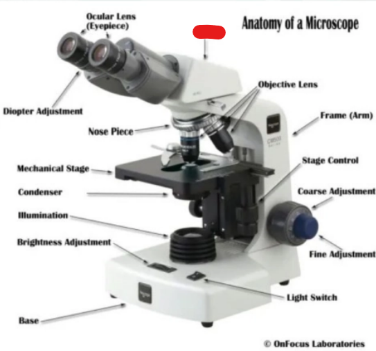
head
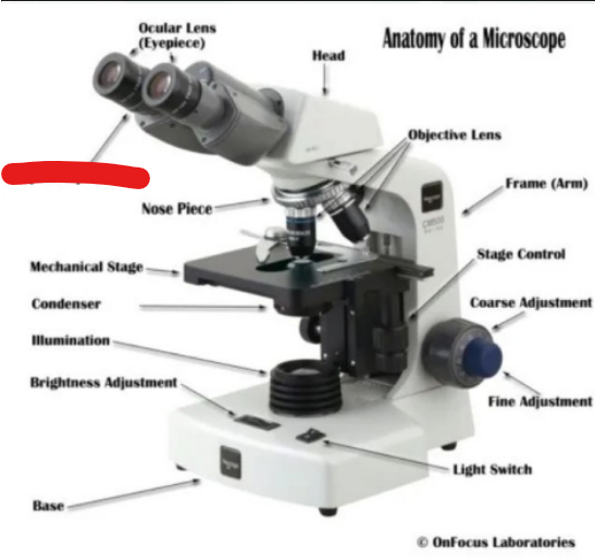
what is it
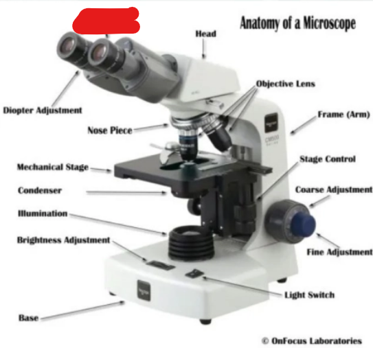
ocular lens (eyepiece)
define Mechanical stage
The platform on which the slide or object is placed for viewing.
define stage bracket
hold the slide or specimen in place on the stage.
define stage control knobs
These knobs move the slide or specimen either horizontally (x axis) or vertically (y axis) when it is being viewed.
purpose of arm/frame
Used to carry the microscope.
what are the binocular microscope adjustments
Interpupillary distance
Individual ocular adjustment
Individual ocular adjustment is aka
diopter adjustment
define Interpupillary distance adjustment
adjusts distance between oculars so that you see a single field.
purpose of Individual ocular adjustment
compensates for any differences between your eyes
functions of ocular eyepieces
Magnifies the real image formed by the objective.
Forms a virtual image of the real image.
Can carry measuring scales for micrometry to measure the specimen
why is measuring important in micrometry
some species are similar so measuring identifies them
define rotating nosepiece
The rotating nosepiece holds objective lenses and allows for quick exchange of objectives.
Most compound microscopes have what 4 objectives
4X
10X
40X
100X.
microscope objectives are the first to
produce a real image
define magnification power
the degree to which the visual of an object has been altered.
4X is for
scanning/park position
10X means
low dry objective
40X means
high dry objective
100X means
oil immersion objective
Higher power objectives are — — for safety
spring loaded
how to calculate magnification
Take the power of the objective (4X, 10X, 40X) and multiply by the power of the eyepiece, usually 10X.
when measuring under 10x multiply by
10
when measuring under 40X multiply
2.5
when measuring under 100X multiply
nothing, it’s already at the actual size
why is oil required on 100X
Oil is required so that the light rays are not refracted when they pass from the slide to air before entering the objective lens.
define condenser
Is a series of lenses mounted in such a way that the object and the slide are completely and evenly illuminated.
define iris disphragm
this controls the amount of light coming up into your scope.
define field diaphragm
controls only the width of the bundle of light rays reaching the condenser.
define illuminator
Built-in low voltage illuminator on the underside of the microscope. Illuminating apparatus is a built-in light source.
define Kohler Illumination
a method of adjusting a microscope in order to provide optimal illumination by focusing the light on the specimen
How to Set up Kohler Microscope Illumination (5)
The Condenser should be all the way up to the stage (unless looking at urine or feces)
Your Iris Diaphragm lever (under the stage) should always be at 70-80% open so light can enter the condenser.
Light intensity should be between 3-5.
The Field Diaphragm should be between the “ C and the “L” , not to the open position.
Make sure that under 10X, your field of view is centered. And if not please do so by using the Centering Screws
define micrometry
The science of measuring objects under the microscope
oculars can be cleaned with (2)
dampened soft cloth or lens paper
how to store microscope (9)
Turn off light
Lower stage completely
Ensure the 4X objective is in place (lowest objective)
Condenser down
Remove slide
Oculars together
Replace dust cover
in cupboard; place arm out
power cord neatly around base
how to carry microscope
one on the arm and one under the base
what happens if you don’t carry a microscope properlly
ocular can fall out
what to do when No light when you switch to oil immersion lens
check to see that you have placed a drop of oil on the slide.
what to do when viewing is blurry or unable to focus under 40x (2)
can mean your slide is upside down
you could have accidentally dipped it in oil causing blurry vision.
what to do when unable to see anything under urine or fecal sample,
your light is too bright - lower condenser
what happened when you cracked your slide under 40x or 100x,
you used the course adjustment knob
define centrifuge (2)
A centrifuge is designed to accelerate sedimentation by taking advantage of centrifugal force.
It is used to separate substances of different densities in a solution.
define Microhematocrit centrifuge (3)
This centrifuge is used exclusively for spinning down microhematocrit tubes
This process is used for determining a patient’s packed cell volume (PCV)
can also provide a plasma sample for protein analysis.
what are the 3 main types of clinical centrifuge (3)
variable-angle centrifuges / horizontal centrifuge
define fixed-angle centrifuges
multi-use centrifuge
define variable-angle centrifuges / horizontal centrifuge
The tube or bottle is held in a shield or cup, which is attached to the head so that it is free to swing from a vertical position (at rest), to a horizontal position when the centrifuge revolves.
define fixed-angle centrifuges
The fixed-angle centrifuge has buckets that are in a fixed position, typically about 50°.
define Multi-use Centrifuge (2)
Many centrifuges are multi-use, able to accommodate urine, blood, and hematocrit tubes.
These centrifuges have specific centrifugal force settings for each specific type of sample and feature removable shields
Relative centrifugal force (RCF) assumes (2) …
a unit mass
uses the force of gravity as a base unit.
what does RCF stand for
Relative centrifugal force
function of RCF
It is a function of the speed of rotation and the radius from the centre of rotation to the point at which the force is to be determined.
what units are RCF in (unit + unit name)
g - gravities
safety precautions of microscope (5)
Check electrical specifications of instrument before plugging in.
Be sure the centrifuge is balanced to avoid excessive vibration, which results from improper balancing.
Never, under any circumstances, open the lid of the centrifuge while the motor is running.
Never stop a centrifuge with your hands. There is a danger of catching hands in revolving parts, remixing of sediment, and causing excessive wear on the machine.
If there is glass breakage inside the centrifuge, do not open the centrifuge for 15 minutes to avoid aerosols. If any glass is broken remove the head and clean the bowl thoroughly.
define refractometer
measures the refractive index of a solution and is an indirect measure of the specific gravity.
how do refractometers work (2)
When a solution (e.g., urine) is measured, light passes through the sample and bends.
The angle of this refraction is visualized as a shadow and correlates with the concentration of the solution.
what do refractometers measure (2)
blood total protein
urine specific gravity
how to calibrate refractometers
take a reading with distilled water that should be 1.000
use a small screwdriver to turn the zero adjustment setting clockwise to — reading
increase
use a small screwdriver to turn the zero adjustment setting counter clockwise to — reading
decrease
MCHT Procedure (7)
make your 2 microhematocrit tube with required whole blood
spin it in the centrifuge.
Now carefully remove it from the centrifuge, and break it in half appropriately (just above the buffy coat)
Now carefully place the protein (unbroken side of MHCT) just overtop of the refractometer prism (rectangular glass section)
And place a small amount of the protein plasma onto it. You may need to gently jar the tube in a forward motion to get the substance out. (note - stay very close to prism but not close enough to touch it)
With the cover plate depressed, gently press it down to hold/ spread the plasma protein as you lift the refractometer up towards the light and look through the eyepiece to view scale.
Using the SP (serum protein) scale, the reading scale is taken at the boundary line separating the light and dark area.
Rinse prism and cover plate with distilled water between samples and dry with Kimwipe.
Urine SG procedure (4)
put a small drop of urine (spun or unspun) onto the refractometer with a pipette
With the cover plate depressed, gently press it down to hold/ spread the urine as you lift the refractometer up towards the light and look through the eyepiece to view scale.
Using the S.G (specific gravity) scale, the reading scale is taken at the boundary line separating the light and dark area.
Rinse prism and cover plate with distilled water between samples and dry with Kimwipe.
define incubator
provides the ability to artificially control the environmental temperature (and humidity, to some extent) for many microbiological procedures.
define Coagulation analyzers (2ish)
prothrombin time (PT)
activated partial thromboplastin time (aPTT)
fibrinogen tests can be performed using fresh or citrated whole blood.
what is IDEXX ProCyte for
CBC
what does IDEXX ProCyte give for results (6)
Detection of band neutrophils
Reticulocyte count
Reticulocyte hemoglobin (RETIC-HGB)
True five-part white blood cell differential
Detection of nucleated red blood cells
The ability to run abdominal/thoracic, synovial fluid analyses
define Laser flow cytometry
delivers an advanced five-part white blood cell differential.
define Optical fluorescence
provides a highly sensitive and accurate reticulocyte count.
define laminar flow impedance
performs the fastest, most precise red blood cell count
Laser Based / Impedance Technology princple
Laser beams determine the density of solid components and their size.
Color Photometry Principle (2)
The photometer receives whatever light is not absorbed by the sample.
A signal is then transmitted to the readout device as a unit of measurement.
what additive in in red/gold/tiger cap (3ish)
serum
with or without clot activator
with or without gel
what additive in in green cap (4ish)
sodium or lithium heparin
with or without gel
what additive in in lavendar/pink cap (2)
potassium
EDTA
what additive in in gray cap (3ish)
sodium fluoride
sodium or potassium oxalate
what function is the additive in blue cap
prevents blood from clotting by binding calcium
what function is the additive in red/gold/tiger cap
clot activator promotes blood clotting with glass or silica particles
gel seperates serum from cells
what function is the additive in green cap
prevents clotting by inhibiting thrombin and thromboplastin
what function is the additive in lavendar/pink cap
prevents clotting by binding calcium
what function is the additive in gray cap (2)
fluoride inhibits glycolysis, and oxalate
prevents clotting by precipitating calcium
common lab tests for blue caps
coagulation
common lab tests for red/gold/tiger caps (3)
chemistry
serology
immunology
common lab tests for green caps (2)
stat
routine chemistry
common lab tests for grey caps (3)
glucose
blood alcohol
lactic acid
common lab tests for lavender/pink caps (2)
hematology
blood bank
what is blood
the body’s fluid connective tissue
what is plasma
is the bloods liquid component
what does blood contain (4)
RBC
WBC
Platlets
Plasma
what does Plasma contain (8)
water
proteins
waste products
minerals
clotting factors
immunoglobins
carbon dioxide
hormones