4: iris pupil and lens copy)
1/83
There's no tags or description
Looks like no tags are added yet.
Name | Mastery | Learn | Test | Matching | Spaced | Call with Kai |
|---|
No analytics yet
Send a link to your students to track their progress
84 Terms
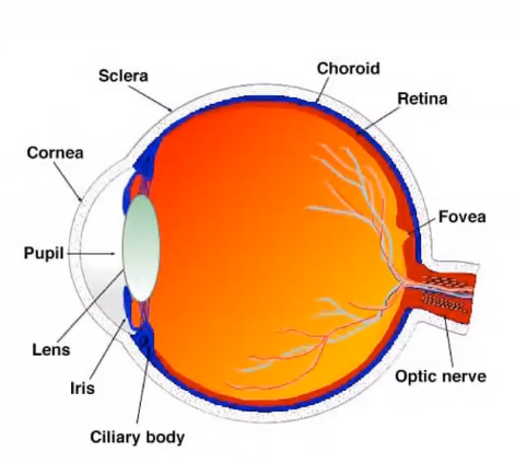
uvea
middle layer of the eye
composed of 3 regions front to back- the iris, ciliary body and choroid
uvea is largest structure
choroid mainly composed of blood vessels
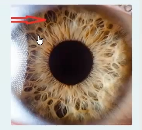
appearance of the iris
pupillary zone closest to the pupil
ciliary zone closest to the ciliary body
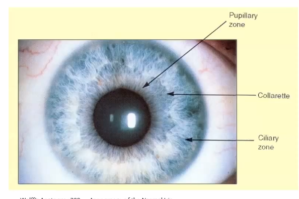
what is the iris
thin circular structure located anterior to the lens
the centre aperture- pupil, located slightly nasal and inferior to the iris centre
pupil size regulates retinal illumination
holes in the iris is crypt
ruff- ring in the iris
function- acts as a diaphragm to regulate the amount of light entering the eye. the two iris muscles are innervated separately.
the pupil
very small in brightly lit conditions and fairly large in dim illumination
it is the thickest in the rgeion of the collarette
collarette encircles the pupil and ciliary zone which extends from the collarette to the iris root
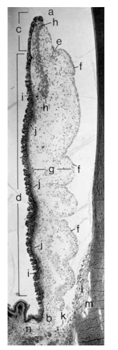
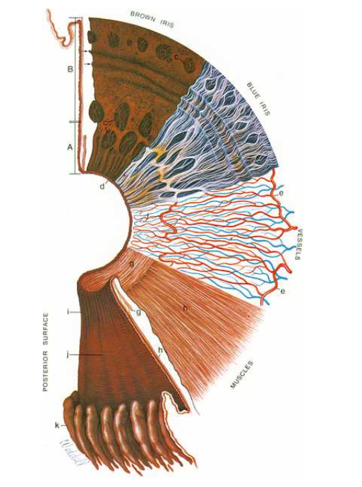
a. pupil and pupillary ruff
b. iris root
c. pupillary portion of the iris
d. ciliary portion
e. collarette
f. cellular anterior border layer
g. stromal tissue
h. sphincter muscle lies in stroma
i. posterior iris
j. anterior epithelium
k. uveal band
l. trabecular meshwork
m. canal of schlemm
n. iris
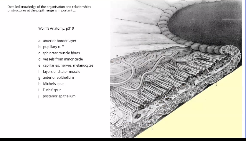
pupillary margin of the iris
rests on the anterior surface of the lens and in profile, the iris has a truncated cone shape such that the pupillary margin lies anterior to its peripheral termination , the iris root
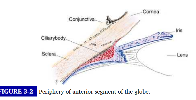
iris root
thinnest part of the iris and joins the iris to the anterior aspect of the ciliary body
attaches to ciliary body
iris divides the anterior segment of the globe into anterior and posterior chambers and the pupil allows aqueous humor to flow from the posterior into the anterior chamber with no resistance
pupillary zone of iris
rests on the lens , lens fibres are fragmented
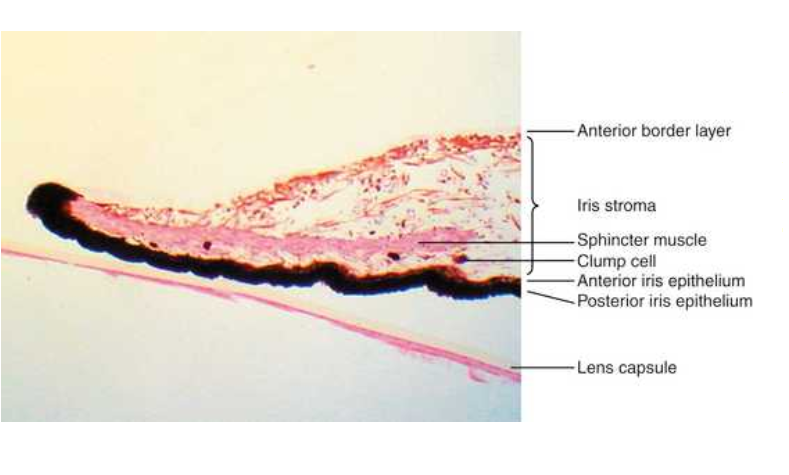
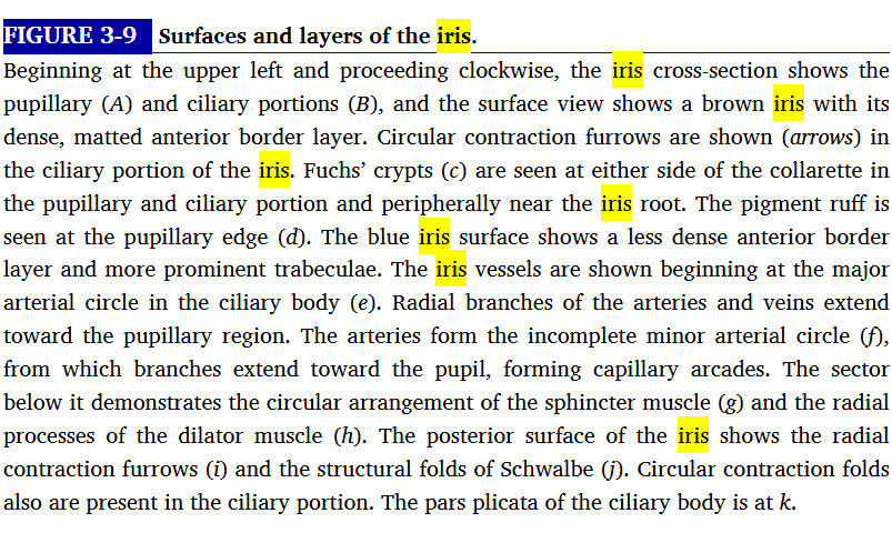
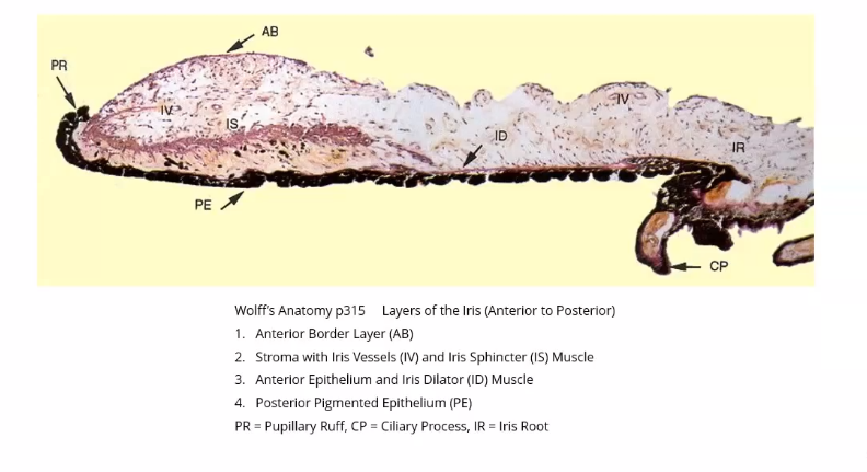
4 layers of the iris
anterior border layer
stroma and sphincter muscle
anterior epithelium and dilator muscle
posterior epithelium
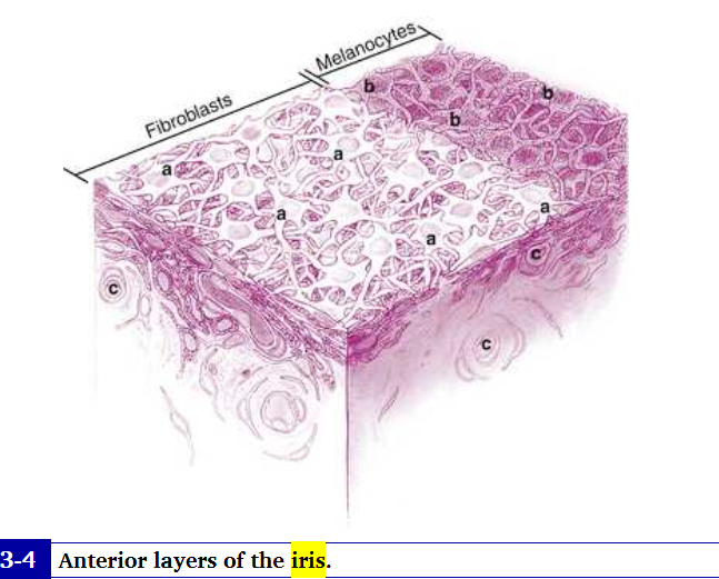
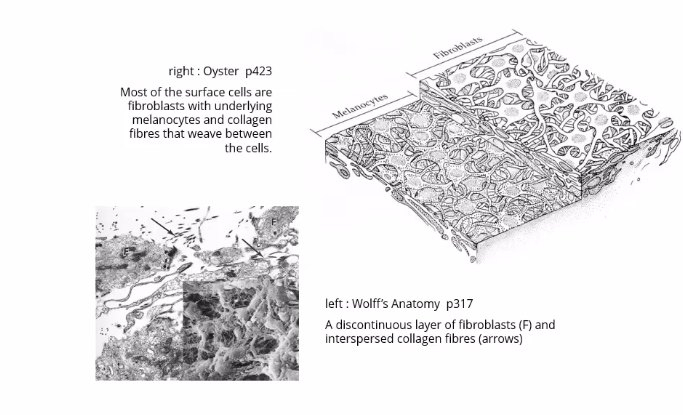
anterior border layer of iris
thin condenstaion of the stroma
forms strands from connective tissue
composed of fibroblasts and pigmented meloncytes. the highly branching porcesses of the cells interweave to form a meshwork in which the fibroblasts are on the surface and meloncytes are located below
loose network of connective tissue- needs to be loose as iris is muscular
density and arrangement of meshwork differ amongst irises and are factors in iris colour

a. fibroblasts - long branching processes which interconnect
b.superficial layer of fibrobtasts revoved
c. capillaries
where is anterior border of the iris absent at
absent at the oval shaped iris crypts
near the root, extensions of this layer form finger shaped iris porcesses that can attach to the trabecular meshwork.
iris stroma
connective tissue is composed of pigmented and non pigmented cells, collagen fibrils, and extensive ground substance
pigmented cells include meloncytes and clump cells
non pigmented cells are fibroblasts, lymphocytes , macrophages and mast cells
meloncytes and fibroblasts do not form a meshwork
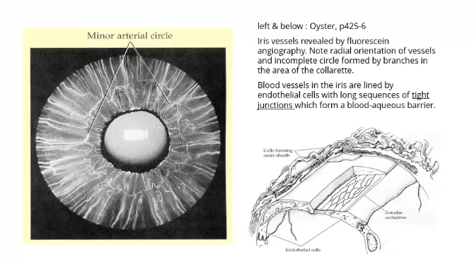
vessels run root to pupil margin forming arterial circle at collarette
have tight junctios
clump cells
large darkly pigmented cells and are likelu altered macrophages and are scavengers of free pigment within the iris. located in the pupillary portion of the stroma near phincter muscle
collagen fibrils arranged in radial columns that are seen as white fibres in light coloured irises
the iris arteries
branches of a circular vessel, the major circle of the iris.
the major circle of the iris is located in the ciliary body near the iris root
iris vessels usually follow a radial course from the iris root to the pupil margin
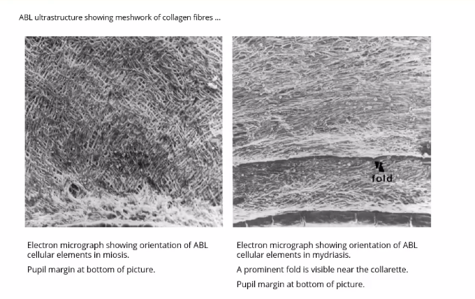
fibril network anchors the vessels in place and protects them from kinking and compression during estensive iris movement that occurs with miosis and mydriasis
the minor circle of the iris
an incomplete circle of the iris is located in the iris stroma inferior to the collarette
iris capillaries are not fenestrated and form part of the blood aqeous barrier
iris stroma is continours with stroma of ciliary body
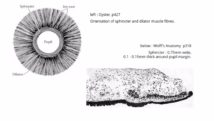
sphincter muscle
lies within the stroma and is composed of smooth muscle cells joined by tight junctions
circular muscle located in the pupillary zone of the stroma - closest to pupil
anchored firmly to adjacent stroma ad retains its function even if severed radially
contraction of sphincter causes the pupil to constrict in miosis- pupil gets smaller
muscle innervated by parasympathetic system
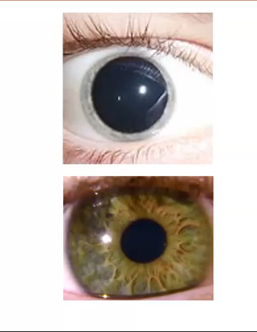
dilator muscle
dilator for pupil -mydriasis
radial muscle is the dilator muscle when dilator muscle contracts the pupil gets bigger
runs around the pupil outwards - runs underneath sphincter
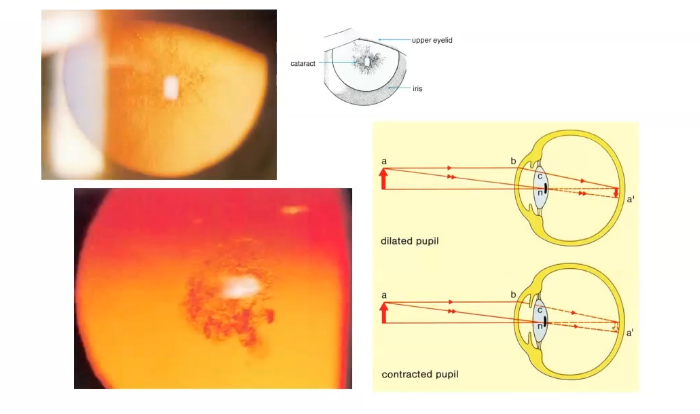
top image- constricted dilator, relaxed sphincter
bottom- contricted sphincter, relaxed dilated
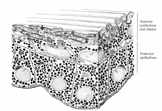
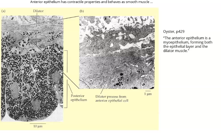
anterior epithelium of the iris
posterior to the stroma are two layers of epithelium; anterior iris epithelium and dilator muscle
anterior iris epithelium is composed of myoepithelial cell. the apical portion is pigmented cuboidal epithelium joined by tight junctions and desmosomes, whereas basal portion is composed of elongated , contractile smooth muscle processes
it is the same as dilator muscle -
anterior epithelial cells are flatter with lower density of pigment, long extesniosn which form strands of dilator muscle
anterior epithelium forms dilator muscle
dilator muscle present from iris root to a point in the stroma below the midpoint of sphincter
stroma separating sphincter and dilator is dense band of connective tissue
anterior iris eithelium continues to pupillary margin as cuboidal epithelial cells and anterior iris epith continues posteriorly as the pigmented epithelium of the ciliary body
posterior epithelium of iris
the second epithelial layer posterior to the stroma is posterior iris surface
single layer of heavily pigmented columnar cells joined by tight junctions and desmososomes
they rest on the anterior surface of the lens
in periphery the posterior iris ep begins to lose pigment as it continues into siliary body as the non pigmented epithelium
thin basement membrane covers basal aspect of this cellular layer which lines the posterior chamber
positioin of anterior and posterior iris epithelial layers
positioned apex to apex , result of embryonic development
apical microvilli extend from both surfaces and desmosomes join the 2 apical surfaces
the epithelial cells curl from posterior iris to anterior surface at pupillary margin forming the pigmented pupillary rudd , which encicles the pupil
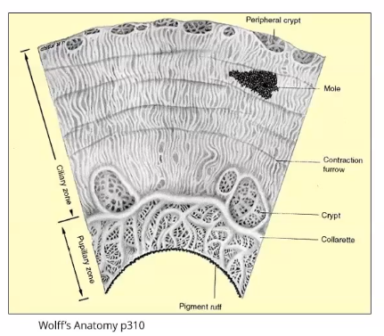
anterior iris surface
thin radial collagenous columns or trabeculae are evident in lightly pigmented irises
thicker, radially oreinted branching encircle depressions or openigns in the surface called crypts
they allow the aqueous quick exit into spaces in the iris stroma as volume of iris changes with contraction and dilation
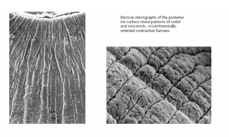
cirucular contraction folds
evident on anterior surface of ciliary zone
result from tissue moving forward toward the iris root during pupill dilation
posterior iris surface
smooth but small circular furros evident near the pupil
radial contraction furrows located in pupillary zone , and the deeper structural furros run throughout the ciliary zone and continue into the ciliary body
colour of the iris
made up of differenr areas of pigment
overall pigment of colour based on wavelengths of light hitting iris, some absorbed, some reflected
iris colour determined by meloncytes in stroma and anterior border layer
when iris is brown, meloncytes are well pigmented , longer wavelenghts are reflected bacl
in blue iris, wavelengths of light that are reflected back are shorter
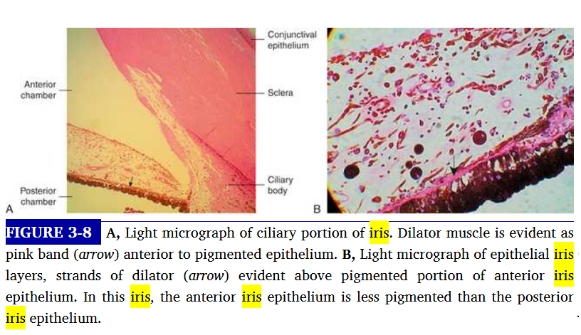
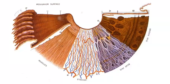
microscopic section of iris
blood vessels flowing through the muscle layers
iris regulates pupil size and keeps regulates light entering the eye
most anterior layer - anterior borfer layer
anterior epithelum- quite posterior
surface and layers of iris
right to left shows how each layers look brown iris ha slots of melalocytes
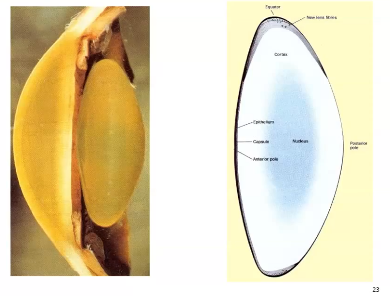
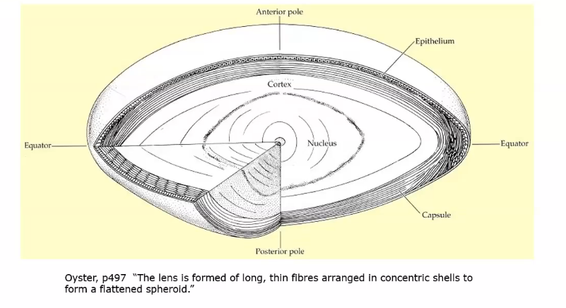
sections of the crystalline lens
posterior surface is more curved
curvature:
anterior radius = approx 10.50 mm
posterior radius= approx 6.00mm
centre thickness- 4mm approx
diameter approx 9mm
refractive idnes approx 1.43
purpose of the crystalline lens
avascular, transparent structure that aids in focusing light rays onto the retina
lens is located within posterior chamber, anterior yo the vitreous chamber and posterior to the iris
suspended from surrounding the ciliary body by zonular fibres
what can cause lens shape to change
its malleable, and the ciliary muscle contraction causes lens shape to change, increasing its dioptric power of the eye
the mechanism that causes increase in lens power is accomodation, which allows near objects to be focused on the retina
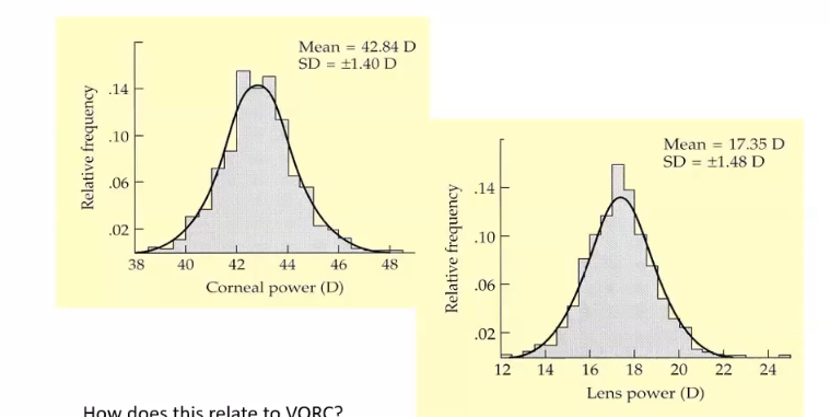
cornea and lens power
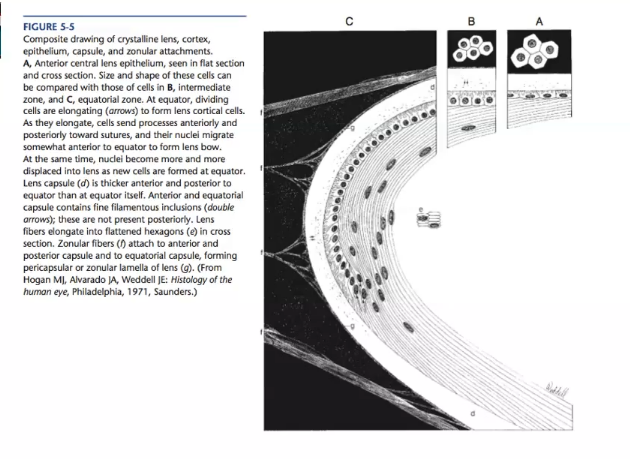
lens structure -external to internal
external capsule
anterior epithelium
equator : zonule (external) and formation of lens fibres ( internal)
cortex - young lens fibres
nucleus - old lens fibres
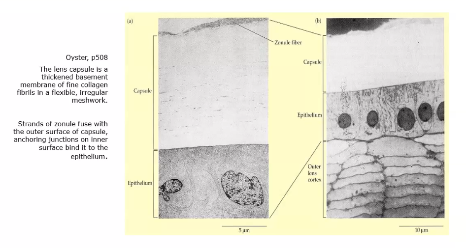
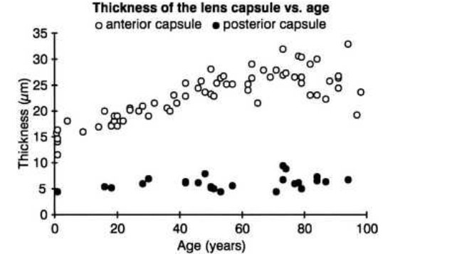
lens capsule
a transparent envelope that surrounds enitre lens
capsule is a basement membrane and with time, becomes the thickest in the body
at posterior pole its the thinnest
thickens at anterior pole increasing with age
capsule primarily collagen, has no elastic fibres high highly elastic due to lamellar arrangement of fibres
capsule shape
helps mold the shape of the lens
it prefers to take on a spherical shape but this tendency is counteracted by the pull from the zonular fibres, these fibres insert into capsule merging with it
lens capsule barrier function
prevents large molecules such as albumin and hemoglobin from entering the lens
anterior lens capsule is produced by anterior epithelium and thickens with age
posterior lens capsule may recieve contribution from basal membrane of lens fibres but thickeness of posterior capsule changes minimall y
more capsule functions
mould lens into a more convex shape in accom
insertion of zonular fibres which attac the lens to cilary body
ciliary body controls accomodation
as capsule gets thicker, it gets less easier to stretch causnig decrease in accom
thickest at equator - very top, very bottom, very nasal/temporal
where zonule attaches to the capsule
at the equator
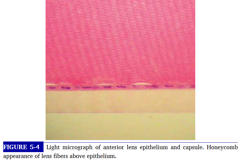
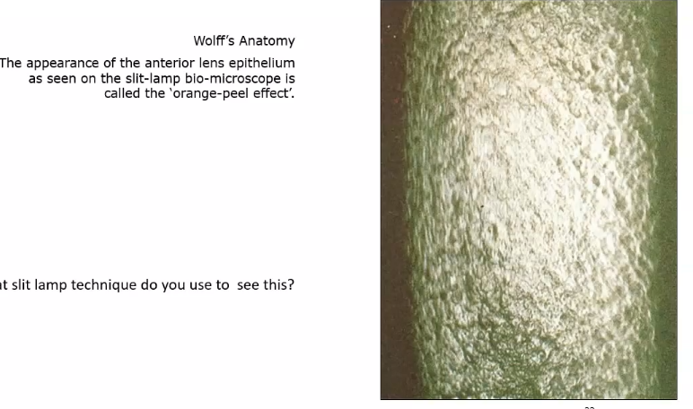
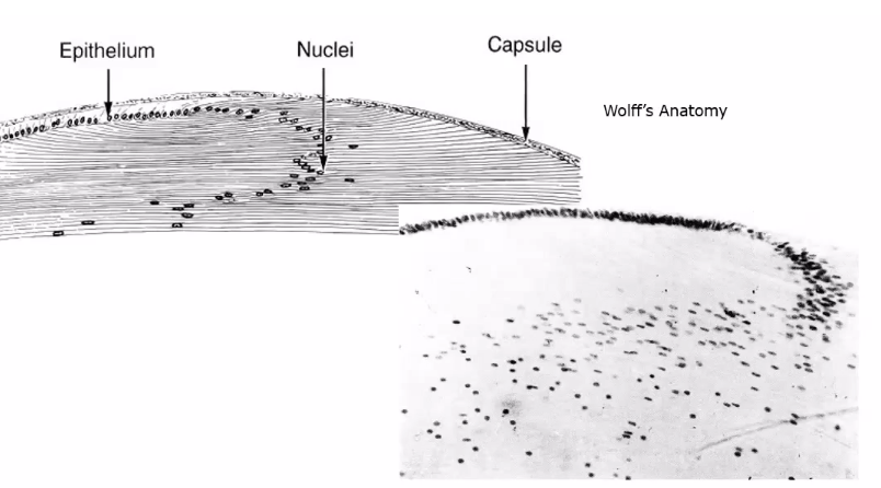
lens epithelium
adjacent to anterior lens capsule is a layer of cuboidal epithelium - anterior lens epithelium
these cells secrete the anterior capsule throughout life and are the site of metabolic mechanisms
they form the lens fibres - constantly elongating to become fibres
to see lens use the parallelepiped
why is there no posterior epithelium present
it was used during embryonic development to form the primary lens fibres
the lateral membranes of epithelial cells are joined by desmosomes and gap junctios
what is germinal zone of lens epithelium
the band of cells in the preequatorial region that lies anterior to the equator
is the location of cell mitosis
fibre formation
lens epithelium elongate and form fibres
epithelial cells have other functions like secretion of capsule, active transport of ions and water for lens transparency
transport of metabolites into the lens from the aq humour
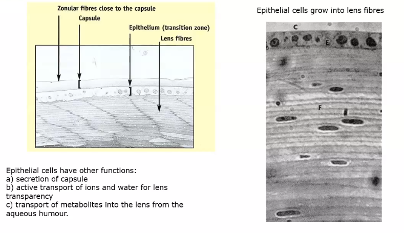
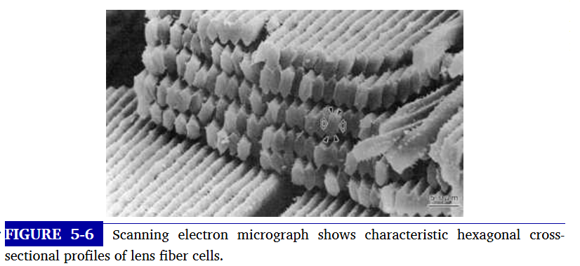
lens fibres
picture shows lens fibres afte elongation
growth results into concentric layers of secondary lens fibres
section through equator of les shows that fibres cut in cross section are hexagonal in shap and arranged in concentric rings
reason why transparent, is because theyre regularly arranged , and cannot change shape
what happens if lens fibres change shape
can form cataracts
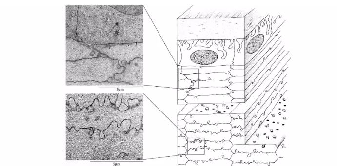
fibres are usually locked together with a ball and socket and tongue and groove formations
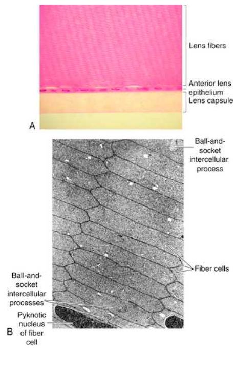
why do the fibres of lens lose their cellular organelles as they age
its because they have no vascular supply
theres an extensive network of gap junctions throughout the lens along the fibre to account for facility with which nutrients and ions move within the lens
gap junctions of lens fibres
have different packing arrangement and different protein connexins , forming the channel
gap junctions are not evenly distributed throughout the lens near the poles, more toward equator
epithelium- fibre interface
border between apical membrane of anterior epithelium and apical mem of elongating fibre known as epithelium fibre interface
nutrients and ions exchange across the EFI.
which iris strucrure is most posterior
anterior epithelium
what is this called
crypt of fuchs
cilliary zone

insertion of lens zonule
they are attached at equator to the capsule.
inbetween cillary body and lens
formed of extracellular matrix that includes fibrillin and elastin
fibres arise from basement embrane of non pigmented ciliary epithelium
zonules are interwoven into components of the capsule
those that attach to the lens are called primary zonles, secondary join the primary zonules with eachother
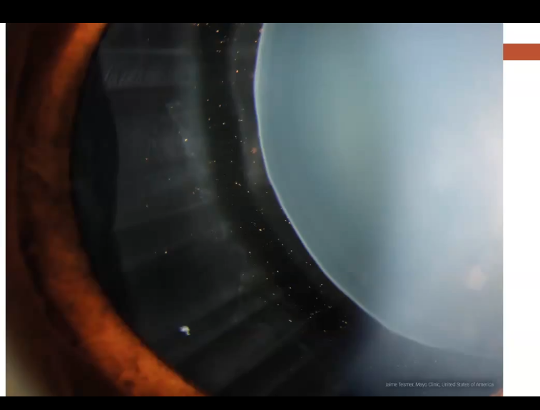
zonules on slit lamp
shows zonule with pigment dispersion
lens divisions
cortex is constantly growing, epithelium turn into cortical cells
nucleus stays same size
nucleus is subdivided - does grow from embryonic stage
growth of lens nucleus
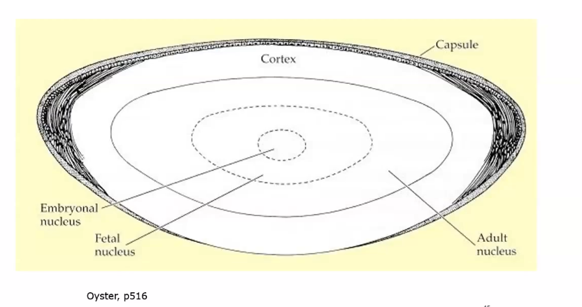
embryonic lens
the primary lens fibres from elongating posterior epithelium form centre of the lens, embryonic nucleus
cell mitosis begins in preequatorial region of epithelium, new cell migrates toward equator and then elongates forming lens fibre
all fibres formed are secondary lens fibres
fetal nucleus includes embryonic nucleus and fibres surrounding it that are formed before birth
regions of adult lens
includes the embryonic and fetal nuclei and the fibres formed from birth to sexual maturation
lens cortex contains fibres formed after sexual maturation
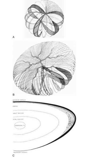
A. fetal nucleus : a is anterior suture b is posterior
B. adult lens cortex
C. adult lens
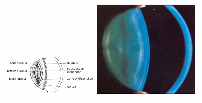
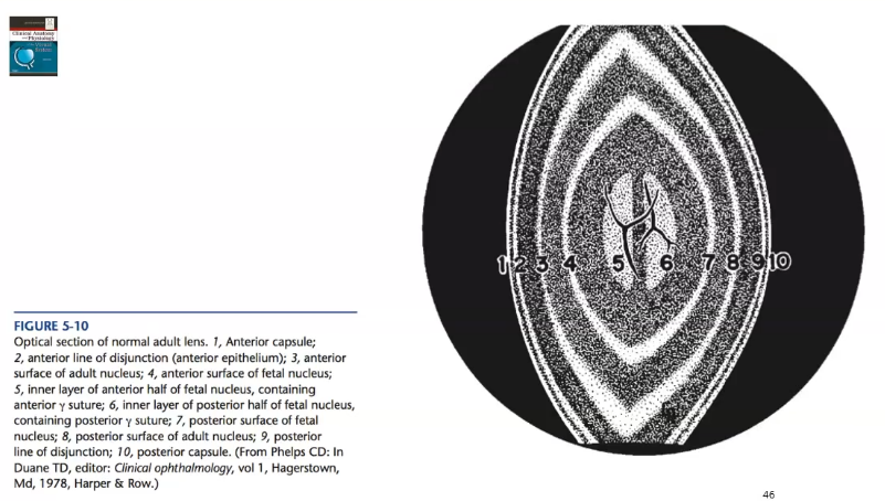
lens fibre zones in vivo
seen using the optic section
light coming from right hand side
lens sutures
as the lens fibres reach the poles they meet with the other fibres in their layer, forming a junction known as a suture
the secondary fibres formed during embryonic meet in 3 branches , forming Y sutures
how is anterior and posterior suture formed
by joining of the apical aspects of the fibres- anterior is an upright Y shape
posterior formed by joining of basal aspects - inverted Y shape
as growth continues and lens becomes larger, the sutures become asymmetric so get less transparent
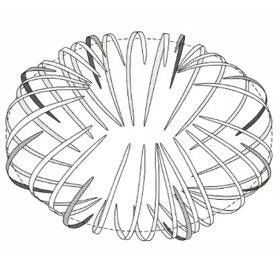
lens fires forming new shells
the fibres elogate around the equator
form a belt around equatory of the lens
this prodces a complete shell of fibres
the anterior and posterior ends of each fibre are attached to sutures near the lens surface
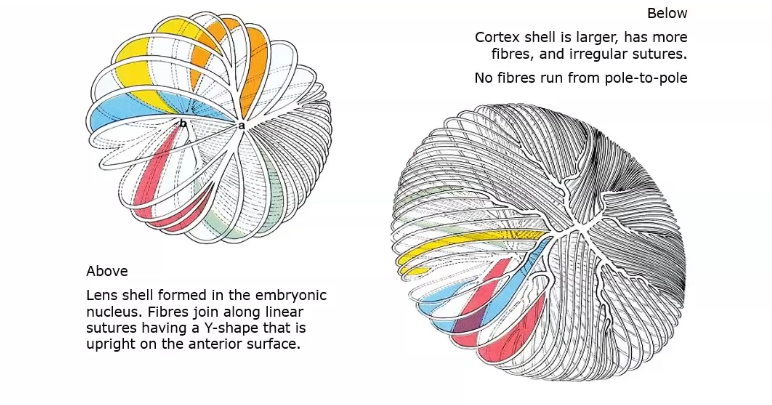
lens nucleus and cortex sutures
when the shell is larger it has more fibres and irregular sutures. no fibres run from pole to pole
all fibres meet in the suture
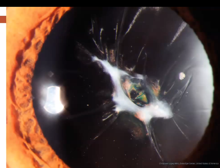
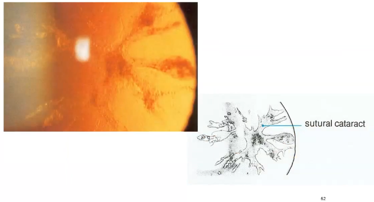
sutural cataract
lens fibres dont meet together neatly so get opacification
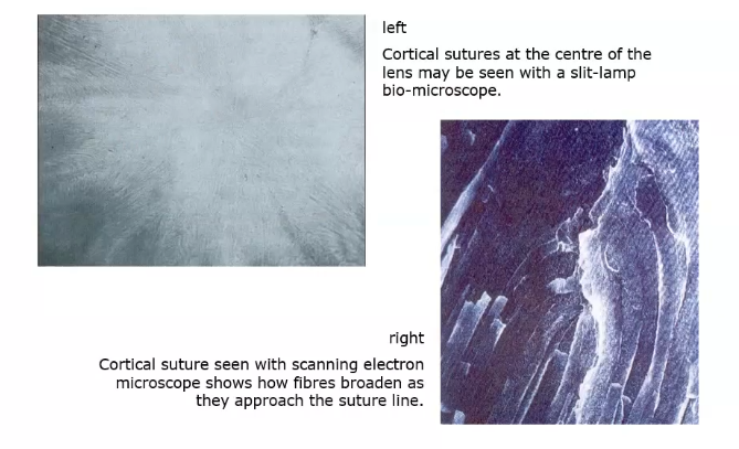
crotex sutures under microscope
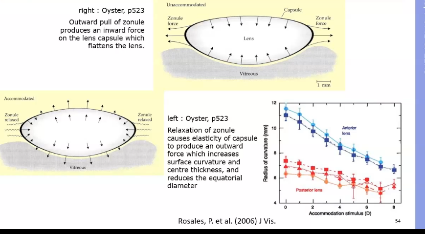
lens- accomodation
unaccomodated state- zonules are tight , pulling on the lens. this flattens the lens
accomodated- ciliary body becomes smaller and contracts, zonules relax. causes elasticity of capsule to produce outward force increasing curvature and centre thickness . anterior lens becomes more convex
what happes during accomodation
lens shape changes by contracting ciliary muscle - increases power when looking at something that is not in the distance
les thickens increasing anterior to posterior
lens thins along the equator
anterior lens surface moves forward and anterior chamber becomes shallower
posterior pole remains in same position
lens shape in accomodation
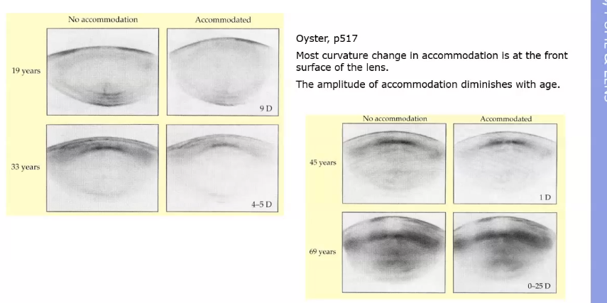
changes as you get older
younger can accomodating more as front surface of lens is becoming a lot more convex
viteeous role in accomodation - when cilary muscle contracts
when ciliary muscle contracts, the choroid is pulled forward slightly,
contraction of ciliary body by decreasing cicumference of sclera, may lead to an elongation of axial length of eye
accom can cause a widening of intertrabecular spaces, causing aq outflow and result in a decrease of IOP
Vitreous role in accom- when ciliary muscles are relaxed
muscle is moved outward, and ciliary body is stretched posteriorly by elastic tissue of Bruchs membrane
ciliary ring expands and tension of zonules stretches capsule , restoring lens to its unaccomodated state
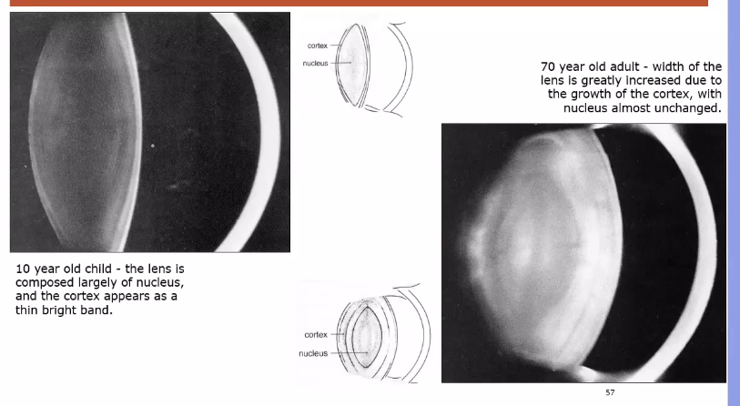
growth of lens
lens is constantly growths.
cortex and capsule become thicker
in adult the cortex is thicker - reason why it restricts accomodation
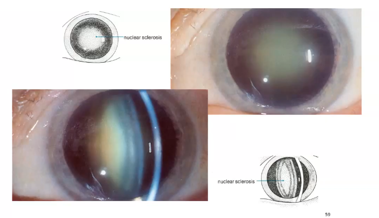
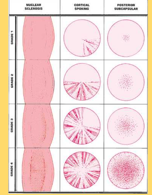
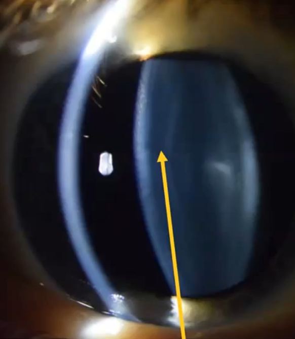
types of cataract
most common is nuclear cataract
cortical cataracts- affect cortex
age related with nuclear cataract
nucleus has opacified
an opacity located in the embryonic, fetal or adult nucleus is called nuclear cataract
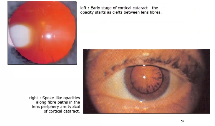
cortical cataract
located in cortex, thicker in periphery and tapering towards the lens centre , it follows the shape of the fibre
fluid accumulatesand membrane rupture in equator can occur
only affect vision when it spreads to centre of the lens
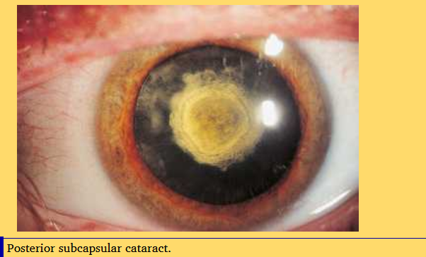
sub- capsular cataract- shown on retro illumination of lens from retina
in the middle at the back
located beneath posterior capsule
impacts vision early and significantly along visual axis
risk factor is high dose steriod use
sutural cataract
affects space where lens fibres are supposed to meet
grading cataracts
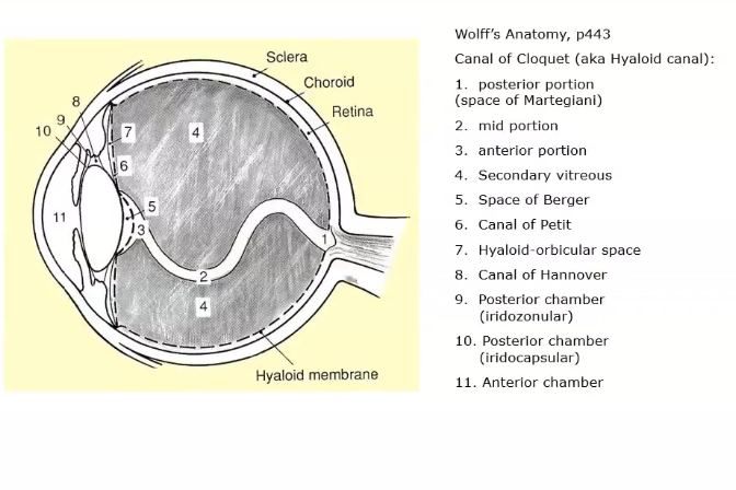
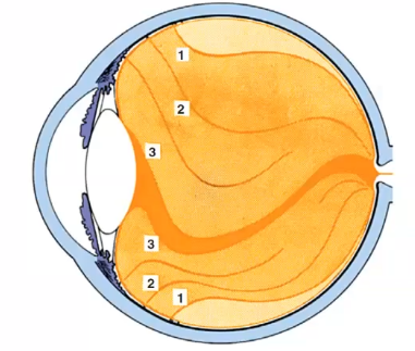
vitreous chamber
filled with the gel like vitrous body and occupies the largest prtion of the globe
all surfaces that interface with vitrous are basemement membrane
helps maintain shape of eye and keeps retina on the inside surface of eye
canal running from anterior to posterior there for embryology
some liquid bits some solid
attachments of vitreous
vitrous is attached to the inside surface of eyeball both anteriorly and posteriorly
attachment of vit at the macula- macula is centre of retina posteriorly
also attached to optic disc posteriorly
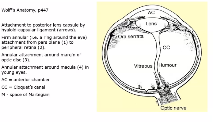
relations of the vitreous
vit base is attached to pars plana
ora serrata - where retina finishes
these are anterior attachmets of vitreous
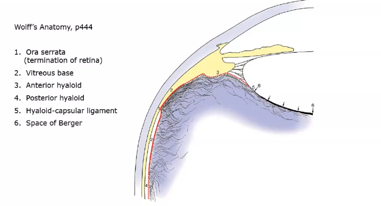
divisions of vitreous space
pre retinal tract : separates tge vit cortex from intermediate substance
median and coronary tracts : running to median and coronary ligaments of the pars plana
hyaloid tract: seperates intermediate substance from central channel
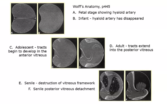
growth and ageing of the vitreous
gradually becomes more liquid as collagen fibres break down
when it gets too liquidly it can detach from retina which can result to a retinal detachment
whats this
anterior cortex
what does the lens epithelial do
secretes capsule
active transport of ions
transport metabolites
elongates to become cortical cells