Human Bio Ch. 2
1/58
There's no tags or description
Looks like no tags are added yet.
Name | Mastery | Learn | Test | Matching | Spaced | Call with Kai |
|---|
No analytics yet
Send a link to your students to track their progress
59 Terms
Broad Ligament
Double layer of peritoneum that supports uterus, uterine tubes, and ovaries
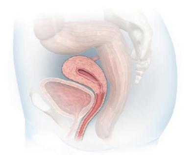
Anteverted
Uterus position for 75% of females
Retroverted
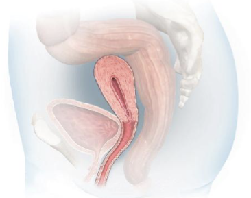
Anteflexed
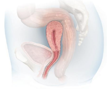
Retroflexed
Oogonia
Starting form for oocytes; peak at ~7 million by 5th month of gestation
Primary Oocytes
Form after oogonia begin meiosis I (end of 7th month), then arrest in prophase I
Atresia
Natura oocyte degeneration before birth/throughout life; reduces to ~1 million eggs at birth
Ovarian Follicles
Oocyte + supportive cells (theca and granulosa cells)
Theca and Granulosa Cells
Produces estrogen
Avg. number of ovulations in a lifetime
450
Follicle Stimulating Hormone (FSH)
Stimulates cohort of primordial follicles to start maturation every cycle
Graafian Follicle
The only follicle that fully matures and ovulates after FSH
Luteinizing Hormone (LH)
LH surge triggers ovulation
Corpus Luteum
Ruptured follicle after ovulation; secretes progesterone/estrogen to maintain uterine lining for implantation
Menstruation Trigger
Corpus luteum degenerates → progesterone/estrogen drop → becomes corpus albicans
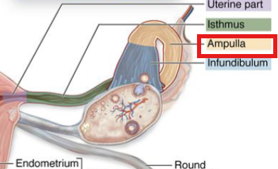
Fimbriae
Finger projections on fallopian tube that capture secondary oocyte after ovulation
Where fertilization occurs
Distal 1/3 of tube (ampulla)
Zygote
Fertilized ovum (mature egg) with 2n = 46 chromosomes
Oogenesis
Process of forming ova; begins before birth until menopause
Ova
Female gametes that are surrounded by follicle cells that release estrogen
Viability Period of Released Secondary Oocyte
12-24 hours
Fertilization
Completes meiosis II and begins cleavage; fertilized ovum divides and conserves 23 pairs of chromosomes
Meiosis I
Homologous chromosomes separate → 2 haploid cells with duplicated chromosomes
Meiosis II
Sister chromosomes separate → 4 total genetically unique haploid cells
Polar Bodies
Non-functional extra cells from 1st meiotic division and 2nd meiotic division (fertilization)
Metaphase II Arrest
Preserves secondary oocyte for ovulation
Menstrual Cycle Pathway
GnRH (hypothalamus) → stimulates ant. pituitary → AP release FSH and LH → FSH triggers follicle development → estrogen and LH surge trigger ovulation → corpus luteum secretes progesterone and estrogen
Endometrium during Menstrual Cycle
Menstrual phase → proliferative phase → secretory phase
Menstrual Phase
Progesterone/estrogen drops → spiral arteries constrict → outer functional layer sheds
Proliferative Phase
Estrogen signal → functional layer starts regenerating → glands are straight and narrow
Secretory Phase
Functional layer thickens → coiled uterine glands secrete glycogen-rich fluid → maintained by progesterone
Follicular Phase
Estrogen levels rise → fallopian tubes move closer to ovary, fimbriae beat faster, ciliated cells increase in fimbriae → egg capture success increase
Egg vs Sperm
Egg is larger with more mitochondria, while sperm can survive for up to 5 days in female reproductive tract
Acrosomal Reaction
Sperm head (acrosome) contains enzymes that digest zona pellucida
Cortical Reaction
Cortical granules inside egg release after fertilization and harden zona pellucida
Zona Pellucida
Egg’s outer glycoprotein layer; changes electrical charge or hardens after sperm contact
Post Menopause Changes
Estrogen drops → increases osteoporosis risk
Zygote Development
Cleavage → morula → early blastocyst → hatching blastocyst → implantation
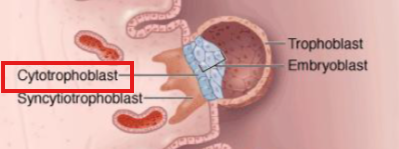
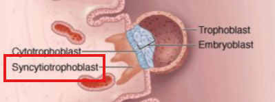
Trophoblast
Outer layer of implanted blastocyst; differentiates into cytotrophoblast and syncytiotrophoblast
Cytotrophoblast
Maintains implanted blastocyst cellular structure
Syncytiotrophoblast
Secretes enzymes to anchor blastocyst to uterine tissue
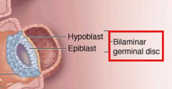
Bilaminar Germinal Disc
Made from hypoblast and epiblast; later develops into germ layers
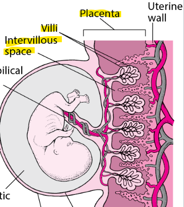
Placenta
Made from maternal tissue and outer layer of trophoblast (chorion); site of nutrient, gas, and waste exchange
Immune System Rejection Prevention
Trophoblast secretes suppression enzymes and lacks surface proteins needed to show antigens
Placenta Hormone Production
Replaces progesterone/estrogen production by corpus luteum after fully forming by end of 1st trimester
Placenta Structure
Villi (anchor), spiral arteries, intervillous space
Amniotic Sac and Fluid
Strong membrane containing fluid for free movement and cushioning
Preeclampsia
Abnormal placental development that causes hypertension and organ damage (kidney and liver)
Ectopic Pregnancy
Implantation occurs outside the uterus (tubal, interstitial, ovarian, cervical), resulting in non-viable and potentially fatal pregnancy
Abdominal Pregnancy and Lithopedion
Implantation occurs in peritoneum, ovary, or intestines; fetus can calcify into “stone baby”
Germinal Period
Weeks 0-2; from fertilization to embryo establishment in uterus
Embryonic Period
Weeks 3-8; critical period containing organogenesis and formation of neural tube, heart, limbs, etc.
Fetal Period
Weeks 9-birth; growth/refinement of organs and rise in viability after ~24 weeks
Primitive Streak
Forms around week 3; forms in epiblast and where epiblast cells migrate through during gastrulation
Trilaminar Embryonic Disc
Formed after gastrulation; contains endoderm, mesoderm, and ectoderm
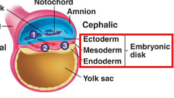
Endoderm
Inner layer → epithelial lining of digestive and respiratory organs, glands
Mesoderm
Middle layer → muscle, bone, connective tissue, blood, kidneys, gonads
Ectoderm
Outer layer → skin, nervous system