Integment SDL
 Call Kai
 Learn
Learn Practice Test
 Spaced Repetition
 Match
Match Flashcards
 Knowt Play
1/14

There's no tags or description
Looks like no tags are added yet.
Name | Mastery | Learn | Test | Matching | Spaced | Call with Kai |
|---|
No analytics yet
Send a link to your students to track their progress
15 Terms

Integument System (your skin)
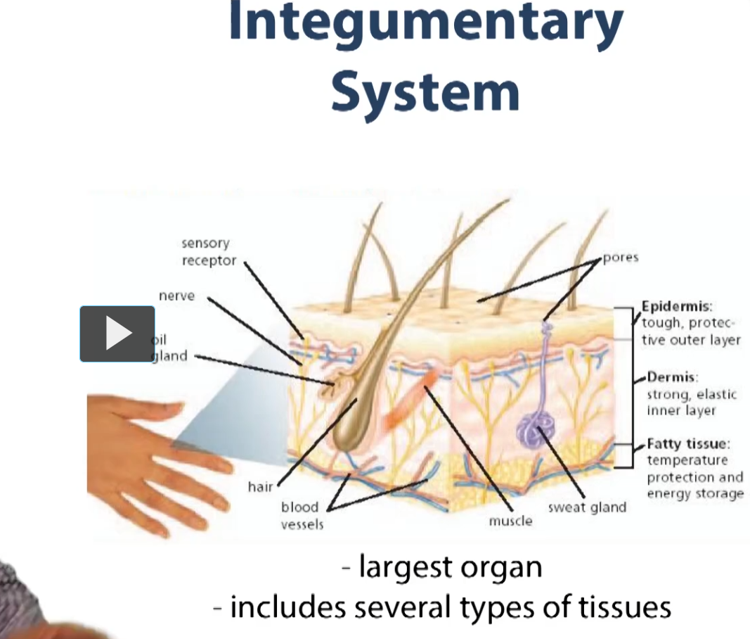
This is a simple schematic presentation of the integumentary system. First of all, what you see is the integumentary system is an organ system. This means we are going to be combing basic tissues that we have already seen and putting them together. And when you combine more than two or more basic tissues, you create an organ.
So, for the skin, which is the largest organ in the body, it includes various types of tissues. So we’re seeing here an epithelium. We’re seeing a connective tissue. We’re seeing specialized connective tissue in the way of adipose tissue, and we also have various nervous system tissues in there as well.
Combined, you’ll see their arrangements as we go through the lecture. They create our organ, the largest organ in the body or one of the largest organs in the body, the skin.
The outer epidermal layer, which is mostly epithelium is tough, and is going to provide our outer protective layer. It’s the first barrier of defense for our body against the environment.
Underneath, we have a dermis, which is mostly connective tissue, which is going to help provide structural support for our epithelial layer sop that it can provide a lot of structural rigidity, and then we see we have other structures, some are epithelial in origin, such as our glands (sweat glands) and sebacious glands, which have various functions such as lubrication and thermoregulation.
We have fatty tissues which also help with temperature protection and energy storage. And, we have some muscle associated with tissue which pulls on the hairs of our skin which allows for that feeling of hair standing on your arms sometimes.
We also have some elements of vasculature and a lot of nervous innervation of the skin, as it is the largest sensory organ in the body.
So, we are going to be looking at most of these features throughout the lectures. Like I said, we are talking now about multiple basic tissues combined to carry out a unified function.
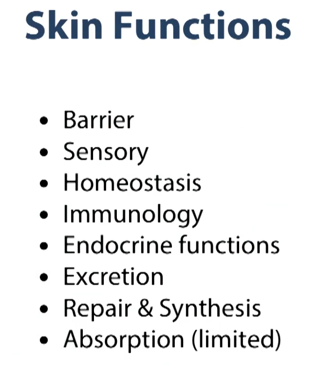
Some of the functions that’s given performs are listed in this slide. So, it’s a barrier, like I said, it’s the first line of defense against our external environment.
Sensory, thanks to the tremendous amount of nervous innervation that is all throughout the skin
homeostasis: skin helps preserve and regulate some basic functions in our body.
Immunology: there’s a lot of activity from the immune system, again, monitoring the very top layers against any foreign pathogens.
Endocrine functions: There are some portions of the skin that carry out endocrine functions
Excretion: via some of the gland
repair and synthesis: so our skin, as you’ll see, has some certain layers that are regenerative.
Absorption: performed by the skin, but done to a lesser degree. carried out by some of our epidermal or integumentary system.
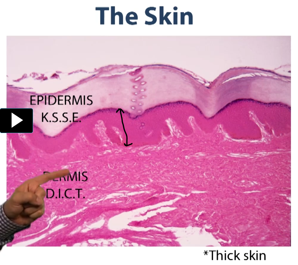
This is an H&E stain section here. Cross section through skin. So you can see that there are various layers.
This is thick skin and we’ll show you how to differentiate thick skin from thin skin. But for the most part, you can see that there is this large layer of dead cells (epidermis) and there is no nuclei or anything associated with these cells. This is the top layer (epidermis).
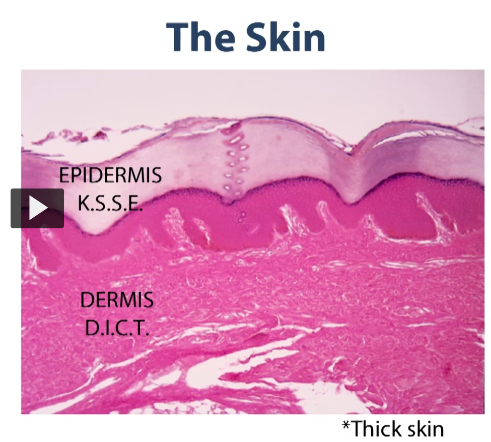
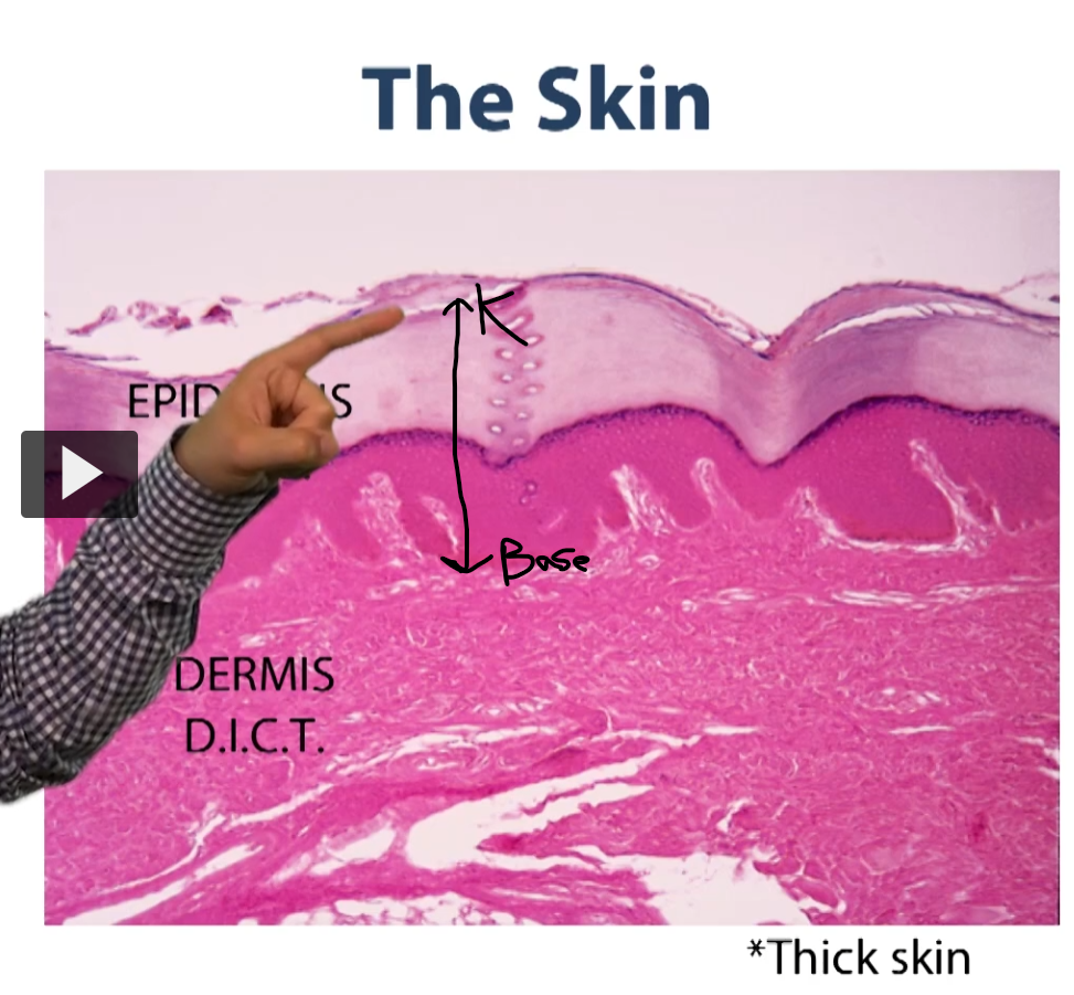
The epithelium of the integumentary system runs from the keratinized layer (K) all the way down to the base of this epithelium.
This is what we classify as a stratified squamous epithelium with keratin.
Notice that all epithelium sit on top of a connective tissue bed, and so all of this is connective tissue in their various layers to this connective tissue, and we will talk about them later.
We have a layer of loose connective tissue immediately under the skin, and we have a layer of dense irregular connective tissue
layer of loose connective tissue immediately under the skin
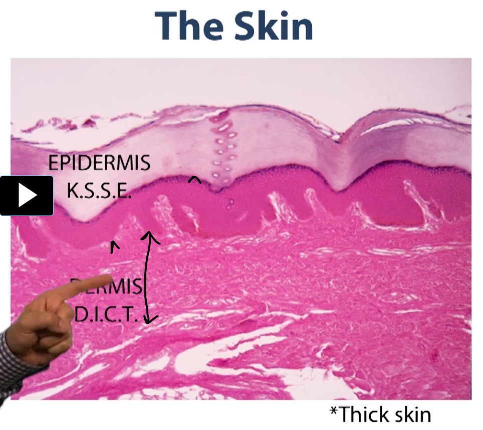
layer of dense irregular connective tissue. Together, these two layers (layer of loose connective tissue and layer of dense irregular connective tissue makeup the dermis).
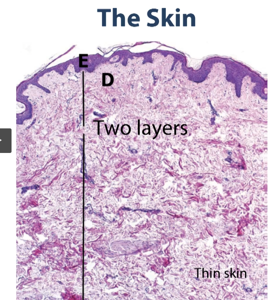
This is thin skin. Here, again, you’ll see those two layers. You’ll see our epithelial layer or epidermal layer (E). The epidermal layer is still a stratified squamous epithelium, but the keratin layer much smaller and thin than it is in thick skin.
Again, we have our dermis, which is made up of two layers, layer of loose connective tissue (under the epidermis) and a rather large layer of dense irregular connective tissue (under the layer of loose connective tissue).
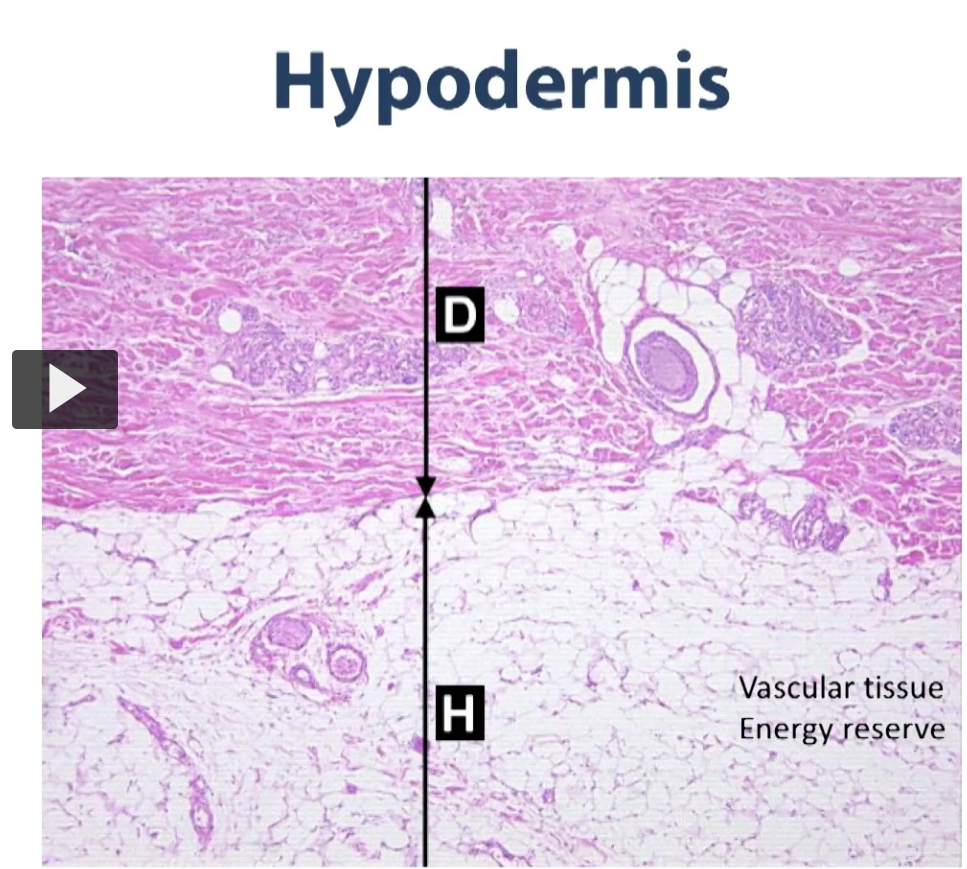
As you go down deep into the dermis, (the darker pink is the dense irregular connective tissue), as we get even deeper down (going towards H), you see that there is yet another transition, as we go from a dense irregular connective tissue to the hypodermis (superficial fascia) (light pink), and this is the layer that also has some connective tissue but it’s also rich in adipose tissue.
So those are the layers of the integumentary system.
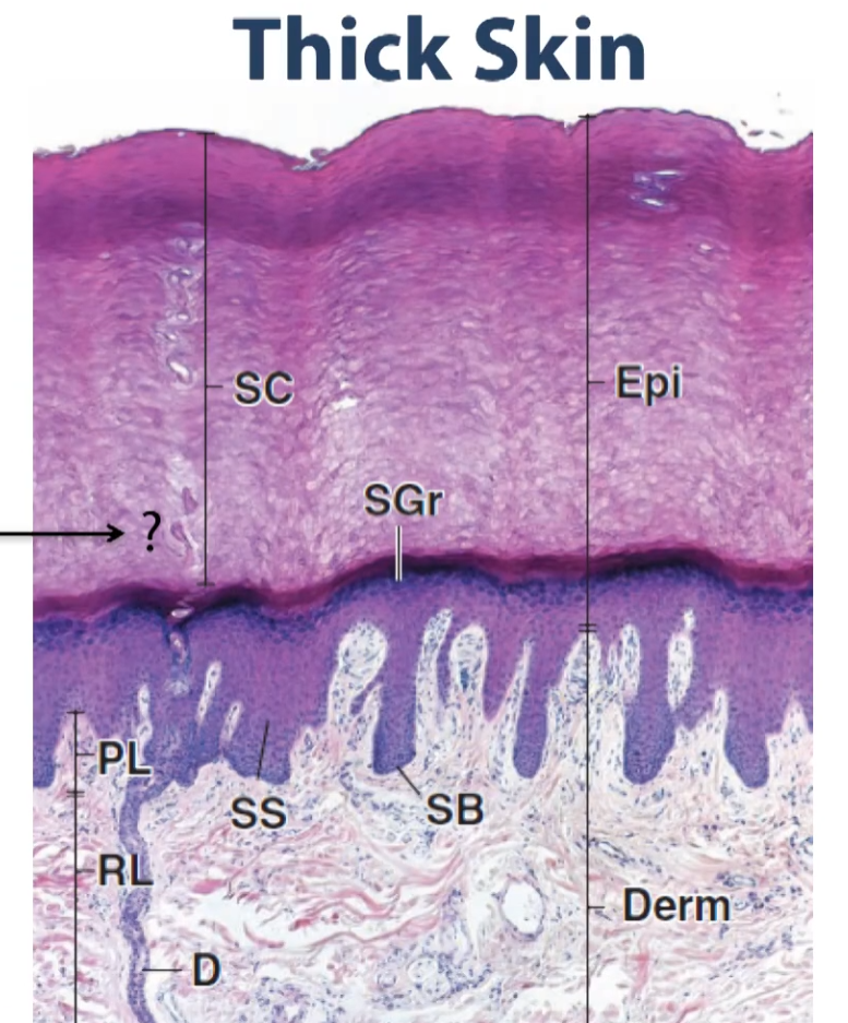
So we have the thick skin again to illustrate all that together. So we have our epidermal layer, and our epidermal layer is divided into multiple sub-layers.
We have our coordified layer (stratum corneum),
then Stratum granulosum (granular layer)
Stratum spinosum (SS, spiny cell layer)
Then the layer that is closet to the basement membrane and the connective tissue, it’s a single layer of cells known as the basal layer or the stratum basale.
This being thick skin, we have an extra layer within the keratinized portion right, above the stratum granulosum, which we call the lucid layer (stratum lucidum) (dark pink over the stratum granulosum), and we will expand on this a little bit later during the lecture.
As you can see then, below the basal layer of the stratum basale, what we have is the dermis, and the dermis is subdivided into two layers, the superficial dermis (the one that is immediately under our skin) and the dense irregular connective tissue.
The layer of the dermis that is loose connective tissue is called the papillary layer, and the layer of the dermis that is dense, irregular connective tissue is termed reticular layer of the dermis.
So those are all the layers that make up the integument in this cross section. So we are going to talk about all the features of all these layers and the specialized cells contained within.
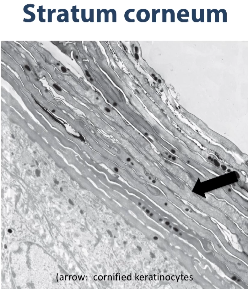
So here we have an electron micrograph of the superficial most layer of the stratified keratinized squamous epithelium.
You can see the corneal layer in this electron micrograph. Notice what we have here are the remnants of the cells that were generated below on this epithelium (the epithelium is the lighter gray towards the bottom of the photo).
As they mature, the cells move up in the epithelium (into the darker grey) and at some point, as you see, they shed their organelles, having made a lot of this intermediate filament protein known as keratin and they become the superficial most layer of our skin offering a lot of protection.
Keratin is a waxy protein that offers some protection against water loss and against foreign pathogens, so that’s what you are seeing in this image.
The dark granules are the remnants up some pigment that are in these empty bags of cells.
so, if we move down from the keratinized layer, the next layer that we would see below would be the granular layer or the stratum granulosa. The stratum granulosum contains these granules, that are called keratohyalin granules, and the material in these granules are exocytosed and it’s this lipid like substance that creates a barrier against moisture adding and moisture prevent and moisture loss within the skin. Those granules, synthesized by these cells, give these layers
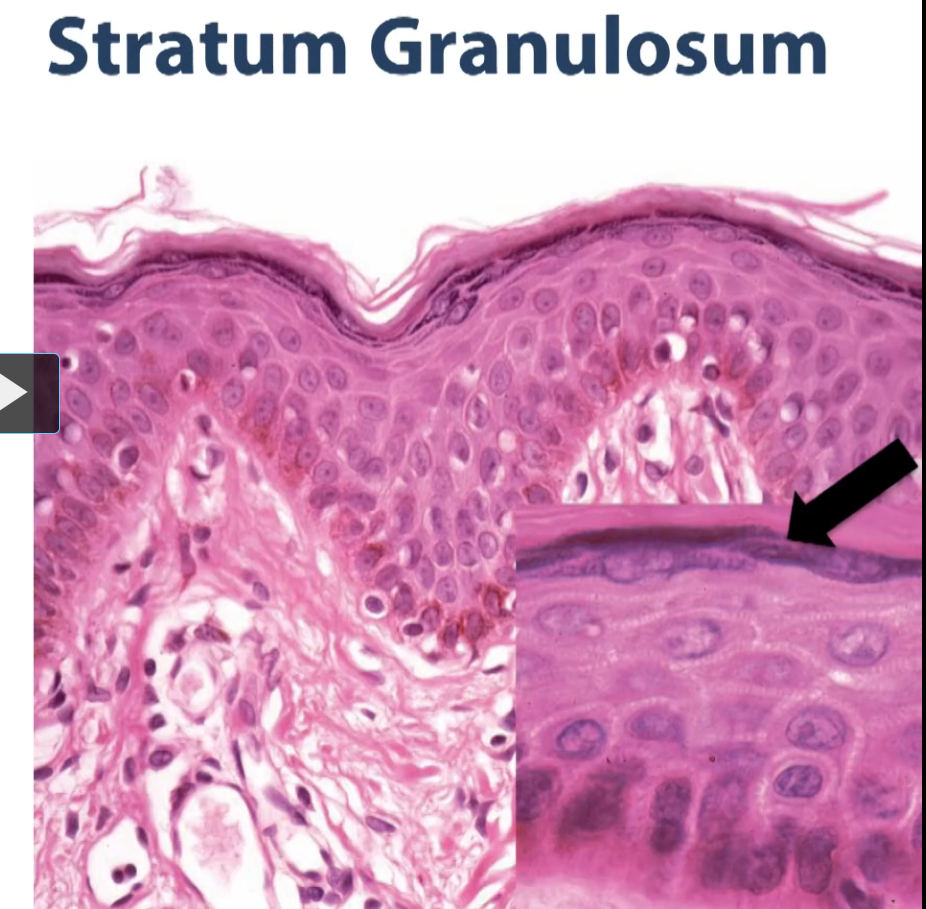
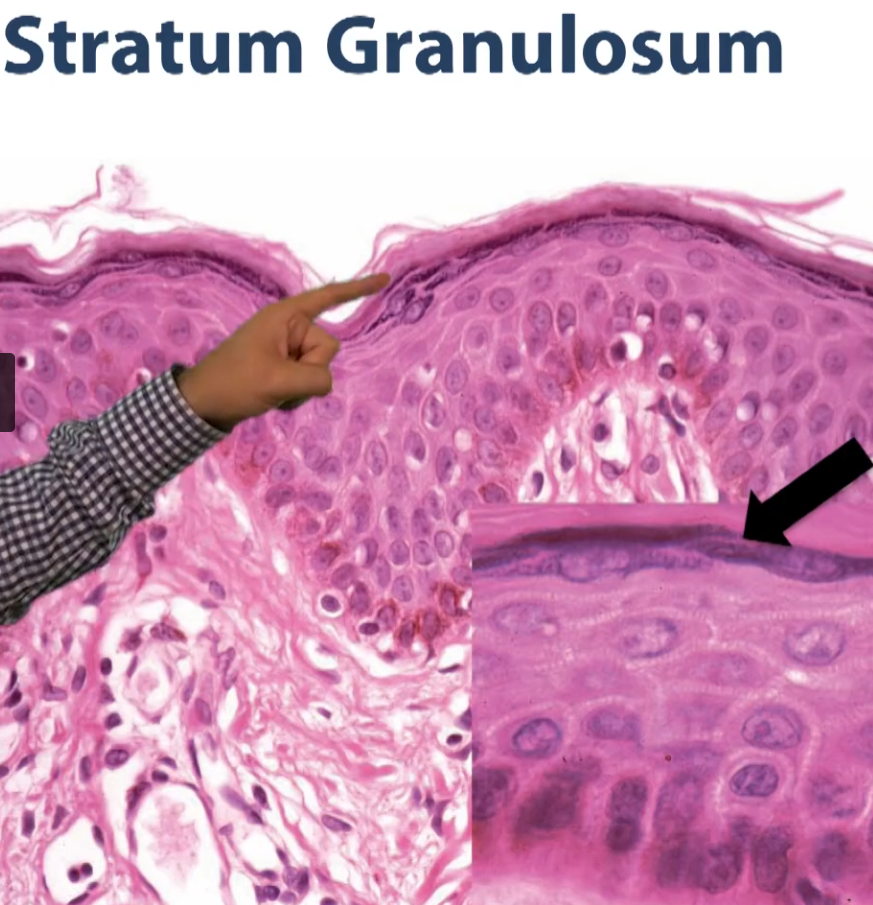
Those granules, synthesized by these cells, give these layers (the one he is pointing to) the stratum corneum this granulated appearance, therefore, it gets the name stratum granulosum.
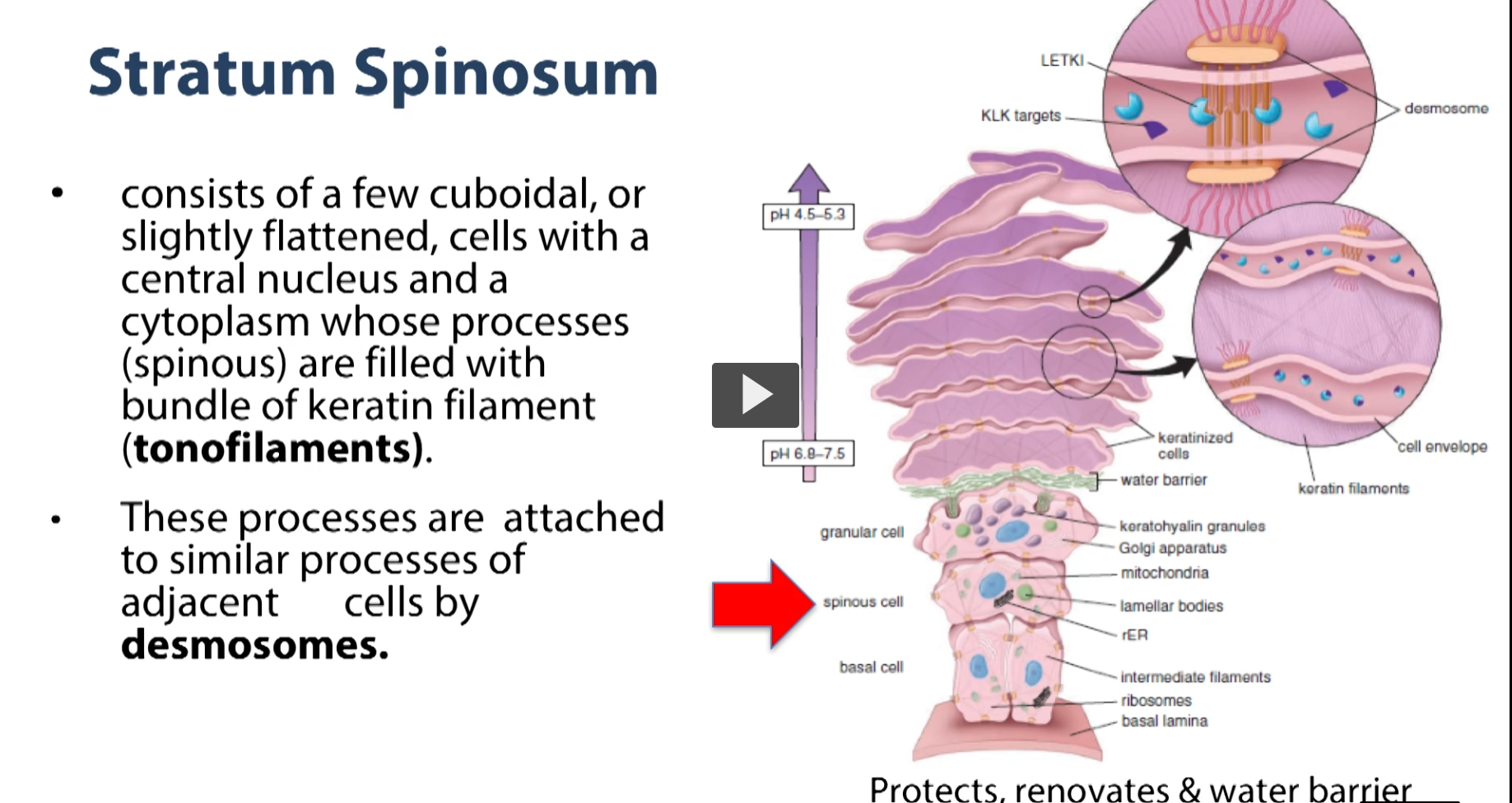
Below the stratus granulosum, we have a layer of cells called the spinous layer. Notice that from the side of each of these cells you see this tiny little spiny processes coming out. What you are seeing here is cytoplasmic extensions from these cells that are contacting similar extensions from neighboring cells, and they are being bound together. They are joining at these spiny processes to one another by way of desmosomes, and they are using the protein keratin to bind adjacent cells to one another.
Then the empty spaces where you see here, where there’s white, what happens is, some of that material, some of those lipids that are being exocytosed by the stratus granulosum, they drip (they not only form a barrier above the granulosum) but they also drip in between these cells and they fill the spaces in-between these cells to provide an extra barrier against moisture loss or moisture gain in our epidermis.
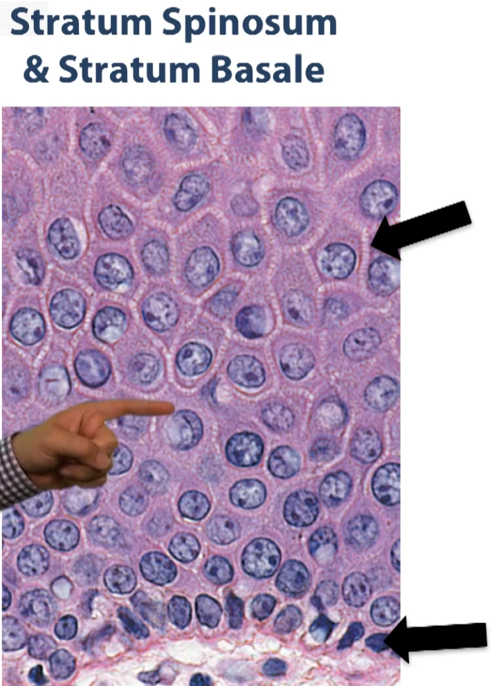
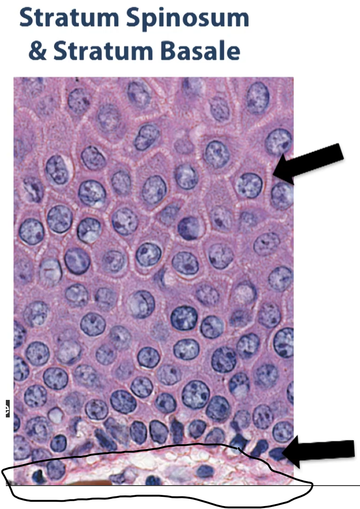
The bottom most layer that you are seeing here, close to the basement membrane, is our basal layer, or the stratum basale.
These are the cells that are responsible for the mitotic activity that replenishes or creates all these cells on the upper surface of that epithelium (he points to the violet cells in the purple).
So there is a lot of mitotic activity going on here. Our pigmented cells are are also located here. So there is a lot of activity going on in this layer (stratum basale).
Then the next layer would be your basement membrane, and underneath would be your papillary dermis.
So, together, there is your spot, your stratum spinosum has some mitotic activity, so together, the stratum spinosum and the basalis is referred to as the germinal layers of the integument or the stratum germinativum.
So those are the layers of the epidermis.
Epidermal cell renewal: the epidermis in humans is renewed about every 15 to 30 days. That rate is going to depend on age and part of the body that we are looking at, as well as some other factors.
The cells of the epidermis consists four different cell types:
Keratinocytes are the majority of the cell (Keratinocytes are the cells responsible for making the intermediate filaments keratin, which is found inside all these cells). In addition
Melanocytes:
Langerhans
Merkel cells
So these are the cells of the epidermis.
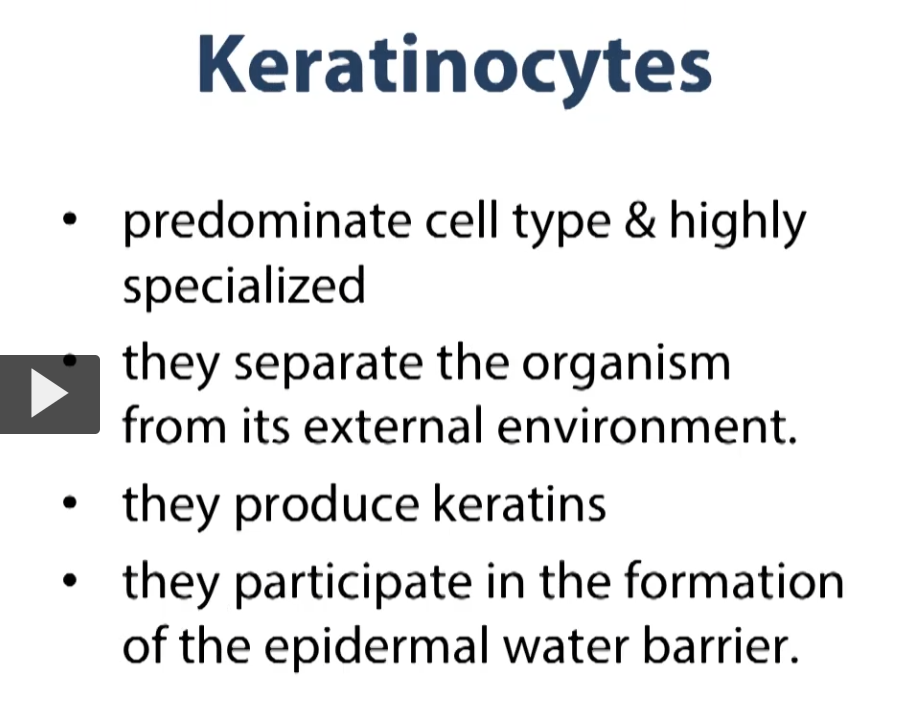
Keratinocytes are a predominant cell type. It is highly specialized, their job is to isolate us from the environment.
The main function of keratinocytes is to produce keratin. These cells synthesize keratin. The keratin is going to ultimately create a water barrier separating out epidermis and protecting us from the environment
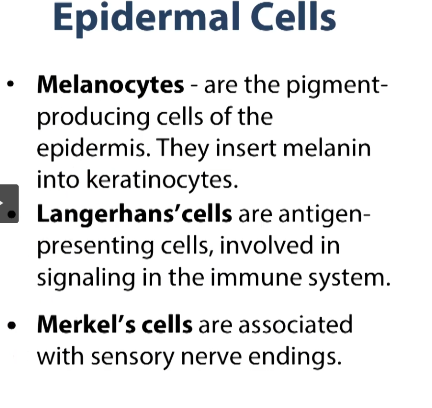
The other cells of the epidermis are the melanocytes, langerhans and merkel cells.
Melanocytes are the pigment producing cells of the epidermis. Melanocytes, along with other factors, such as the amount of vascular capillaries that are found in skin, helped to give you the pigmenation or the particular shade of pigmentation to your skin.
So your Langerhans cells are cells that are related to the immune system, they are antigen-presenting cells. So when they find an invader (pathogen) in our skin, these cells will help breakdown this pathogen, and they will take those pathogen particles and present it to the immune system underneath in the connective tissue can mount the proper immune response.
And you have sensory nerve endings in the shape of Merkel cells as well, which are somewhat difficult to see in the skin.
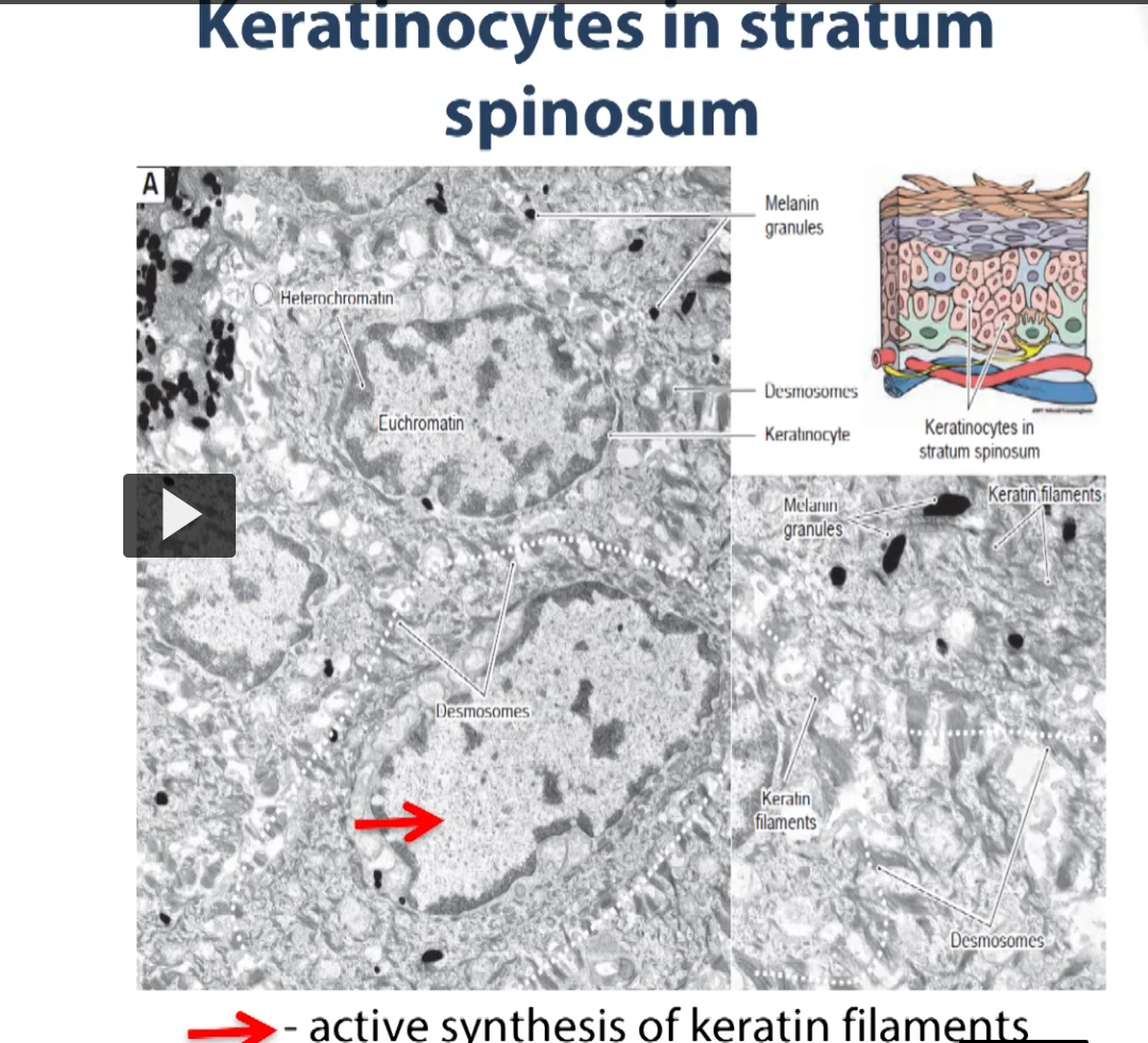
Keratinocytes in stratum spinosum
Here we have an electron micrograph illustrating various aspects of the stratum spinosum. That layer above the basalis, which has all cytoplasmic processes, which you can see here. So these are the extensions, dendrites. These are the smallest spinous extensions of the cell body, that contact other similar extensions from adjacent cells. Notice that the chromatin inside the nucleus of the cells is euchromatic for the most part. What that means is these cells are active in protein synthesis (keratin protein synthesis).
There is a higher magnification insert on the bottom right so you are able to see the spiny extensions from the cells, giving the layer of these cells stratum “spinosum”.
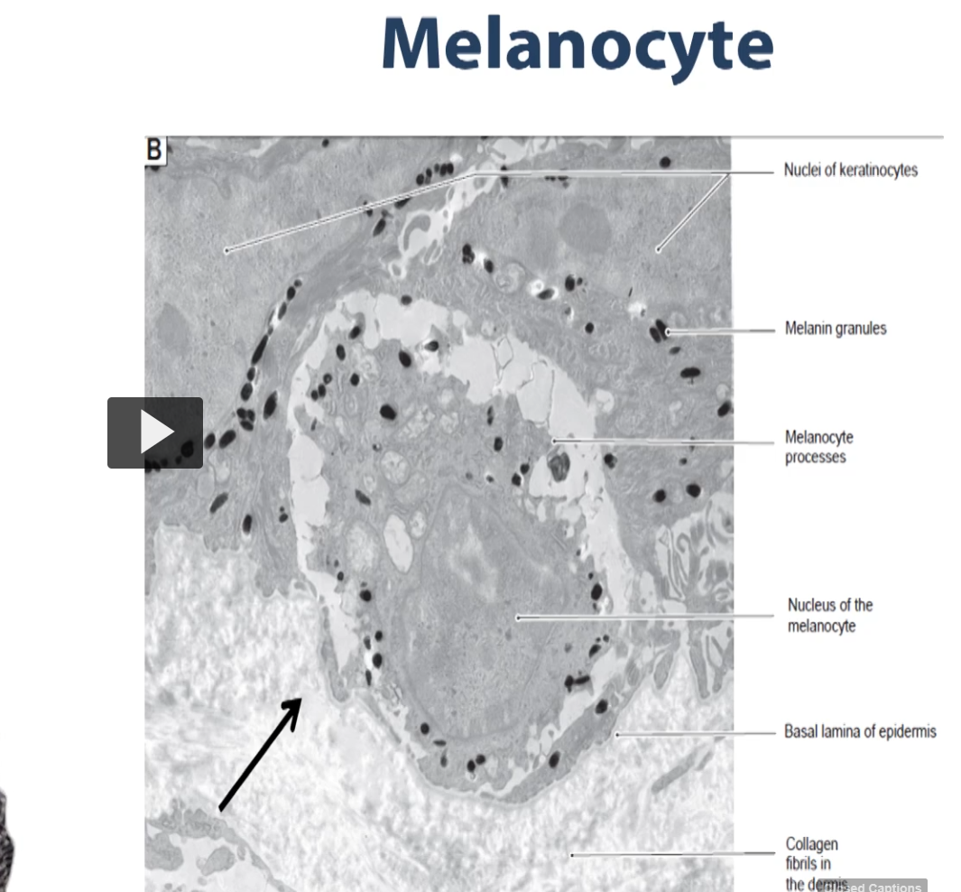
So here we have another electron micrograph, this time illustrating the melanocytes. These are the cells that are in charge of making these electron dense granules which contain the pigment melanin.
These melanocytes will pack that melanin, and we will show in in a second how that melanin is distributed throughout the epidermis.
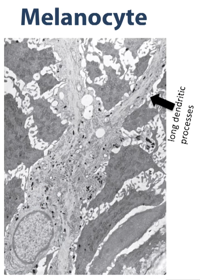
The long dendritic processes represent the apical portions of the cell. The long dendritic processes extend upward into the spinous layer. (the darker grey is some of the cells of the stratus spinosum). You can see that what is going to end up happening is a lot of the melanin granules packed in these vesicles called melanosomes, will be transported into these dendritic extensions and cells from the stratus spinosoum will take up parts of these dendrites and incorporate the melanin granules into their cytoplasm.
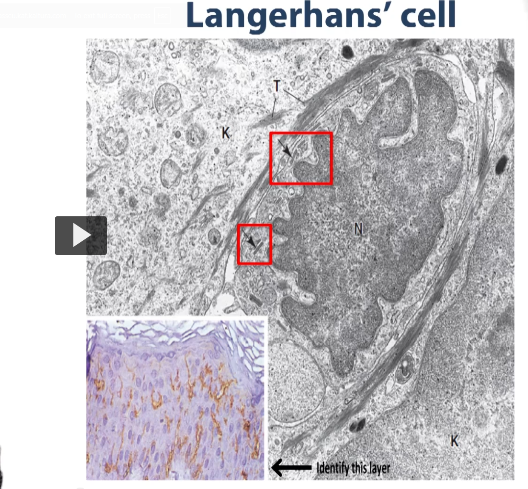
Here you have a Langerhan cell. These are the cells of the skin that monitor the skin, and they are part of the antigen presenting system of the skin, we’ve used an antibody found on the surface of the Langerhans cell to illustrate them here in light microscopy and we’ve counterstained this cross section of skin with a H & E so you can see where the rest of the layers are where the Langerhans cells. You can see they are abundantly distributed throughout the stratum spinosum. monitoring for the presence of foreign pathogens, which is there function.
Then you have the Merkel cell which closely resembles a nerve terminal
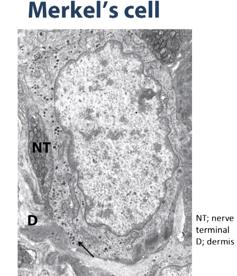
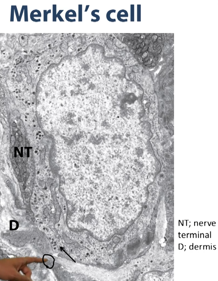
These are neurotransmitters that are packed in a langerhan cell that are packed into these vesicles (dark dots) on this electron micrograph.
So, in the next couple of slides, we are going to go through the different layers of the epidermis and we are going to show the different functions that are going on in the different layers of the epidermis and we are going to show the function or what events are going on in the different layers of the epidermis.
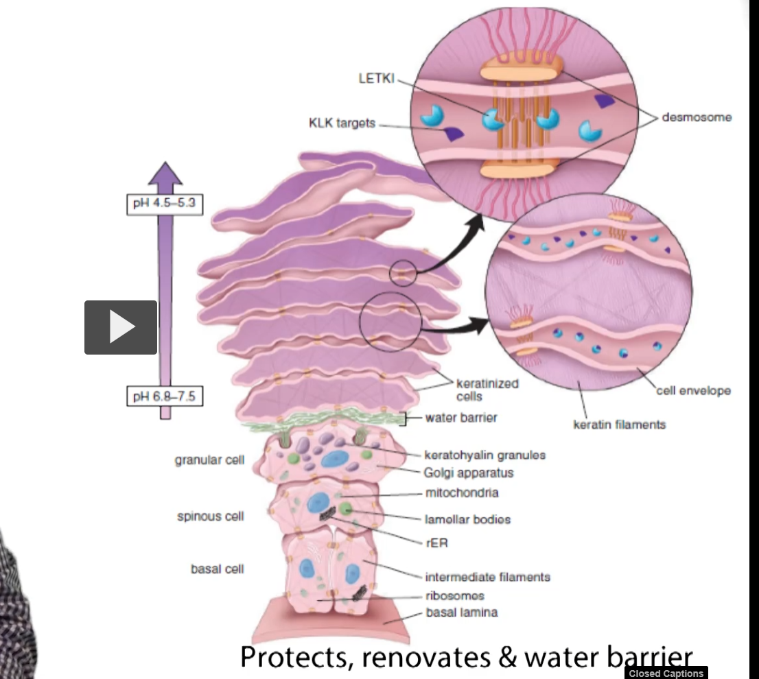
So here we have a single cell representation of each layer of the epidermis with the exception of the stratum corneum, which has multiple layers here.
You can see we have the basal layer, the spinous layer, the granulosum, the stratum, lecidum in thick skin, and the corneal stratum.
So those are the five layers of thick skin.
The stratum lucidum is absent in thin skin,
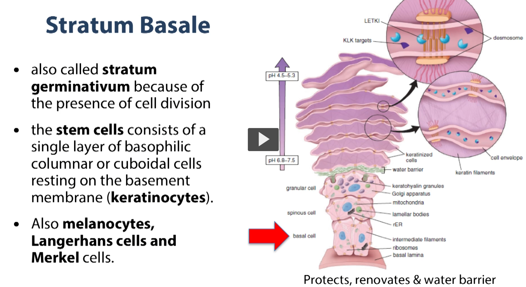
So we are going to start at the bottom where you see the red arrow pointing to the basal layer. This is a part of what’s called the stratum germinativum because there is active mitosis happening at this layer. So the epithelium/epidermis is constantly renewing, and the mitotic activity that replenishes our epithelium happens in this layer. So what we have in this layer are stem cells. So it’s a single layer of basophilic columnar to cuboidal cells that rest immediately on top of the basement membrane. So, in this layer, we have out keratinocytes, our melanocytes (the cells that give out skin its pigment), and we will also see Langerhans cells and Merkel cells in this layer.
We move up to the next layer above the stratum basale is the stratum spinosum.
The stratum spinosum consists of layers of cuboidal cells (slightly flattened, not quite squamous cells with a central nucleus and a cytoplasm whose processes are filled with keratin filaments). These keratin filaments are arranged into bundles, which when observed under electron microscopy, we call tonofilaments.
These tonofilaments attach to the protein plaques of desmosomes on both sites of adjacent cells to link the cells together by way of intermediate filaments.
stopped at 20:14