fn git 2
1/126
There's no tags or description
Looks like no tags are added yet.
Name | Mastery | Learn | Test | Matching | Spaced | Call with Kai |
|---|
No analytics yet
Send a link to your students to track their progress
127 Terms
Cardia, Fundus, Body, Pylorus
What are the four stomach regions?
Duodenum
First portion of small intestine.
Muscularis
A thick, primarily involuntary smooth muscle layer in the gastrointestinal (GI) tract responsible for segmentation and peristalsis.
Longitudinal, Circular, Oblique
The muscularis consists of these three layers.
Left Upper Quadrant
What quadrant does the stomach mostly lie in?
Inferior
Stomach is _____ to the diaphragm.
Anterior
The stomach is _____ to the spleen and pancreas.
Left lower
The stomach is tucked under the _____ margin of the liver.
Mobile
The stomach is anchored at both ends but _____ in between.
1.5, 4
The stomach capacity is _____ L food with max of _____ L.
Stomach
A J-shaped organ that is the widest part of alimentary canal.
Chyme
The substance formed after 4 hours of temporary storage and mixing
Pepsin
A powerful stomach enzyme (protease) that breaks down dietary proteins into smaller peptides and amino acids, essential for digestion. It needs an acid environment.
Hydrochloric acid
The stomach acid that helps kill bacteria.
Heartburn (GERD)
Pain caused when acidic contents reflux into the esophagus, which does not tolerate high acid content.
Water, Electrolytes, Aspirin, Alcohol
These nutrients are absorbed directly in the stomach.
Surface mucous cells, mucous neck cells
Cells that secrete mucus to form a protective barrier that prevents digestion of the stomach wall
Surface mucous cells, mucous neck cells
Cells that allow a small quantity of water, ions, short-chain fatty acids, and some drugs to enter the bloodstream.
Parietal cells
Cells that secrete intrinsic factor and hydrochloric acid.
Intrinsic factor
Needed for absorption of vitamin B12 (used in red blood cell formation, or erythropoiesis).
Hydrochloric acid
Kills microbes in food; denatures proteins; converts pepsinogen into pepsin.
Chief cells
Cells that secrete pepsinogen and gastric lipase.
Pepsinogen
Secreted by chief cells; its activated form breaks down proteins into peptides.
Gastric lipase
Splits triglycerides into fatty acids and monoglycerides.
G cells
Cells that secrete gastrin.
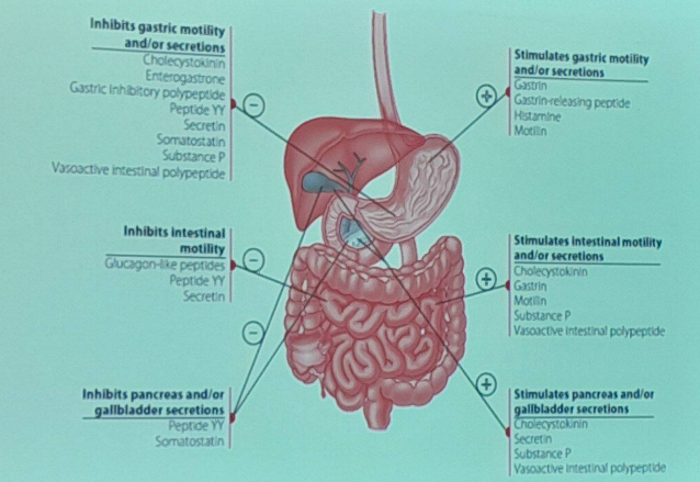
Gastrin
Stimulates parietal cells to secrete HCl and chief cells to secrete pepsinogen; contracts lower esophageal sphincter, increases motility of the stomach, and relaxes pyloric sphincter.
Muscularis
Creates mixing waves that churn and physically break down food and mix it with gastric juice; forces chyme through pyloric sphincter.
Pyloric sphincter
Opens to permit passage of chyme into the duodenum; regulates its passage and prevents backflow from the duodenum to the stomach.
Cardia
The section containing the orifice where the esophagus enters, acting as the initial receiving area for food.
Fundus
The dome-shaped superior section located just beneath the diaphragm, often serving as a temporary storage area for undigested food and gases.
Body
The large central section bordered by the convex and concave borders, functioning as the primary site for mixing and churning food.
Greater curvature
The long, convex, outer curve extending along the left lateral surface.
Lesser curvature
The shorter, concave, inner curve extending along the right medial surface.
Pylorus
The funnel-shaped distal section that connects the main central area to the small intestine.
Antrum, Canal, Sphincter
The pyloric region is divided into these three parts.
Greater omentum
A fatty, apron-like peritoneal fold that hangs down from the outer lateral curve, covering the intestines.
Simple columnar epithelium
The single layer of tall cells lining the stomach surface that secretes a protective, bicarbonate-buffered mucus.
Gastric pits
Shallow indentations on the stomach's mucosal surface that serve as openings for deeper secretory structures.
Gastric glands
Deep, tubular secretory structures that branch off from the surface indentations and contain various specialized cells.
Mucous neck cells
Secretory cells located in the upper, constricted region of the glands that produce mucus.
Parietal cells
Specialized gland cells that secrete hydrochloric acid (HCl) and the glycoprotein needed for B12 absorption.
Intrinsic factor
A specific substance secreted by parietal cells that is strictly required for the body to absorb vitamin B12.
Chief cells
Gland cells stimulated by gastrin that secrete the inactive enzyme precursor for protein digestion.
Pepsinogen
An inactive enzyme precursor that requires an acidic environment (exposure to HCl) to convert into its active, protein-digesting form (pepsin).
Gastrin
A stomach hormone that stimulates cellular activity, such as triggering chief cells to release their secretions.
Enteroendocrine cells
Specialized hormone-secreting cells situated at the very base of the gastric glands.
Mucosa
The innermost tissue layer of the stomach wall, comprising the surface epithelium, lamina propria, and muscularis mucosae.
Submucosa
The connective tissue layer beneath the innermost lining that houses the submucosal nerve plexus.
Muscularis externa
The thick smooth muscle layer responsible for stomach churning, consisting of oblique, circular, and longitudinal layers, and containing the myenteric nerve plexus.
Serosa
The thin, outermost protective membrane layer of the stomach wall.
oki yohan u cool
idk how u can study this but do it anyway. type “oki yohan u cool”
Small Intestine
Optimized for absorption through a huge surface area achieved by its great length and internal structural modifications
Circular folds
Deep macroscopic ridges in the intestinal wall that increase the absorptive area
Villi
1 mm high fingerlike projections covered in simple columnar epithelium that give the lining a velvety texture and increase the absorptive area.
Microvilli
Microscopic cellular projections on absorptive cells that further maximize surface area.
Lacteal
A core network of blood and lymph capillaries located inside the fingerlike projections (villi)
Fat
_____-soluble substances (e.g. pesticides) circulate systemically throughout the body before reaching the liver for detoxification.
Rugae
Longitudinal folds on the internal mucosal surface that allow the stomach to stretch and expand
Muscularis
Features an additional innermost oblique smooth muscle layer, alongside the standard circular and longitudinal layers, for powerful churning
Duodenum (5%), Jejunum (~40%), Ileum (~60%)
The three subdivisions of the small intestines and their corresponding length subdivision.
Retroperitoneal
The duodenum is _____ (stuck down under the peritoneum), unlike the other sections which are loose.
Bile
The duodenum receives _____ from the liver and gallbladder.
Pancreas
The duodenum receives enzymes from which organ?
Ileocecal sphincter
A valve that regulates the flow of material from the last segment of the small intestine into the first portion of the large intestine
Cecum
The first section of the large intestine.
Common bile duct
The tube that delivers secretions from the gallbladder into the first section of the small intestine
Pancreatic duct
The tube that carries digestive enzymes, eventually joining the common bile duct
Sphincter of Oddi
A muscular valve that regulates the flow of combined biliary and pancreatic secretions into the duodenum
Mucosa
The innermost lining containing the mucosal epithelium, lamina propria, and muscularis mucosae.
Submucosa
The tissue layer beneath the mucosa that houses blood vessels, submucosal glands, and the submucosal nerve plexus.
Muscularis
The smooth muscle layer responsible for movement, consisting of an inner circular muscle layer, an outer longitudinal muscle layer, and the myenteric nerve plexus.
Serosa
The outermost protective membrane of the digestive tract also referred to as the visceral peritoneum.
Mesentery
A supportive membranous fold that attaches the digestive tract to the body wall and houses the mesenteric artery and vein.
Plica
A large mucosal fold within the digestive tract that helps increase surface area.
Cecum, Appendix, Colon, Rectum, Anal canal.
The five main divisions of the small intestine.
Large Intestine
Receives digested residue to primarily absorb water and electrolytes.
Ascending, Transverse, Descending, Sigmoid
The four segments of the colon.
Tenia coli
Distinct longitudinal bands of smooth muscle running along the exterior length of the colon.
Epiploic appendages
Small, fat-filled pouches attached to the external surface of the large intestine.
Crypts of Lieberkühn
Glands located between villi containing rapidly dividing cells (every 3-6 days) that renew the epithelium and secrete watery intestinal juice.
Intestinal flora
The permanent, normal bacteria residing in the gut that manufacture certain vitamins, such as vitamin K, for the body to absorb.
Duodenal glands
Glands that produce mucus to counteract stomach acidity and secrete hormones like Cholecystokinin and Secretin.
Rectum
A pelvic segment of the large intestine lacking teniae coli but featuring a strong longitudinal muscle layer and internal valves.
Pectinate line
A dividing line in the anal canal; the area inferior to this line is highly sensitive to pain.
Hemorrhoids
Enlarged veins in the anal canal that are classified as internal (located superior to the pectinate line) or external (located inferior to the pectinate line).
Internal anal sphincter
An involuntary smooth muscle ring that helps keep the anal opening closed.
External anal sphincter
A voluntary skeletal muscle ring that controls the closing of the anal opening.
Hepatic flexure
A sharp, right-angle turn in the colon located in the right upper quadrant (RUQ).
Splenic flexure
A sharp, right-angle turn in the colon located in the left upper quadrant (LUQ).
Sigmoid colon
The distinct S-shaped final segment of the colon that leads into the rectum.
Vermiform appendix
A small, blind-ended tube attached to the cecum (the first part of the large intestine).
Mass peristaltic movements
Powerful contractions occurring a few times a day to force feces toward the rectum, overcoming the otherwise sluggish baseline movement of the colon.
Teniae coli
Three distinct bands of longitudinal smooth muscle located within the muscularis layer.
Haustra
Pocket-like sacs in the colon wall created by the continuous resting tension (tone) of the longitudinal muscle bands.
Epiploic appendages
Small, fat-filled pouches of visceral peritoneum that hang from the external surface of the large intestine
Right hypochondriac, epigastric
The liver is situated on which abdominopelvic regions?
Liver
The largest gland in the human body.
Enterocytes
Are covered with small projections called microvilli which project in the intestinal lumen.
Microvilli
The _____ of enterocytes make up the brush border.
Crypts of Lieberkühn
Cells in these crypts will migrate up to eventually become absorptive cells in the villus.