Test 2 Anatomy
1/249
There's no tags or description
Looks like no tags are added yet.
Name | Mastery | Learn | Test | Matching | Spaced | Call with Kai |
|---|
No analytics yet
Send a link to your students to track their progress
250 Terms
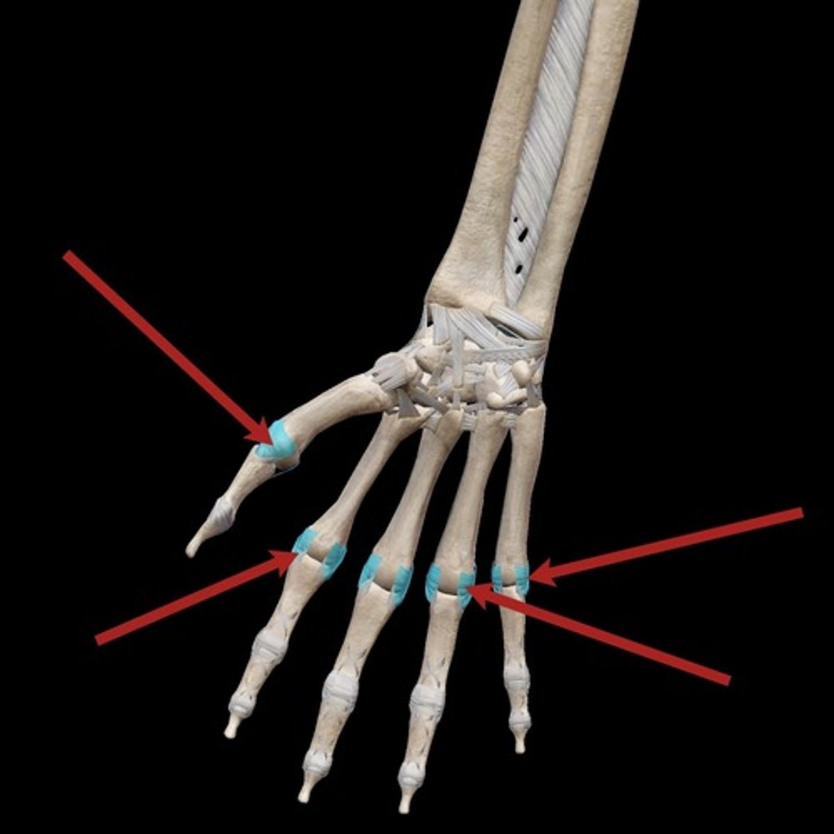
Carpometacarpal joints
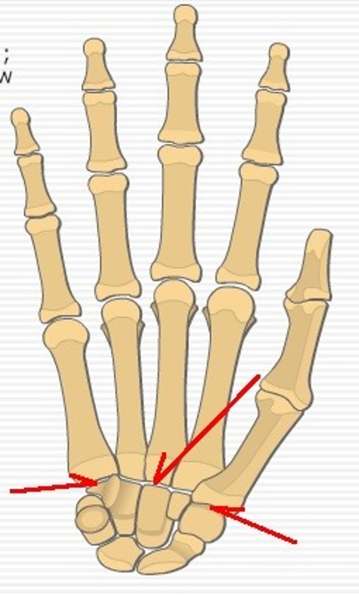
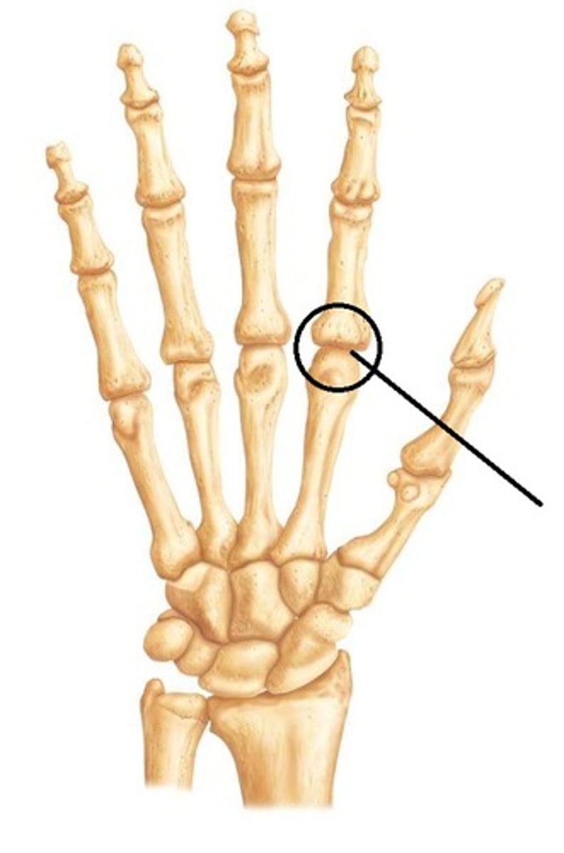
Metacarpalphalangeal joints
Pollux
Thumb
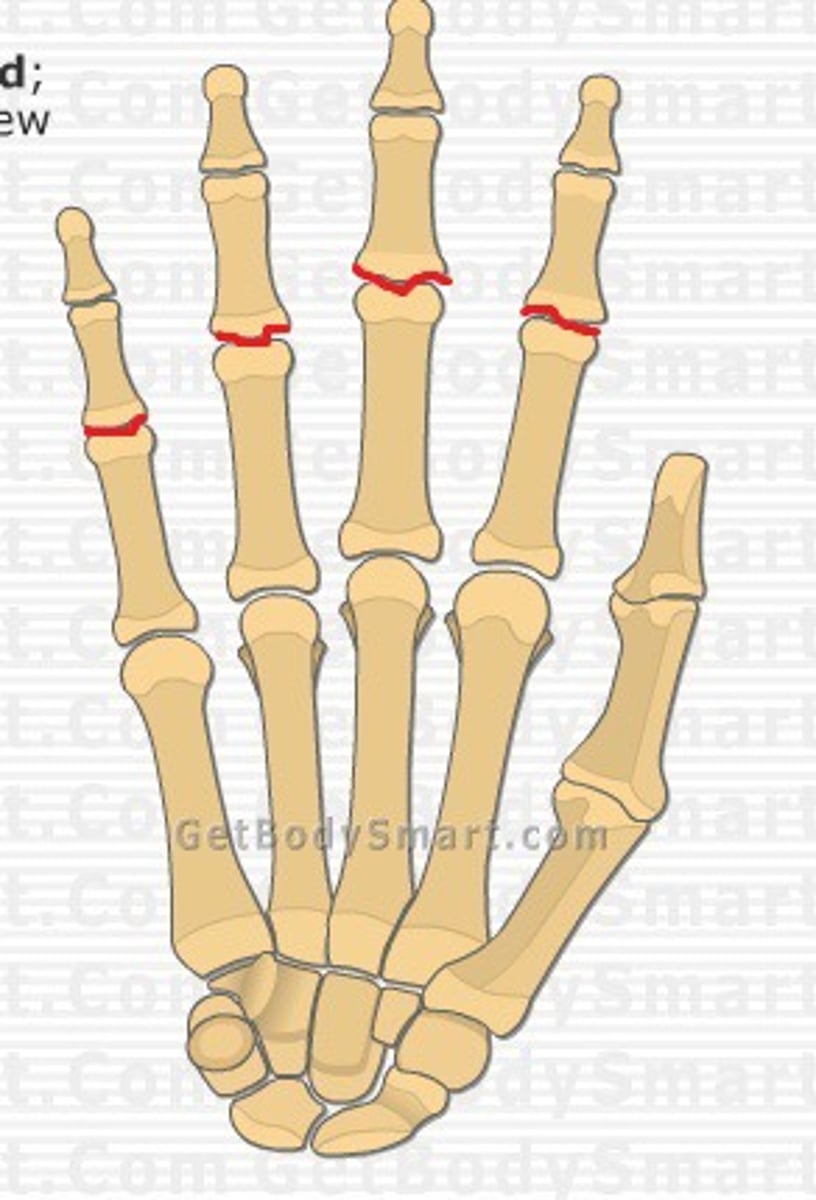
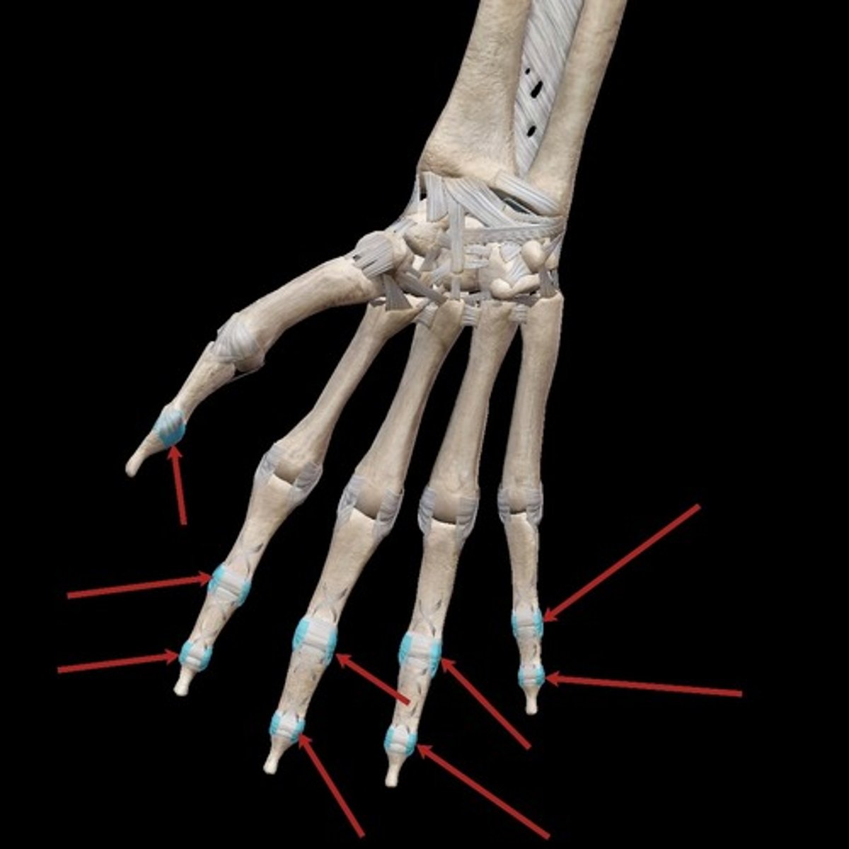
Proximal interphalangeal joints
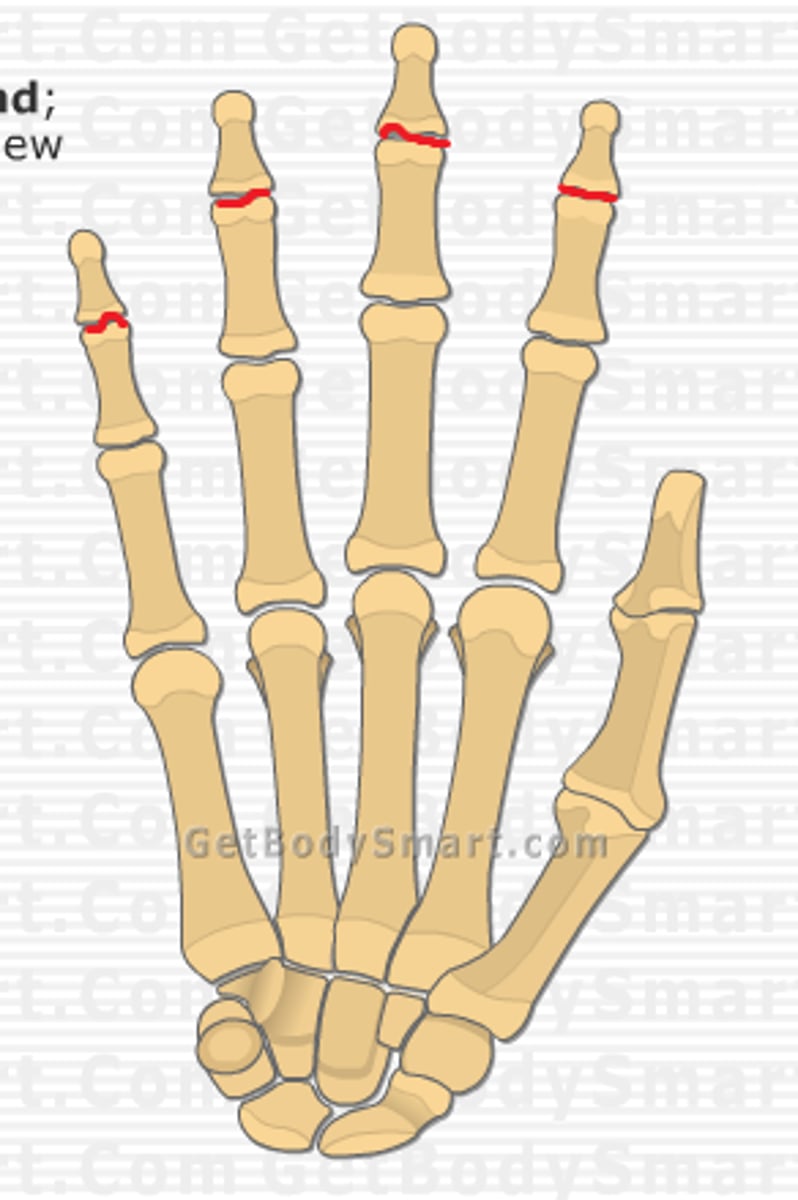
Distal interphalangeal joints
Metacarpophalangeal ligaments
Interphalangeal ligaments
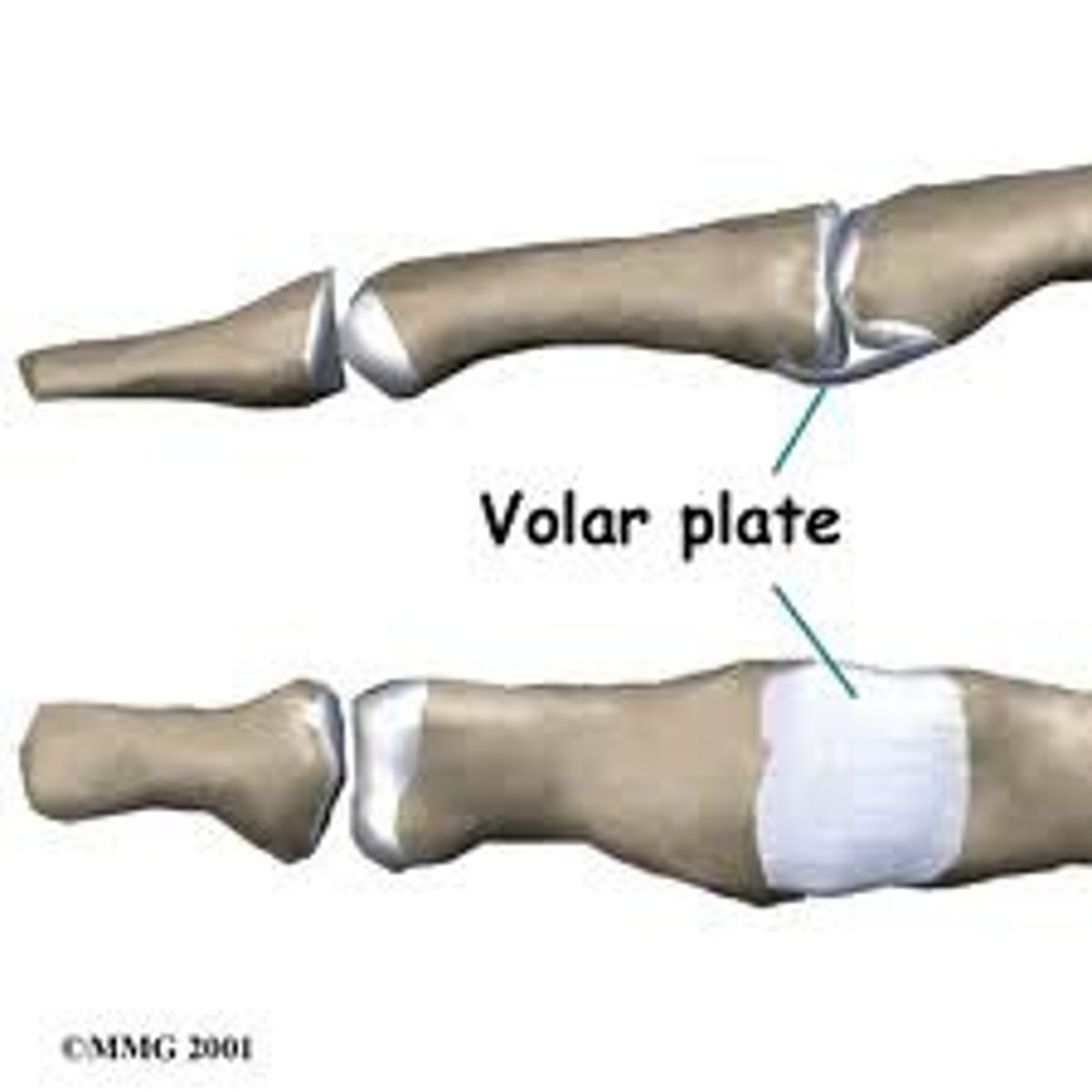
Volar plate
Prevents hyperextension
Median nerve
Innervates some flexors, pronators and intrinsics, provides sensation to the lateral 3 1/2 fingers
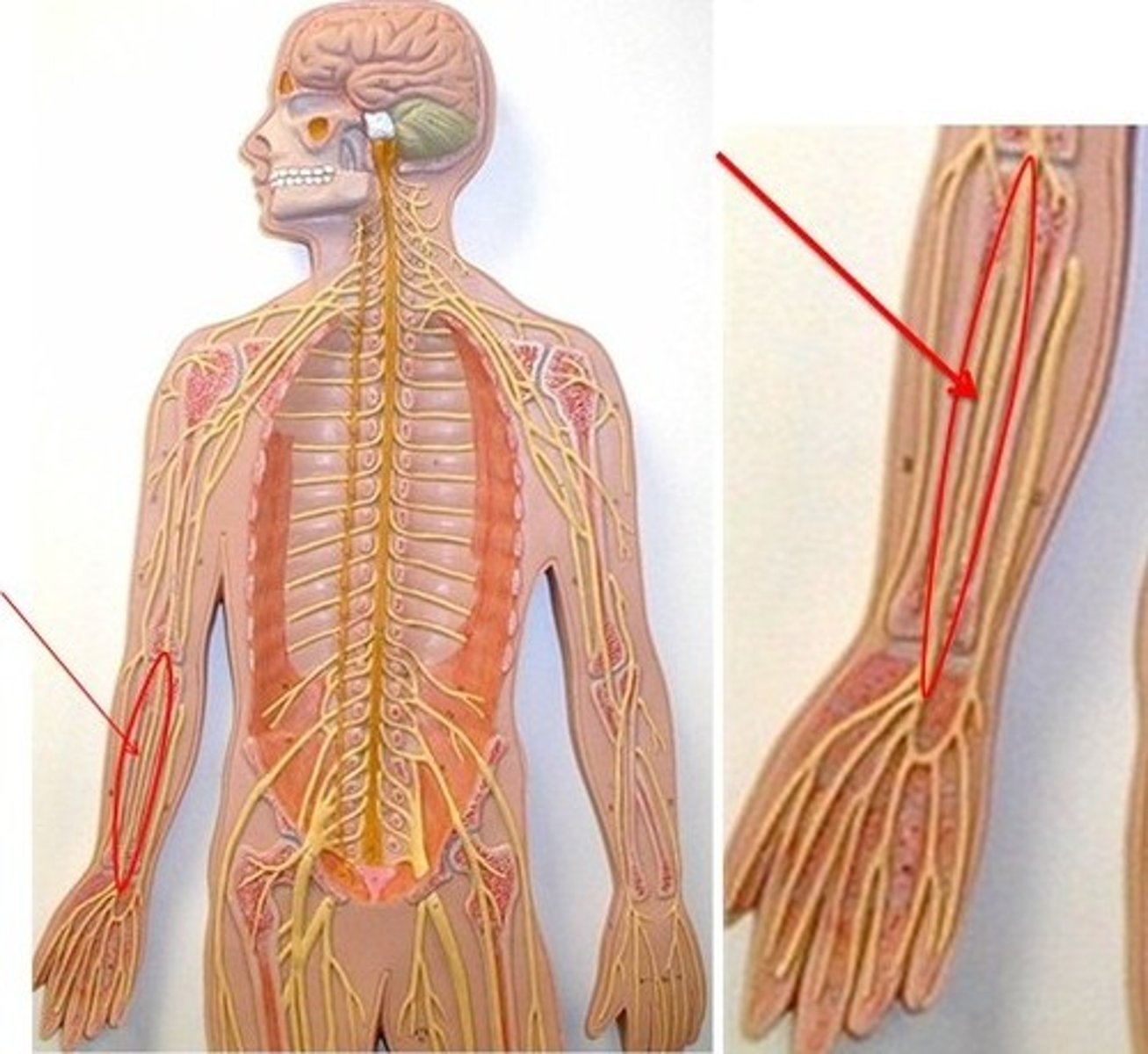
Ulnar nerve
Innervates some flexors, intrinsics, provides sensation to the medial 1 1/2 fingers
Synovial sheath
Covers the tendons in the fingers
Annular and cruciate ligaments
Hold flexor tendons onto the bones
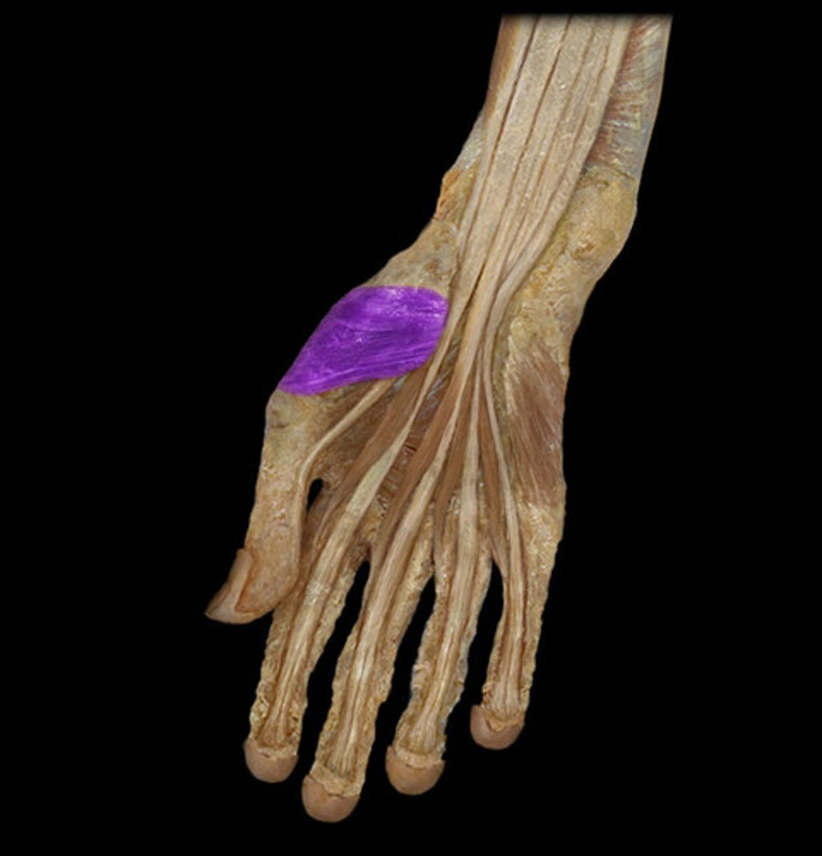
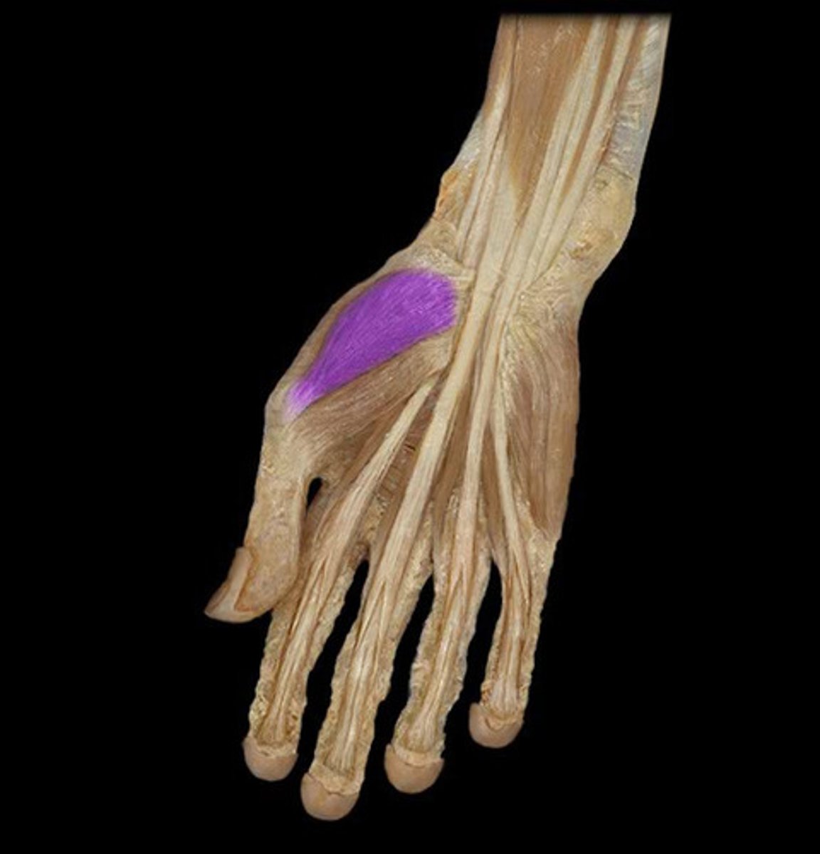
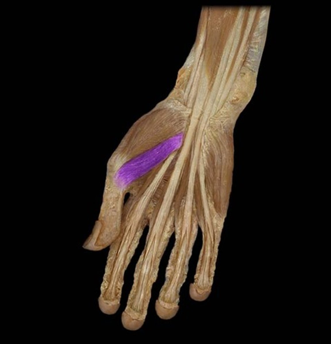
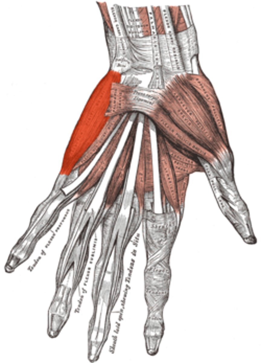
Thenar muscles
Opponens pollicis, abductor pollicis brevis, flexor pollicis brevis
Opponens pollicis
Opposes the thumb
Abductor pollicis brevis
Abducts the thumb
Flexor pollicis brevis
Flexes the thumb
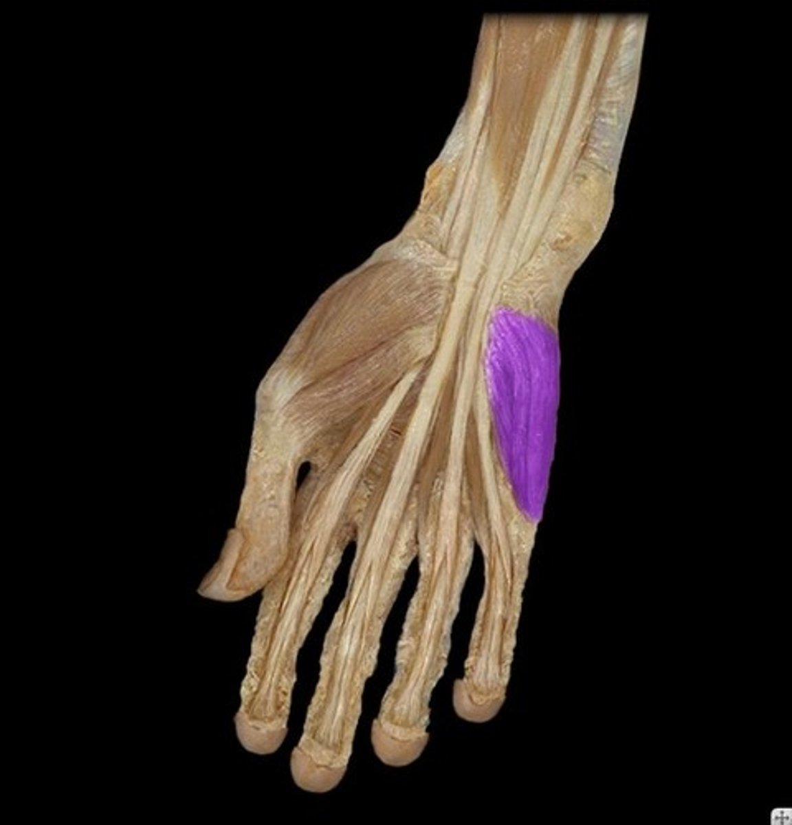
Hypothenar muscles
Opponens digiti minimi, abductor digiti minimi, flexor digiti minimi
Opponens digiti minimi
Opposes the little finger
Abductor digiti minimi
Abducts the little finger
Flexor digiti minimi
Flexes the little finger
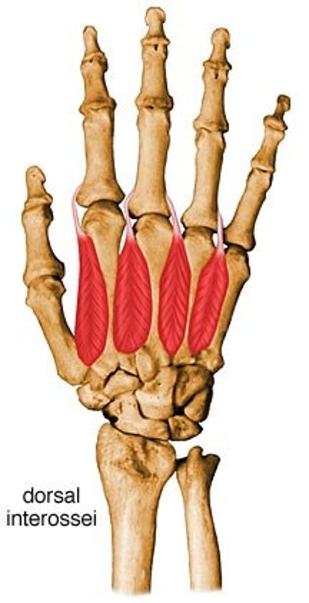
Interossei
Dorsal abduct, palmar adduct
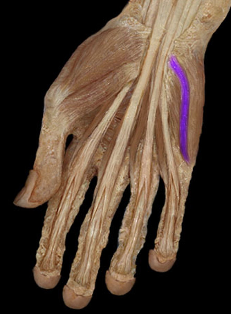
Lumbricals
Flex metacarpophalangeal joints, extend interphalangeal joints
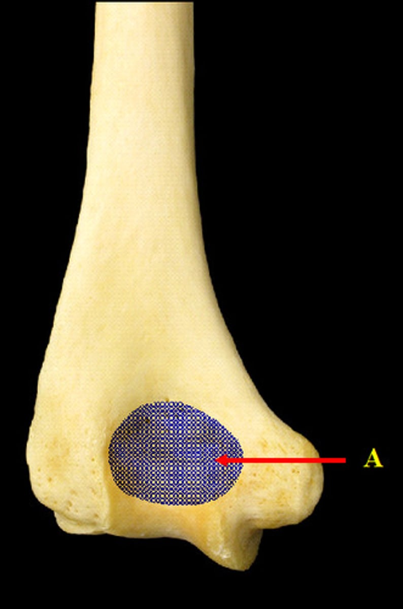
Capitulum
Lateral condyle of the humerus
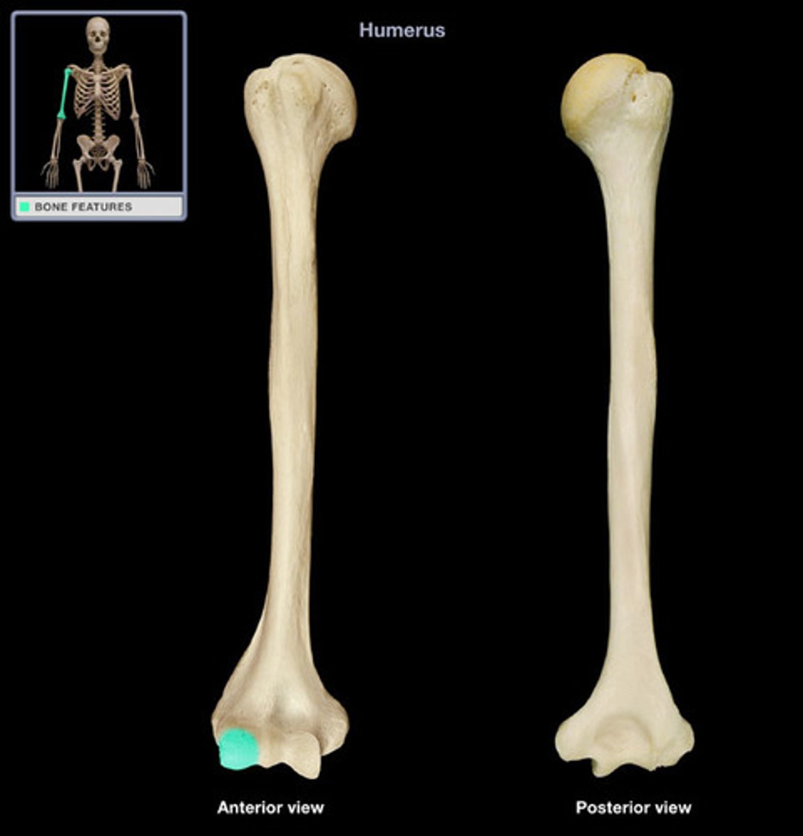
Trochlea
Medial condyle of the humerus
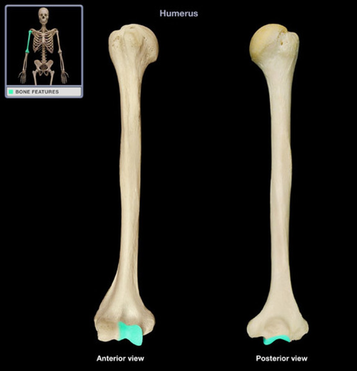
Medial epicondyle of the humerus
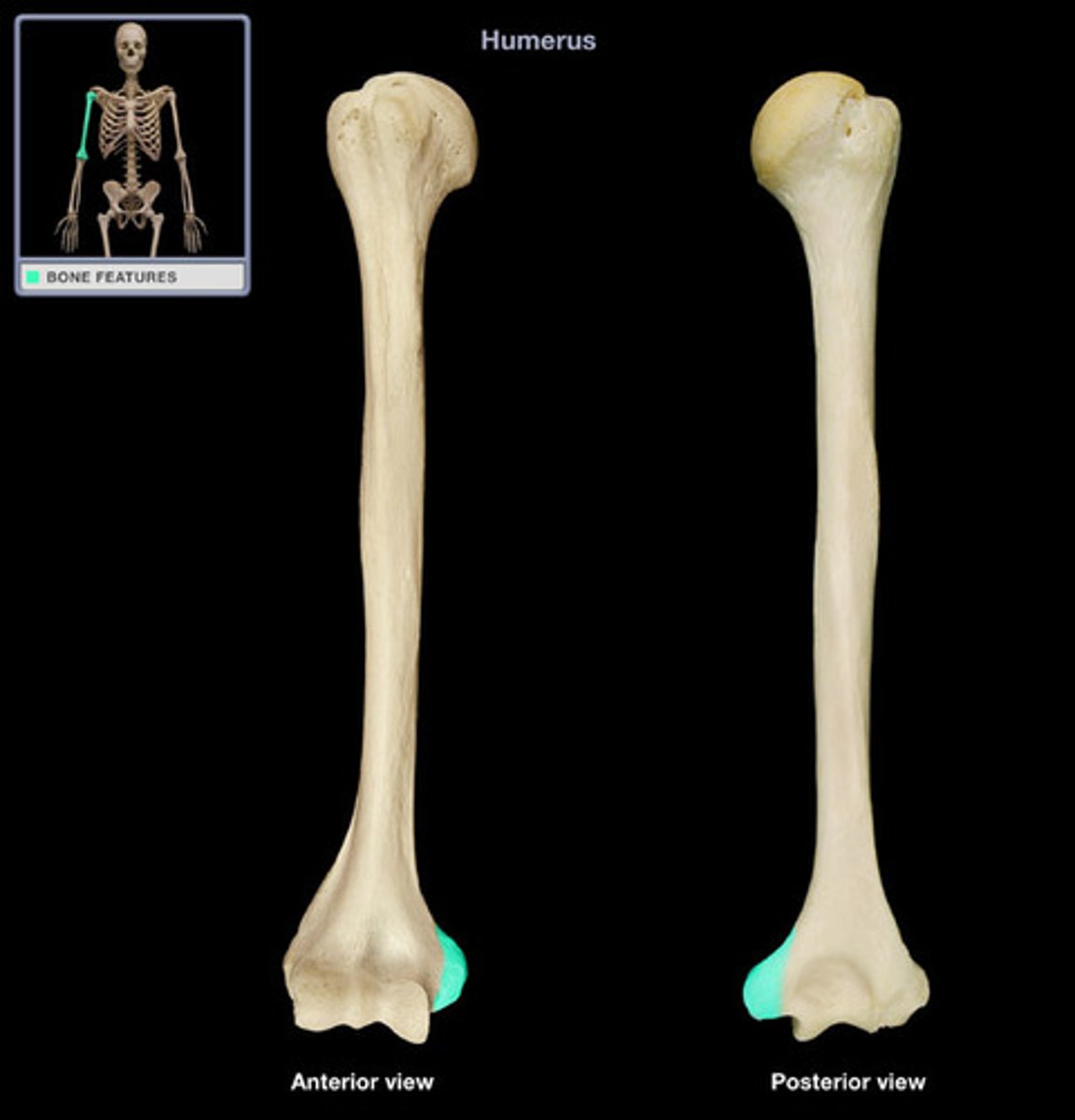
Lateral epicondyle of the humerus
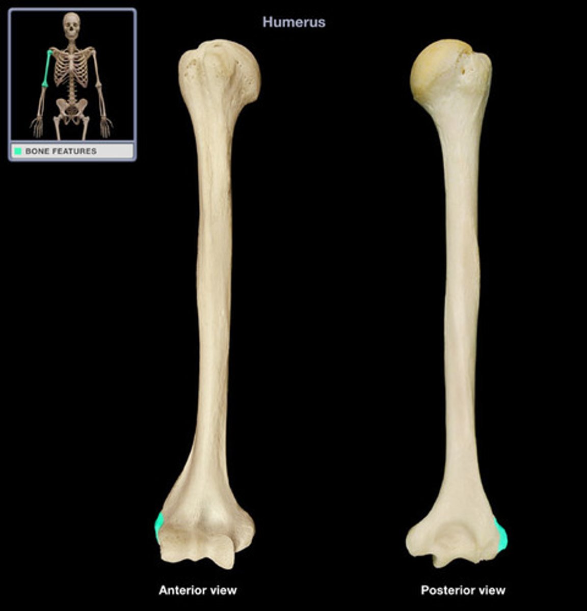
Olecranon process
Olecranon fossa
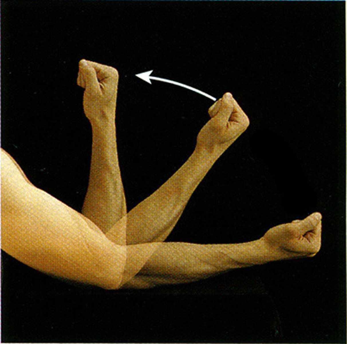
Elbow extension
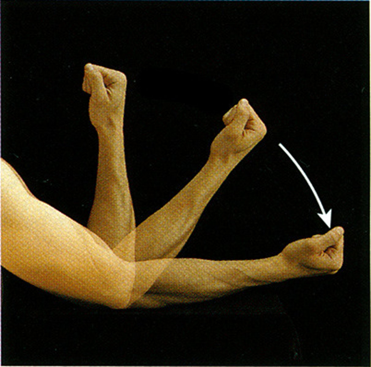
Elbow flexion
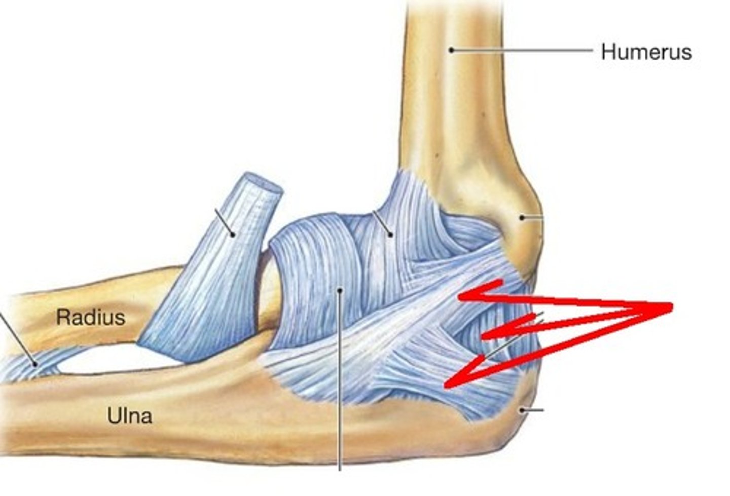
Radial collateral ligament
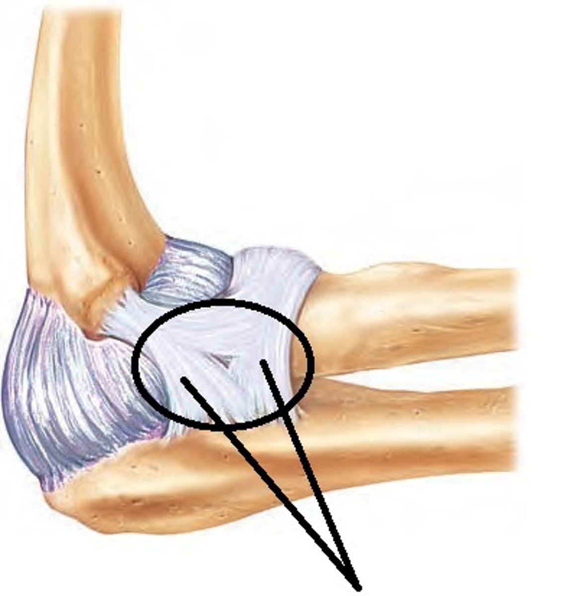
Annular ligament
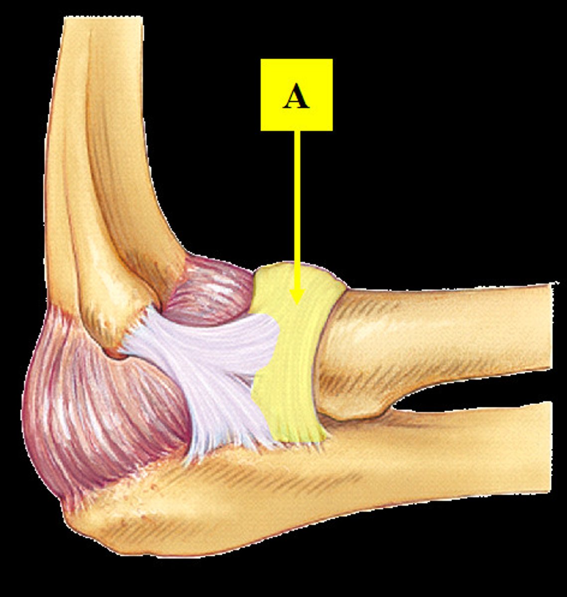
Ulnar collateral ligament
Cubital fossa
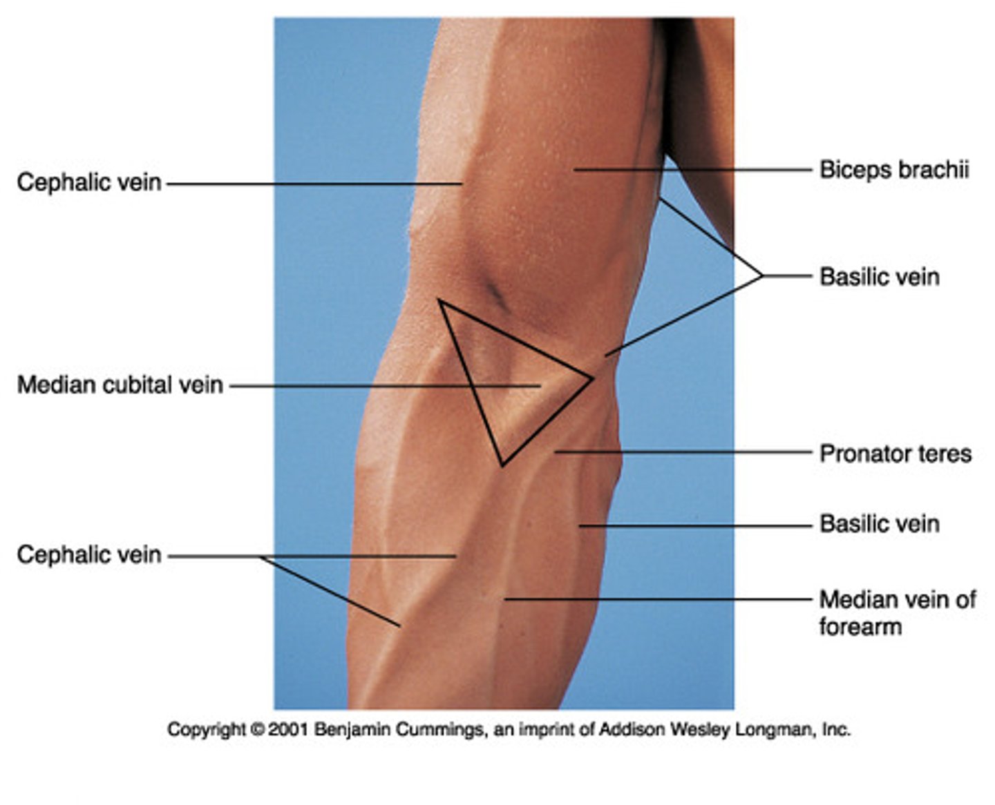
Contents of the cubital fossa
Brachial artery and vein, median nerve
Borders of the cubital fossa
Brachioradialis and pronator teres
Cubital tunnel
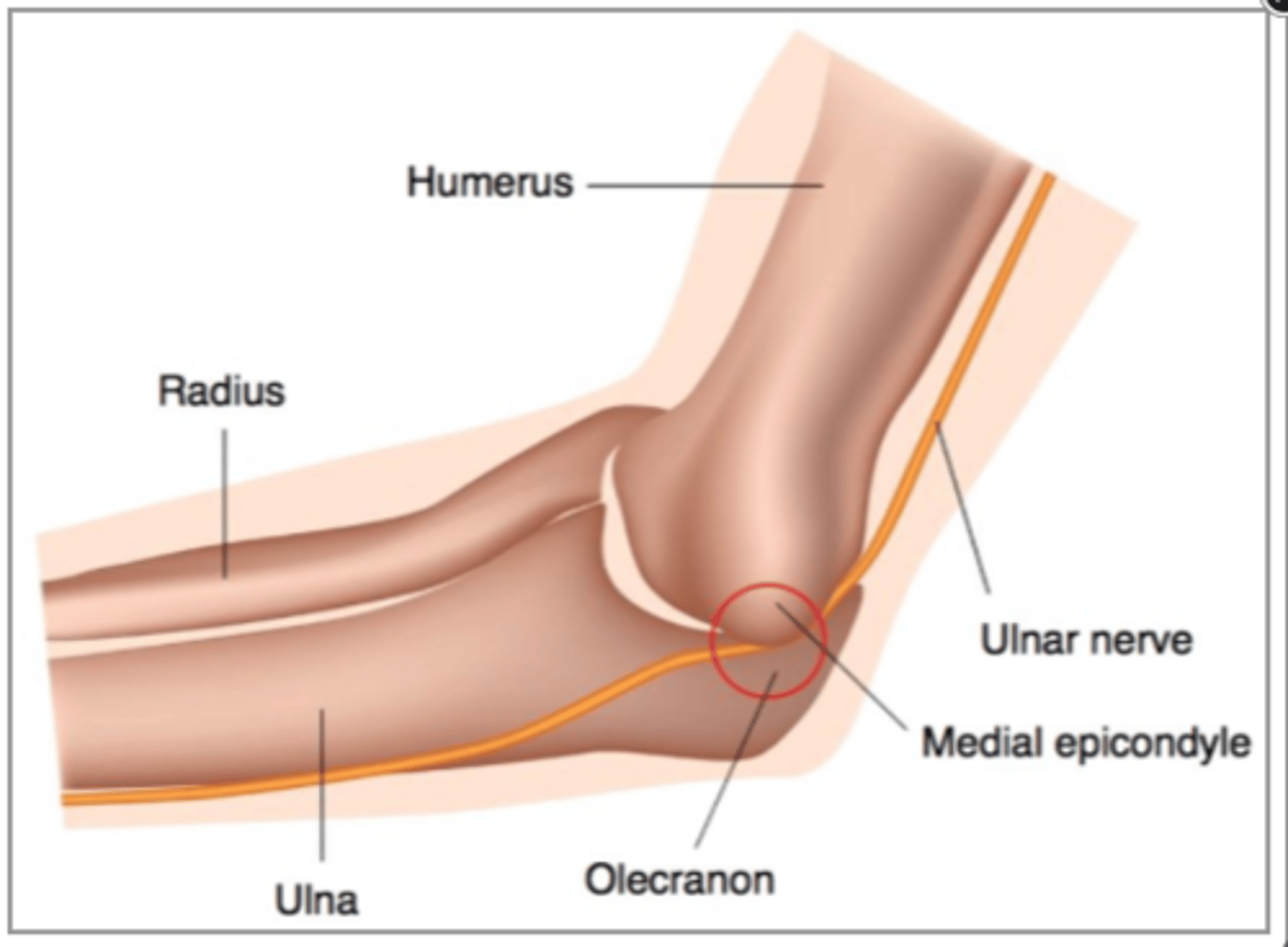
Contents of cubital tunnel
Ulnar nerve
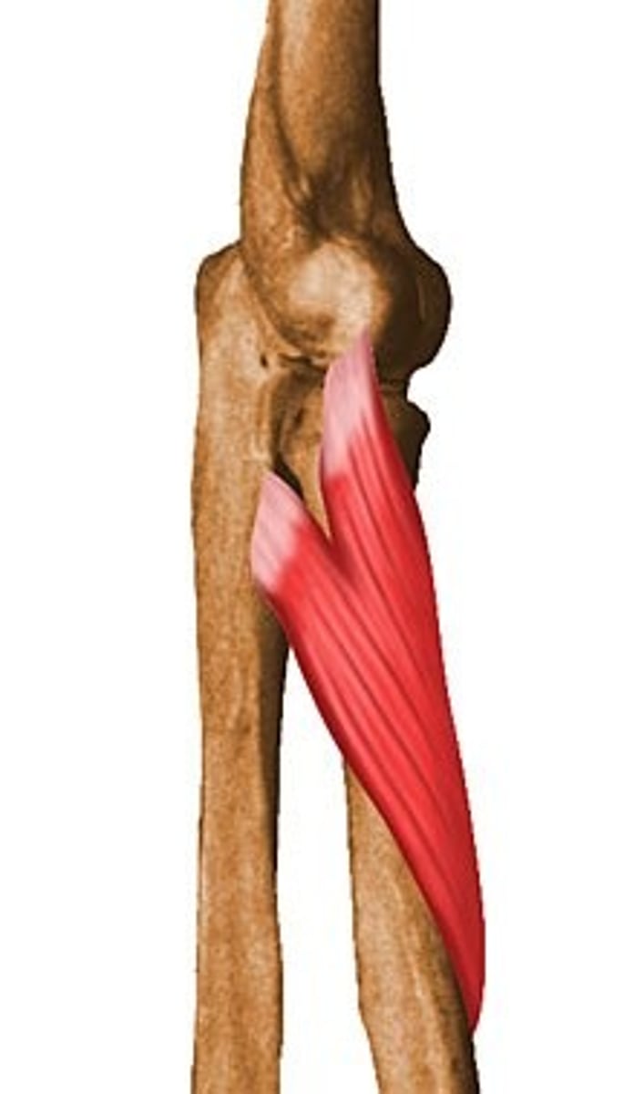
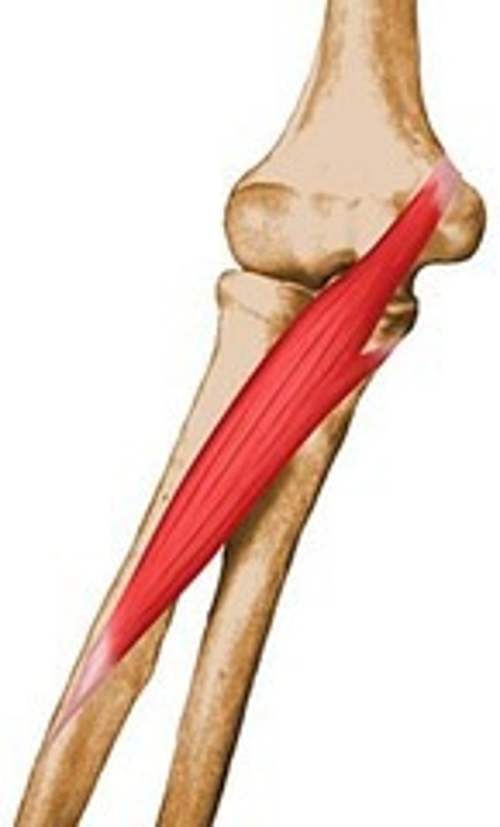
Triceps brachii
Extends elbow
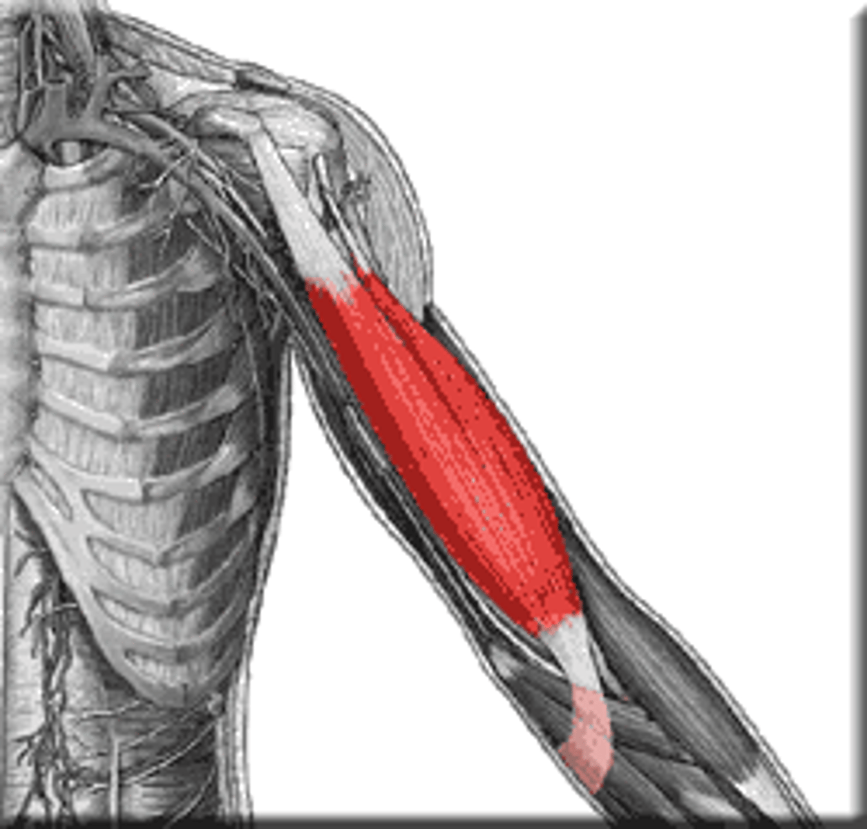
Biceps brachii
Flexes elbow
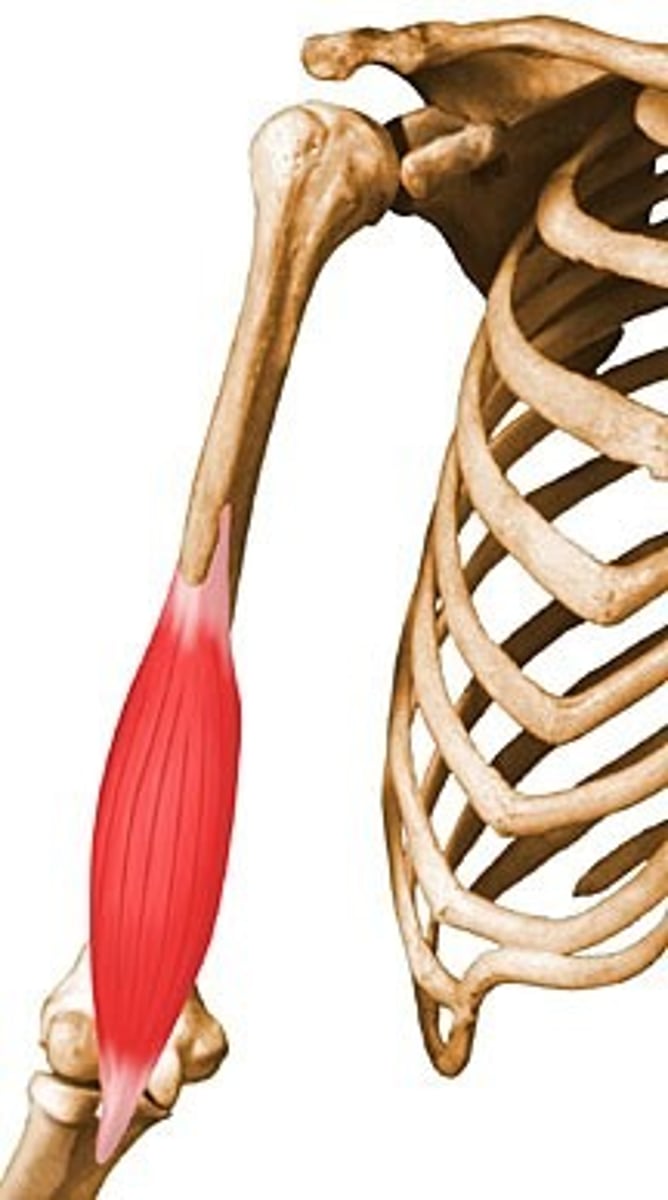
Brachialis
Flexes elbow
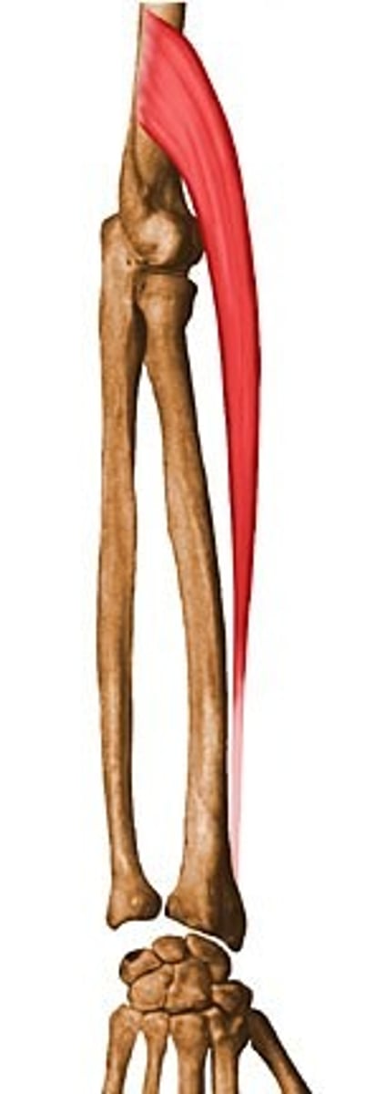
Brachioradialis
Flexes elbow
Proximal radio-ulnar joint
Distal radio-ulnar joint
Supinator
Supinates forearm
Forearm supination
Forearm pronation
Pronator teres
Pronates forearm
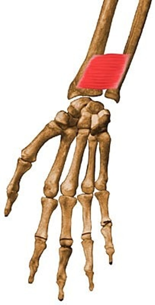
Pronator quadratis
Pronates forearm
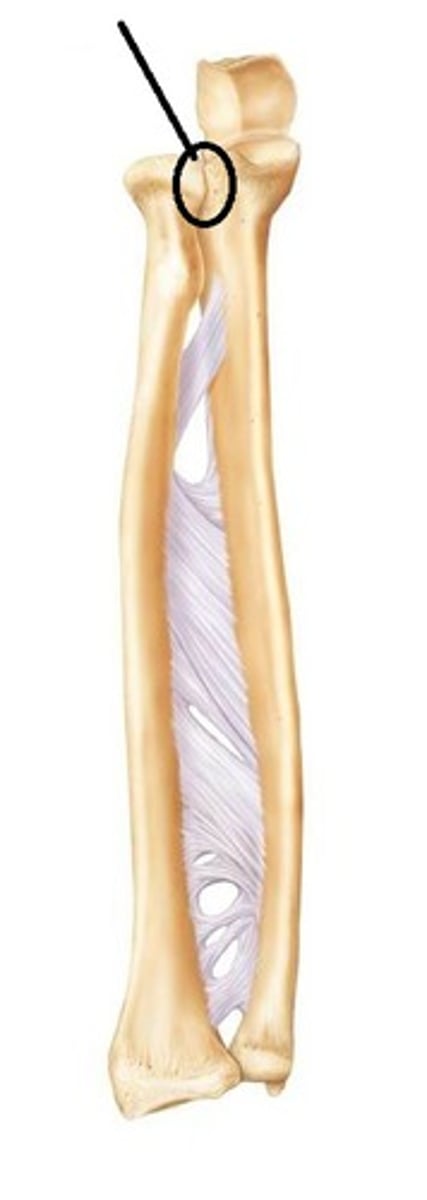
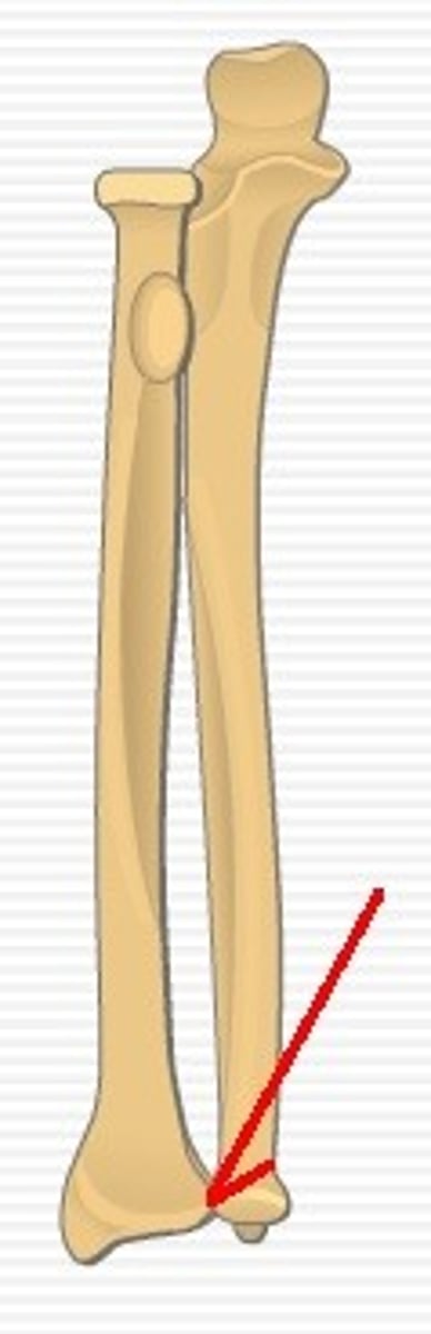
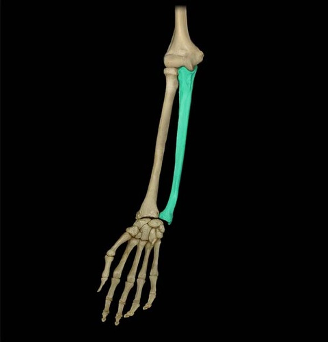
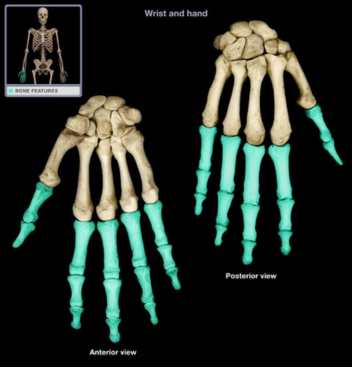
Radius
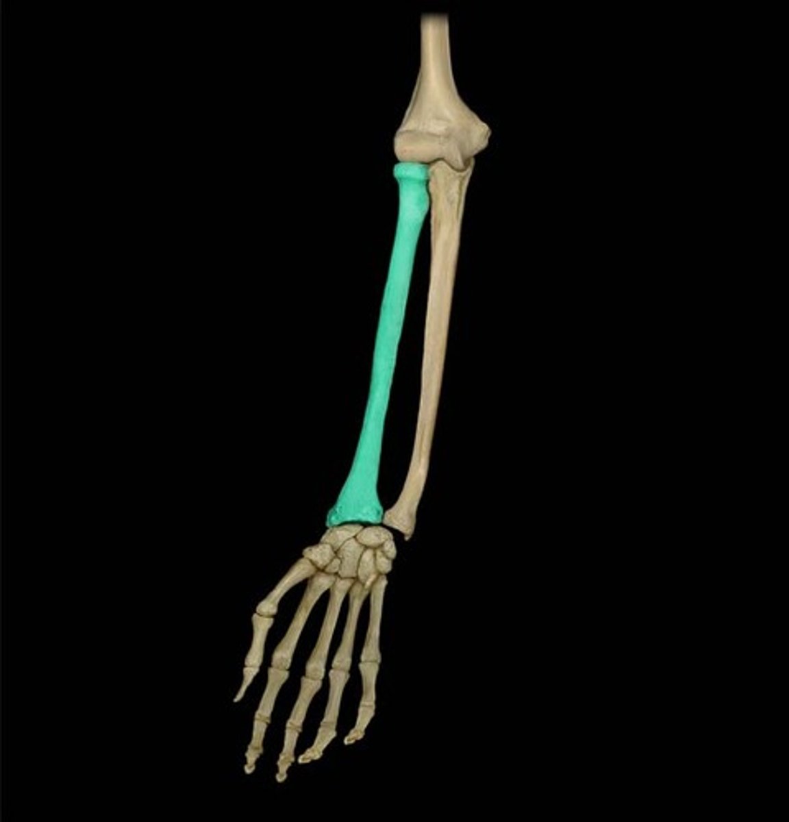
Ulna
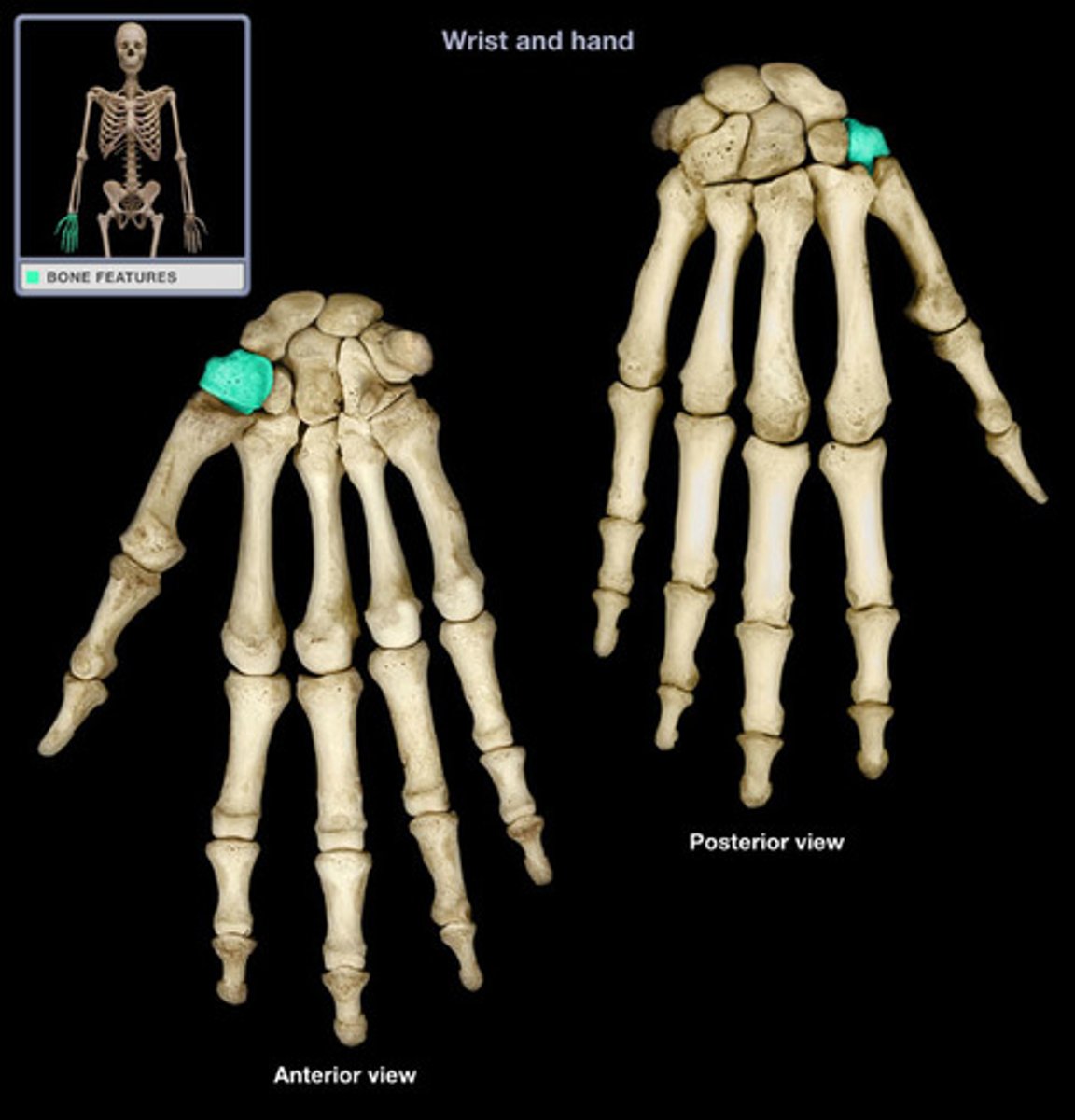
Scaphoid
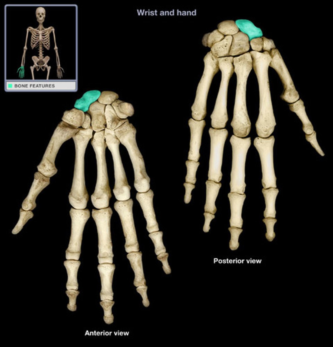
Lunate
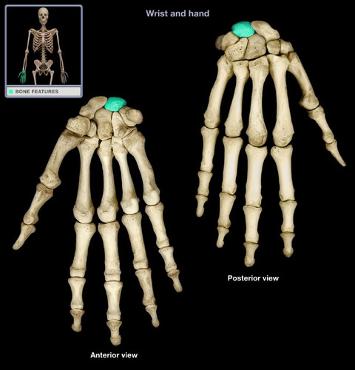
Triquetrum
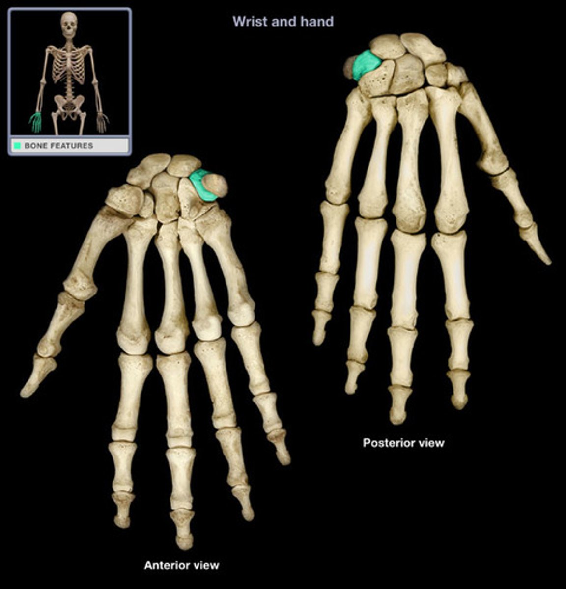
Pisiform
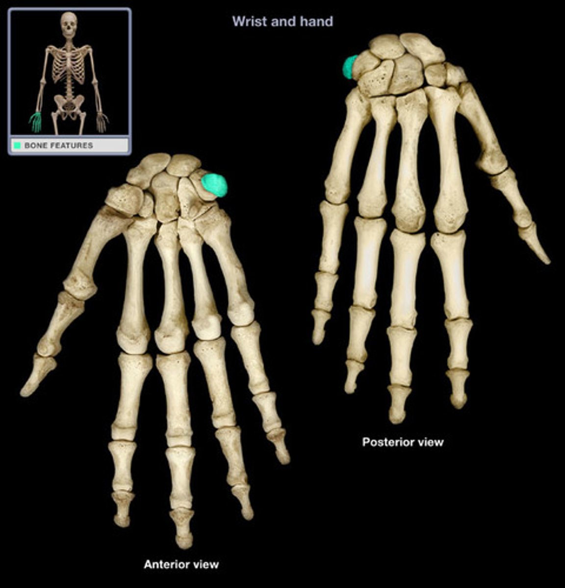
Hamate

Capitate
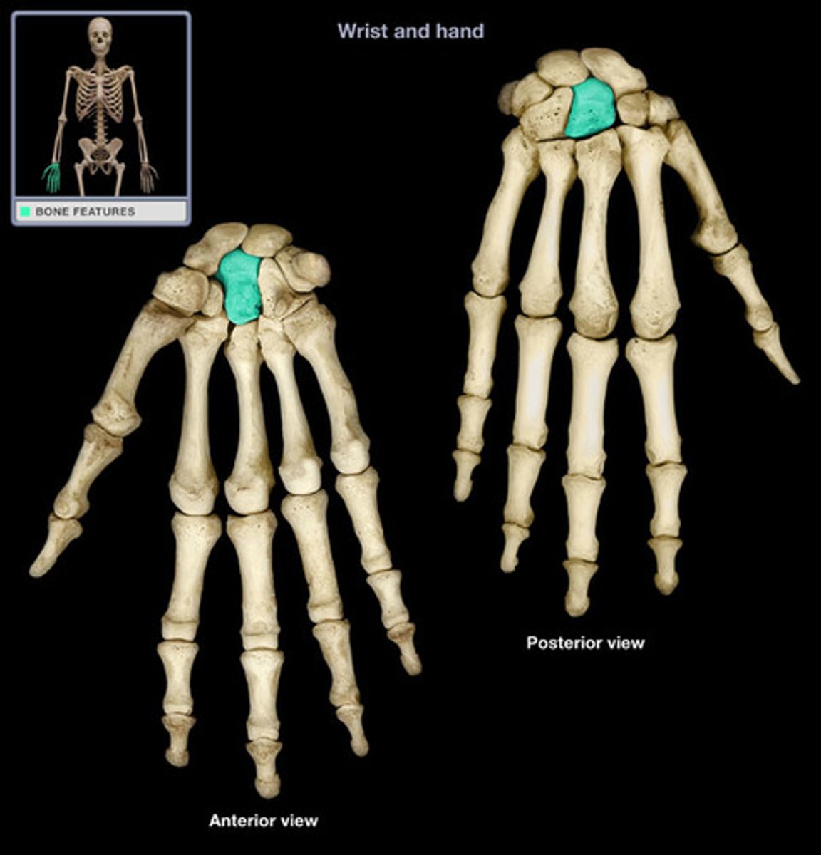
Trapazoid

Trapezium
Metacarpals
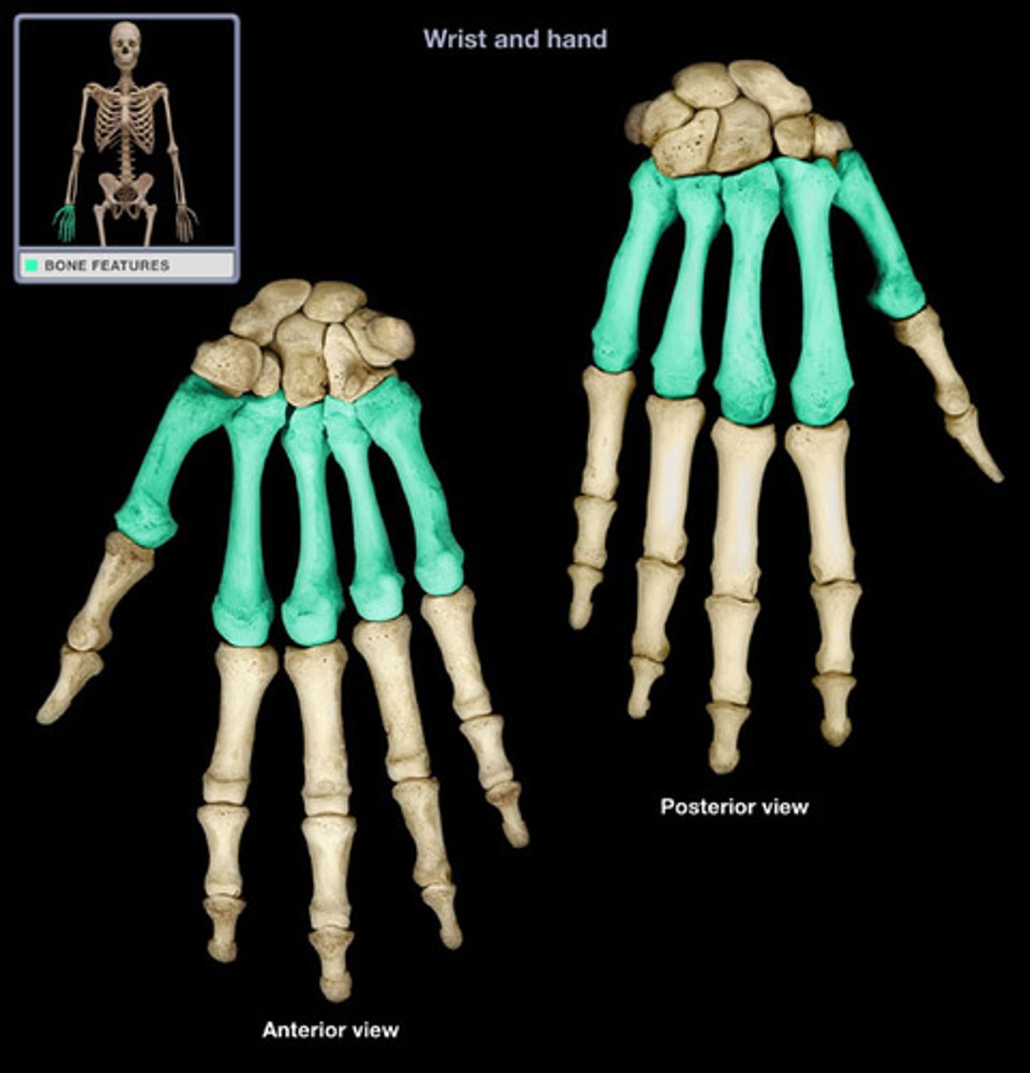
Phalanges
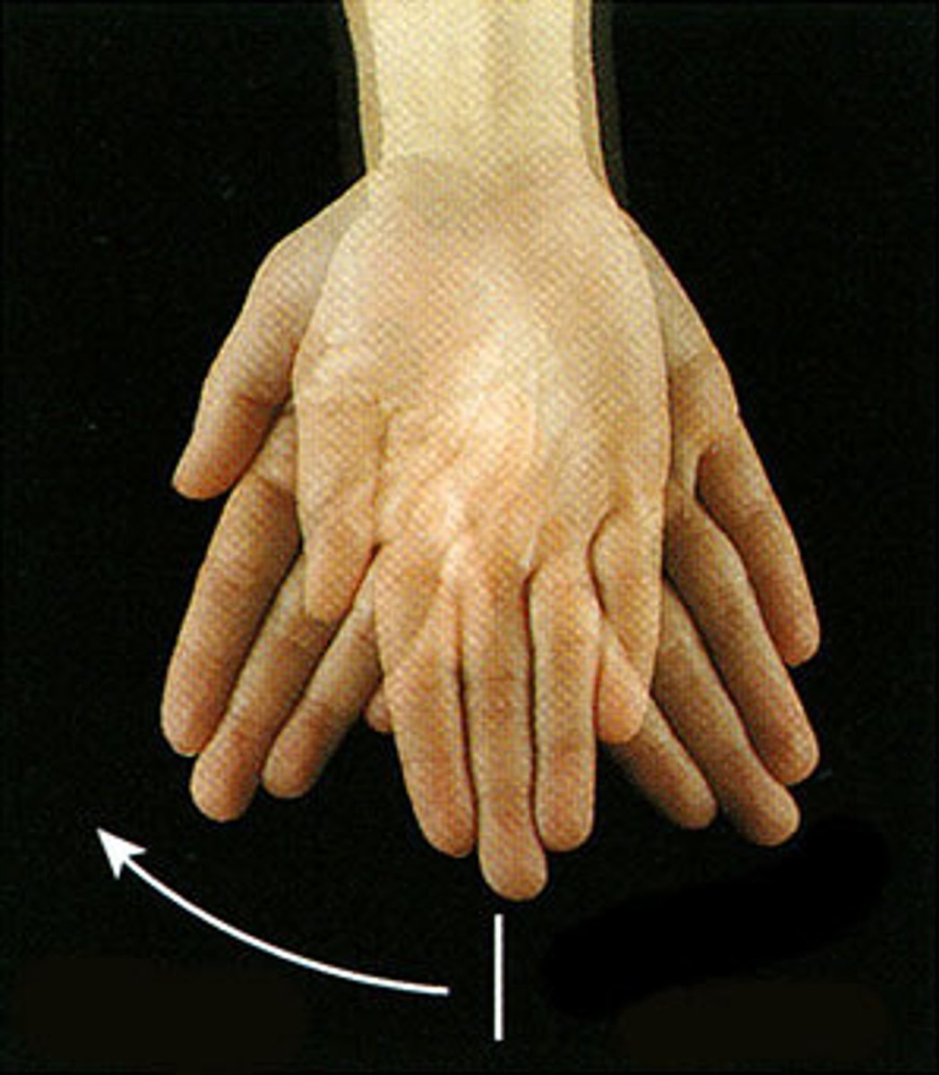
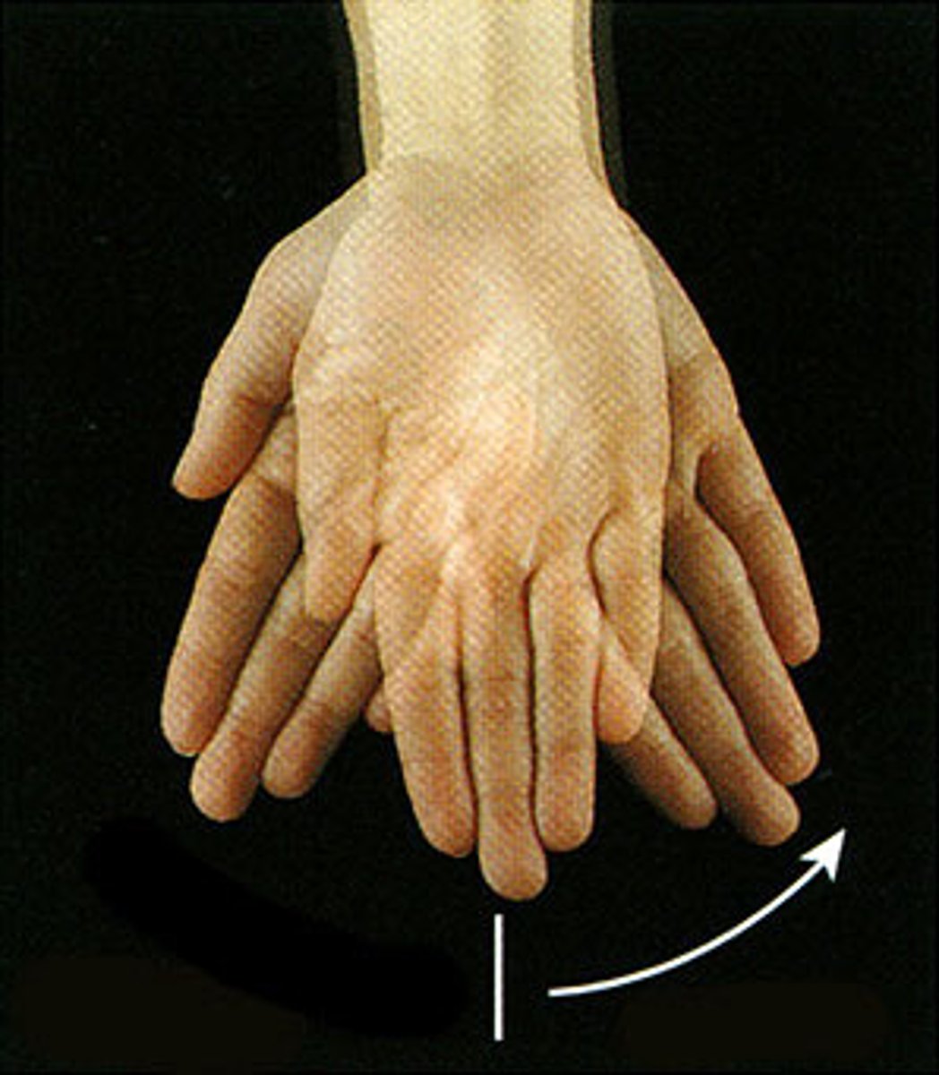
Wrist flexion
Wrist extension
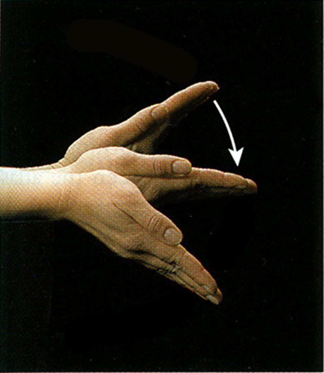
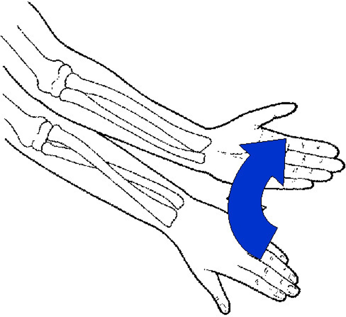
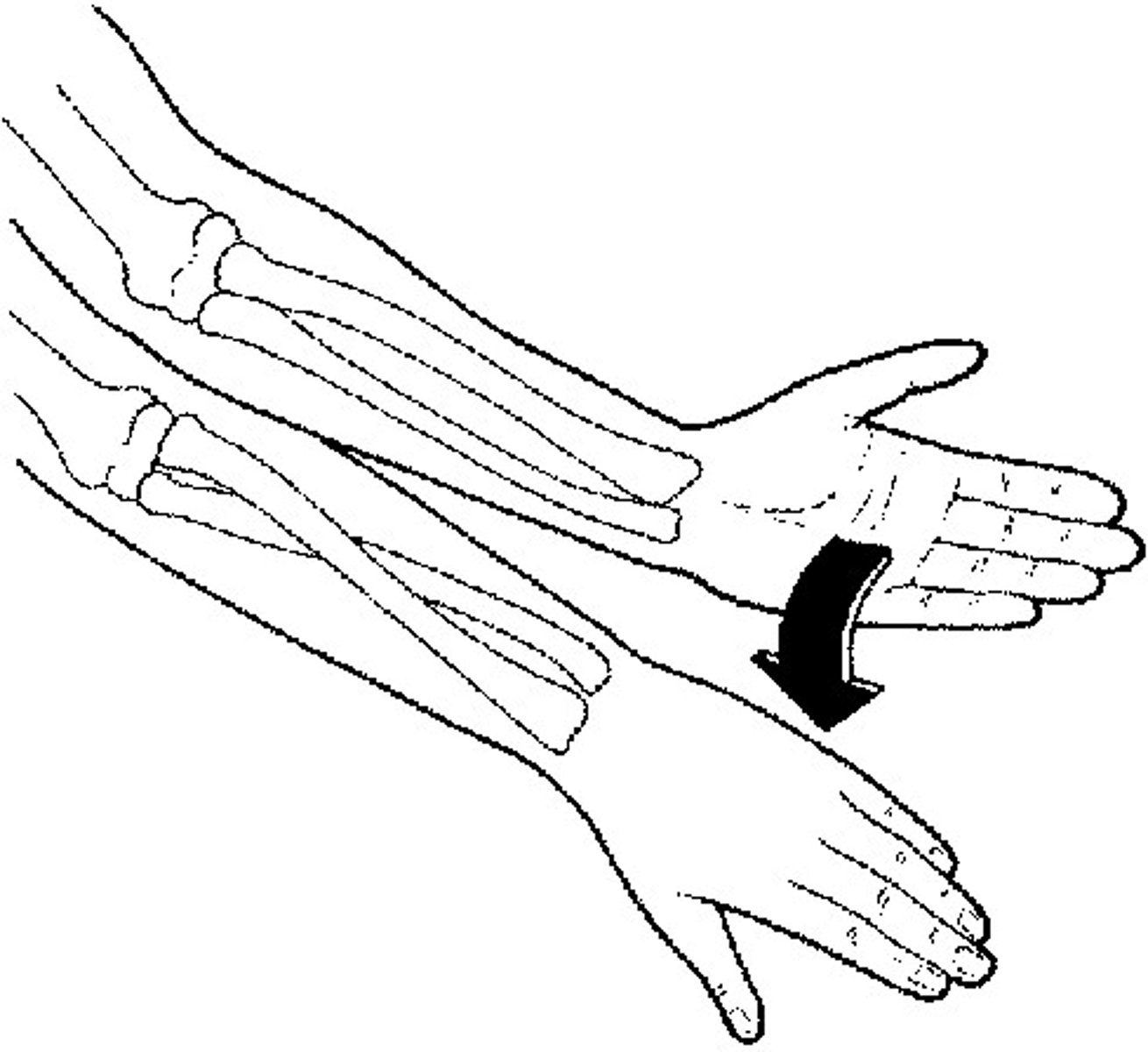
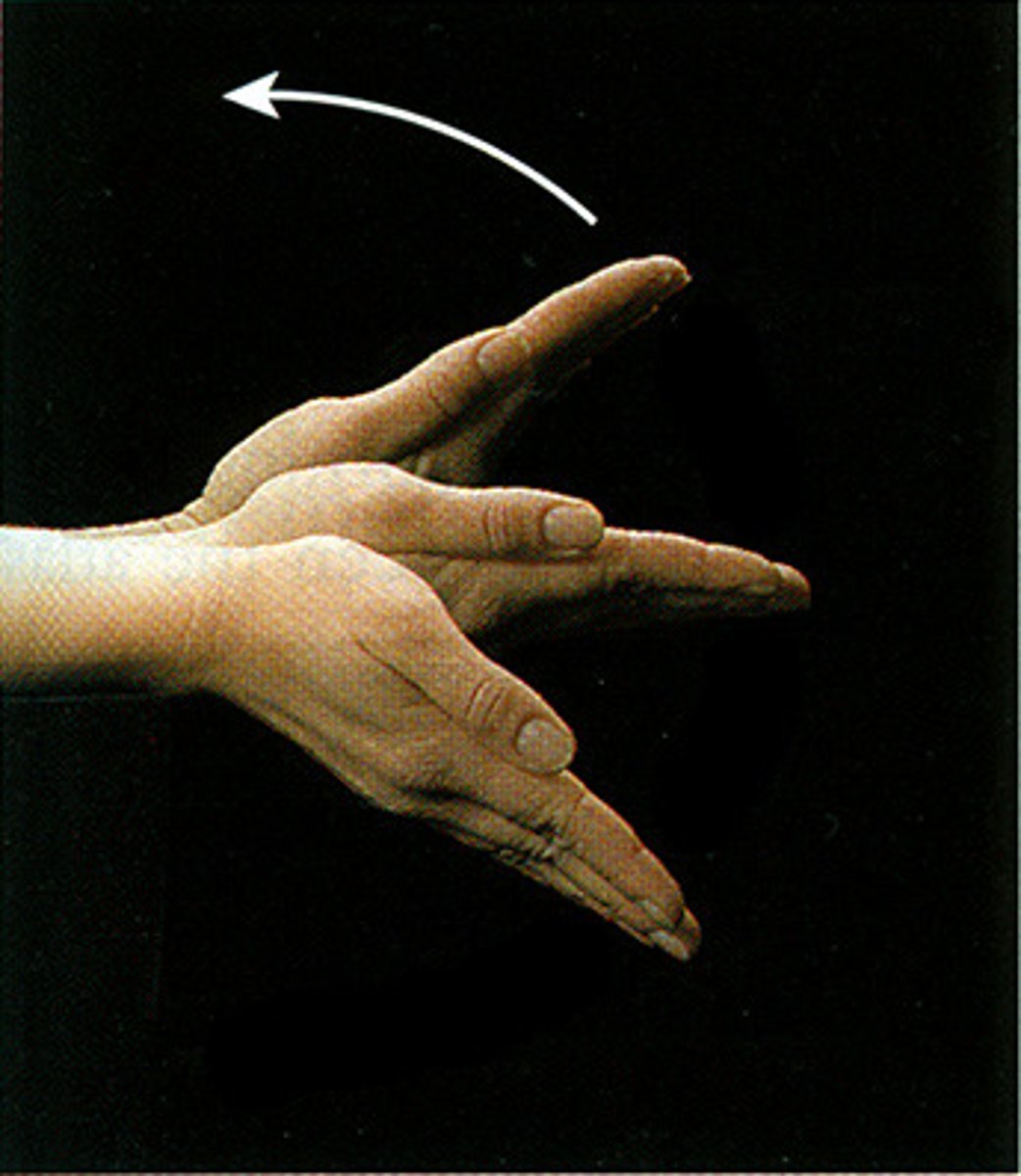
Radial deviation of the wrist
Ulnar deviation
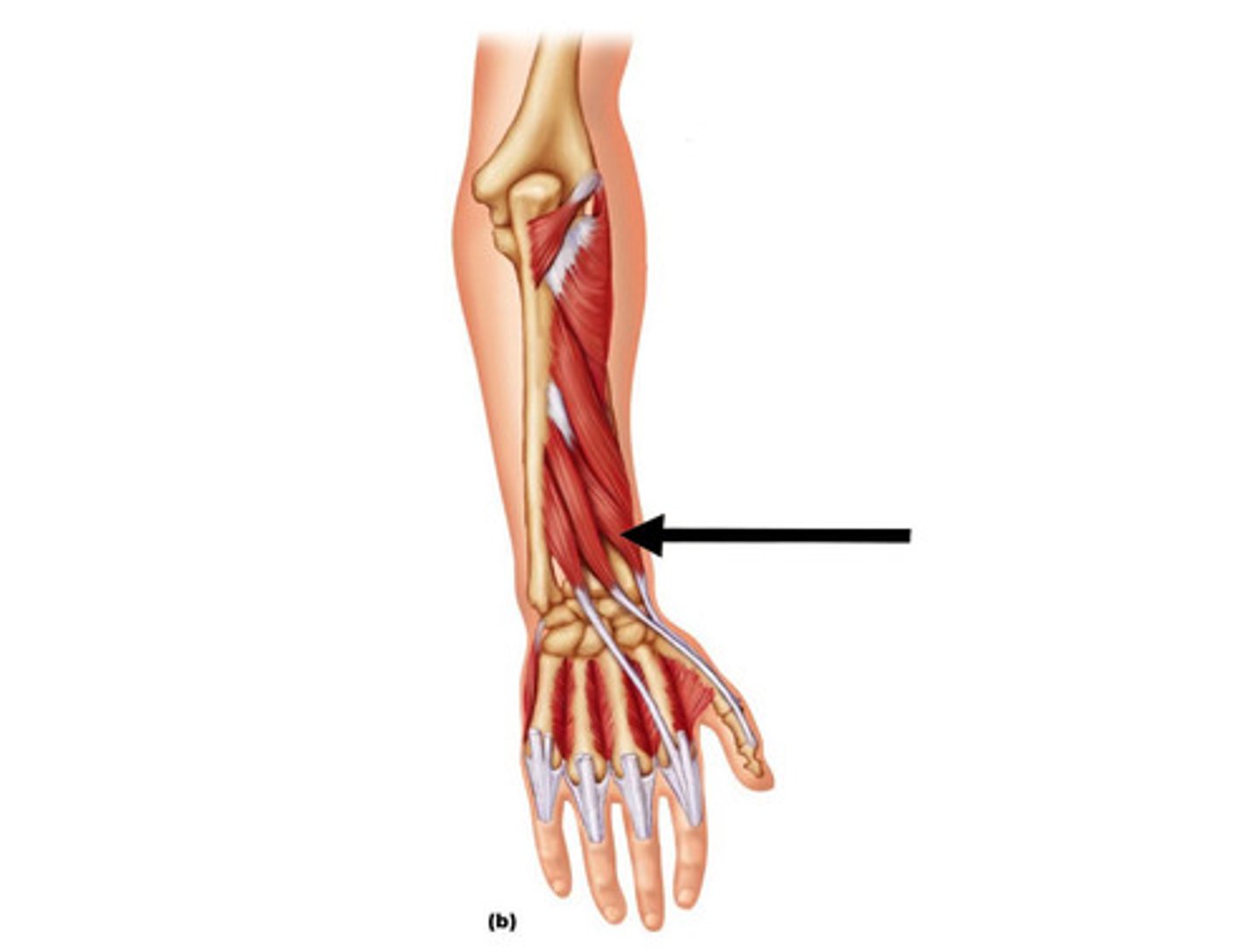
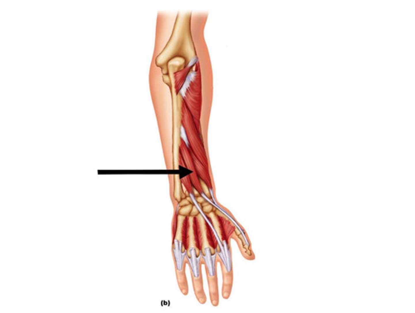
Extensor carpi radialis longus
Extends wrist
Extensor carpi radialis brevis
Extends wrist
Extensor carpi ulnaris
Extends wrist
Extensor digitorum
Extends four fingers
Abductor pollicis longus
Abducts thumb
Extensor pollicis brevis
Extends thumb
Extensor pollicis longus
Extends thumb
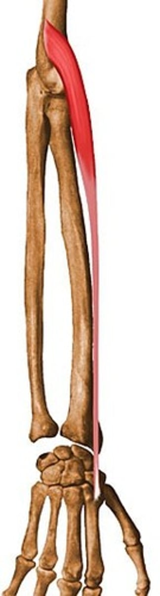
Flexor carpi ulnaris
Flexes wrist
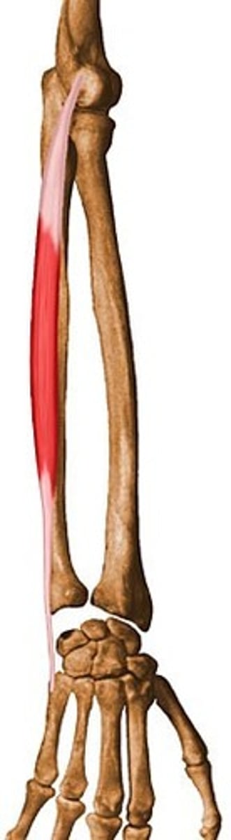
Flexor carpi radialis
Flexes wrist
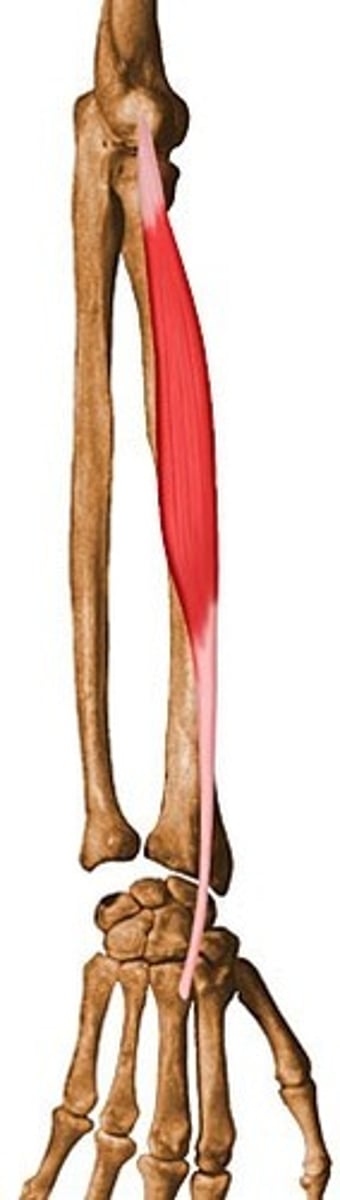
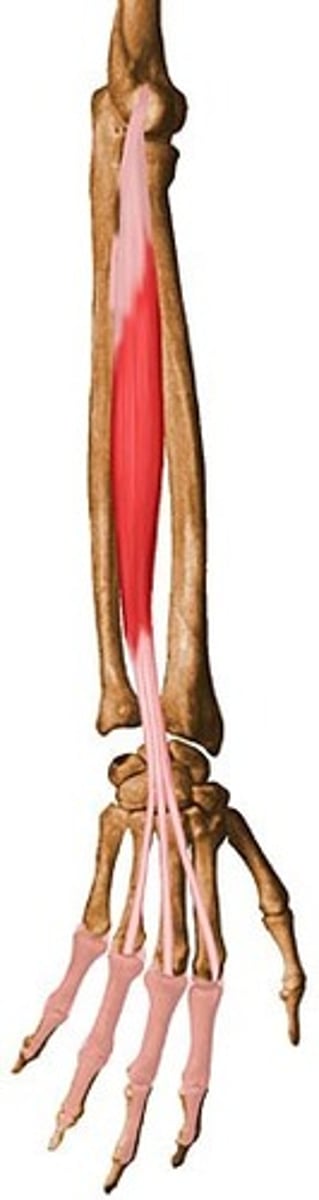
Flexor digitorum superficialis
Flexes fingers
Flexor digitorum profundus
Flexes fingers
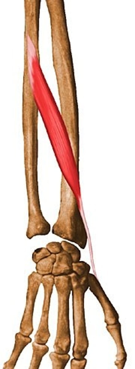
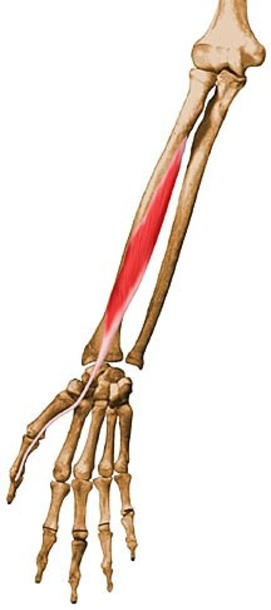
Flexor pollicis longus
Flexes thumb
Muscles that flex the elbow
Biceps brachii, brachialis, brachioradialis
Muscles that extend the elbow
Triceps brachii
Muscles that supinate the forearm
Supinator, biceps brachii
Muscles that pronate the forearm
Pronator teres, pronator quadratus
Muscles that extend the wrist
Extensor carpi ulnaris, extensor carpi radialis longus and brevis
Muscles that extend the fingers
Extensor digitorum
Muscles that extend the thumb
Extensor pollicis longus, extensor pollicis brevis
Muscles that flex the wrist
Flexor carpi ulnaris, flexor carpi radialis
Muscles that flex the fingers
Flexor digitorum superficialis, flexor digitorum profundus
Contents of the carpal tunnel
Median nerve and extrinsic flexor muscles
intrinsic
both originates and inserts in the foot
Foot/ankle functions
Allows stability (lever) and mobility (shock absorption)
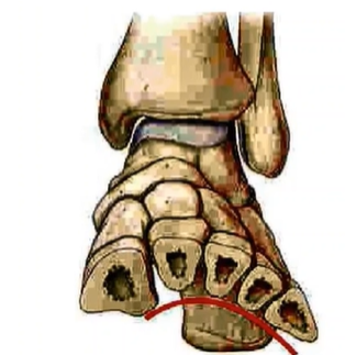
Metatarsophalangeal joints
Joints between metatarsals and the phalanges. Ball of the foot
Talocrural joint
Synovial hinge joint between the talus and distal end of tibia and fibula (medial and lateral malleolus)
talocrurual joint function
provides stability due to large area of bone contacts, the shape, and collateral ligaments.
Inversion
Sole of foot facing upwards medially
Eversion
sole of foot facing upwards laterally (outwards)
Deltoid ligaments
Span from navicular bone to the talus.
Support the talocrural joint
Can be damaged by eversion injury. (rolling sole of foot outwards)
What supports the foot in the longitudinal arch superficially?
Plantar fascia/aponeurosis
What supports the foot in the longitudinal arch deeply?
Long and short ligaments, spring ligament, and anterior/posterior/lateral msucle compartments of the leg
How do muscles in the leg support the longitudinal arch of the foot?
Tibularis anterior and tibularis posterior pull upwards to hold up and medially support the arch.
Fibularis longus from lateral side reaches under the lateral side of the foot, extending its tendons to the medial side of the foot (to the medial cuneiform and first metatarsal)
Transverse arch of the foot
Arch is formed due to wedge-shaped metatarsals, which prevent falling downwards when there is weight/forces from above
Where is the tarsal tunnel located?
Between medial malleolus and the calcaneus