Poultry exam
1/16
Earn XP
Description and Tags
part 2
Name | Mastery | Learn | Test | Matching | Spaced | Call with Kai |
|---|
No analytics yet
Send a link to your students to track their progress
17 Terms
17. Non-infectious, infectious and parasitic diseases of the beak, beak cavity and crop - etiology, symptoms, diagnostics, dif. dg, therapy and prevention.
Non-infectious:
Beak deformities (congenital, rickets)
Beak necrosis (fine feed accumulation)
Vitamin A deficiency → metaplasia
Foreign bodies → crop impaction
Chemicals, trauma
Infectious:
Bacteria: Pasteurella, Pseudomonas, Klebsiella
Viruses: fowl pox
Fungi: Candida albicans (candidiasis)
Parasitic:
Trichomonas gallinae
Capillaria spp.
1. Beak disorders
Congenital deformities: brachygnathia superior/inferior
Acquired deformities: rickets / osteomalacia
👉 Key mechanism: Ca + P + Vit D3 imbalance → poor bone mineralisation
Necrosis: due to fine feed accumulation (Finely ground feed → accumulation → pressure necrosis)
2. Stomatitis
Inflammation of oral mucosa
Causes: infectious + chemical
fowl pox
Candida
Trichomonas
bacteria (Pseudomonas, Klebsiella)
CS: swelling, lesions, reduced feed intake, weight loss, lethargy
3. Ingluvitis (crop inflammation)
Causes: feed (high fiber), chemicals, infections (pasteruella, candida, pox, trichomonas, Capillaria anulata, C. Contorta (flubendazole), Gongylonema ingluvicola.)
Types: catarrhal, hemorrhagic, necrotic
CS: anorexia, weight loss, foul regurgitation
4. Crop disorders
Impaction: foreign material → obstruction
Pendulous crop: enlarged, fluid-filled caused by nerve/muscle damage
Metaplasia → Vit A deficiency
5. Candidiasis (yeast)
Opportunistic (Candida albicans)
Often after antibiotics, dysbiosis and poor hygiene
CS:
Young: anorexia, crop stasis →regurgitation, white plaques
Adults: mild signs
Lesions: white pseudomembranes/plaques in crop → “sour crop”
Dx: cytology from crop swab (smears)
Tx: antifungals (nystatin)
6. Trichomoniasis
Protozoa (Trichomonas gallinae)
Transmission: contaminated water
CS:
yellow caseous plaques (‘yellow buttons’) in oral cavity
can block esophagus → starvation
Rapid weight loss, death
Tx: metronidazole
Diagnosis: crop swab → cytology
Prevention: hygiene, clean water
Clinical Signs (general):
Anorexia
Weight loss
Regurgitation
Oral plaques/lesions
Dysphagia
Diagnostics:
Clinical signs
Oral/crop examination
Cytology (Candida)
Detection of parasites
👉 Key line: diagnosis based on lesions + cytology/parasitology
Differential Diagnosis:
Vitamin deficiencies (A)
Infectious diseases (pox, bacterial infections)
Foreign body/obstruction
Therapy:
Cause-specific:
Antifungals (Candida)
Antiprotozoals (Trichomonas)
Supportive care
Prevention:
Good hygiene
Proper nutrition (Vit. A!)
Clean water
Avoid overcrowding
💡 Exam high-yield lines:
Candidiasis = white plaques in crop/oral cavity
Trichomoniasis = yellow caseous lesions blocking esophagus
Vit A deficiency = metaplasia
Crop disorders = regurgitation + foul smell
18. Non-infectious, infectious and parasitic diseases of the proventriculus and gizzard - etiology, symptoms, diagnostics, dif. dg, therapy and prevention.
Diseases of the proventriculus
1. Proventriculitis (inflammation)
Infectious cause:
Proventriculitis can be caused by viral infections (e.g. Newcastle disease), fungi, bacteria, and mycotoxins
Infectious: Candida, Newcastle disease
Fungal-like organism: Macrorhabdus ornithogaster (mainly pet birds, rare in poultry)
Toxic: mycotoxins (Fusarium, Aspergillus metabolites)
CS:
Weight loss
Regurgitation
Diarrhea
Poor feed conversion
Non-infectious cause:
Intoxocation (Mycotoxicosis)
Fusarium toxins → necorsis and ulceration of oral mucosa, GI reddening, visceral hemorrhages, lymphoid and spleen atrophy
Cyclopiazonic acid (Aspergillus flavus) → lesions in proventriculus, gizzard, liver, spleen, proventricular dilation + ulceration and thickend mucosa
PM: Enlarged, mottled proventriculus with thickened mucosa
CS: imparied feed conversion, decreased weight, mortality
3. Proventricular dilation syndrome
Cause: poor diet (high fiber, mash feed)
CS: enlarged proventriculus, thin wall, gizzard atrophy due to lack of food
4. Parasite: Tetrameres americana
Located in proventricular glands and visible as red spots on serosa
CS: anemia, weight loss, thickened proventriculus
Heavy infection: thick, edematous proventriculus + possible partial obstruction
Diseases of the gizzard
1. Impaction
Cause: litter ingestion (turkeys), indigestible material
CS: sudden death in young birds
2. Dilation of the gizzard
Cause: soft feed, lack of grit
→ inflammation + dysfunction
👉 “Lack of grit → poor mechanical digestion → dilation and dysfunction”
3. Parasite: Amidostomum anseris
Located under keratin layer of gizzard, proventriculus or esophagus in waterfowl
Causes hemorrhage + necrosis
Severe infections → anemia, blood loss
Clostridiosis
Clostridium spp. can cause necrosis in the proventriculus–gizzard junction
Macrorhabdus (megabacteria)
rare, mainly in companion birds, causes weight loss
🧠 Clinical Signs (general):
Weight loss
Regurgitation
Diarrhea
Poor growth
Anemia (parasites)
Sudden death (impaction)
🔬 Diagnostics:
Post-mortem examination (key!)
Detection of parasites in proventriculus/gizzard
Lesions: dilation, hemorrhages, thickening
❌ Differential Diagnosis:
Candidiasis (crop involvement)
Infectious enteritis
Mycotoxicosis vs bacterial enteritis
Foreign body obstruction
💊 Therapy:
Anthelmintics (parasites)
Supportive care
No specific treatment for toxicosis (remove feed source)
🛡 Prevention:
Good feed quality (no mold)
Provide grit
Proper litter management
Biosecurity
Deworming in free-range systems
💡 EXAM HIGH-YIELD POINTS:
Proventriculus diseases → toxins + parasites + diet problems
Gizzard impaction → turkeys + litter ingestion
Tetrameres → red spots in proventriculus
Amidostomum → gizzard hemorrhage in waterfowl
19. Non infectious and infectious diseases of the intestines and cloaca - etiology, symptoms, diagnostics, dif. dg, therapy and prevention.
Enteritis = inflammation of the intestines
- Main etiological groups: Bacteria, Viruses, Parasites, Intoxications, Non-specific causes (management, nutrition, stress)
🦠 1. INFECTIOUS DISEASES
A. Bacterial diseases
1. Necrotic enteritis
Etiology: Clostridium perfringens (toxins)
Predisposition: gut damage, coccidiosis, diet changes
Symptoms:
Sudden death, Depression, ruffled feathers, Diarrhea
Lesions:
Necrosis of small intestine (“Turkish towel” mucosa), Liver lesions
Diagnostics:
Necropsy + bacteriology
Differential diagnosis:
Coccidiosis
Salmonellosis
Therapy:
Antibiotics (penicillin, bacitracin)
Prevention:
Good hygiene, Control of coccidiosis, Feed management
2. Pullorum disease
Etiology: Salmonella pullorum
Transmission: vertical + horizontal
Symptoms:
High mortality in chicks, Weakness, diarrhea
Lesions:
Unabsorbed yolk sac, White nodules in organs, Cecal cores
Diagnostics:
Serology, bacteriology
Differential diagnosis:
Fowl typhoid
Colibacillosis
Therapy:
Usually not treated (eradication programs)
Prevention:
Testing breeder flocks, Biosecurity
3. Fowl typhoid
Etiology: Salmonella gallinarum
Symptoms:
Depression, diarrhea, Pale comb, dehydration
Lesions:
Similar to pullorum, but in older birds
Diagnostics:
Culture, serology
Differential diagnosis:
Pullorum disease
Therapy:
Antibiotics (limited use)
Prevention:
Vaccination + eradication
4. Colibacillosis (E. coli)
Etiology: Escherichia coli
Predisposition: poor hygiene
Forms:
Enterocolitis
Diarrhea, dehydration
Coligranuloma (Hjarre’s disease)
Granulomas in intestine, liver, mesentery
Diagnostics:
Culture
Differential diagnosis:
Tuberculosis
Therapy:
Antibiotics
Prevention:
Hygiene, ventilation
5. Avian tuberculosis
Etiology: Mycobacterium avium
Course: chronic
Symptoms:
Weight loss, emaciation, Lameness (bone involvement)
Lesions:
Granulomas in the intestine, liver
Diagnostics:
Necropsy, histology
Differential diagnosis:
Coligranuloma
Therapy:
Not practical → culling
Prevention:
Biosecurity, remove infected birds
Other bacteria
Pasteurella multocida (fowl cholera)
Campylobacter spp.
Yersinia enterocolitica (usually non-pathogenic)
B. Parasitic diseases (important examples)
Eimeria spp. → coccidiosis (bloody diarrhea)
Ascaridia spp. → obstruction, poor growth
Capillaria spp. → enteritis
Histomonas meleagridis → blackhead disease
Trichomonas spp. → upper GIT, sometimes intestines
Key signs:
Diarrhea, Weight loss, Poor performance
Diagnostics:
Fecal exam (oocysts, eggs)
Therapy:
Antiparasitics (coccidiostats, anthelmintics)
Prevention:
Hygiene, Litter management
C. Viral enteritis (brief)
Rotavirus, coronavirus, adenovirus, parvovirus
Symptoms: diarrhea, poor growth, mortality in young birds
Diagnosis: PCR, histopathology
Prevention: biosecurity, vaccination (when available)
Newcastle disease
Causes enteric + respiratory + neurological signs
Highly contagious, high mortality
2. NON-INFECTIOUS DISEASES
A. Intoxications
Heavy metals
Copper (CuSO₄ overdose):
Catarrhal enteritis
Mucous exudate
Mercury:
Caustic burns, ulcers
Phosphorus:
Diarrhea, weakness, anorexia
General signs:
Diarrhea, Depression, Weight loss
Diagnostics:
History + toxicology
Therapy:
Remove toxin, Supportive care
Prevention:
Proper dosing, Avoid contaminated feed/water
B. Non-specific causes
Poor nutrition
Sudden feed change → enteritis
Stress
Dysbiosis
C. Hypomotility - nervous origin
D. Volvulus – due to gas → obstruction
CLOACAL DISEASES
1. Cloacitis
Etiology:
Infection, irritation, enteritis
Symptoms:
Dirty vent, Foul odor. Ulceration, Stop laying eggs
Therapy:
Clean area, Antibiotics (if bacterial)
Prevention:
Hygiene
2. Cloacal prolapse
Causes:
Enteritis (straining)
Egg-laying problems
Symptoms:
Protruding cloaca
Therapy:
Reposition + supportive care
Prevention:
Proper nutrition (Ca, fiber)
Avoid obesity
🧠 Differential diagnosis (important exam part)
Coccidiosis vs necrotic enteritis
Salmonellosis vs colibacillosis
Tuberculosis vs coligranuloma
Intoxication vs infectious enteritis
✅ Key prevention principles
Biosecurity
Hygiene & litter management
Proper nutrition
Vaccination (Salmonella, others)
Parasite control
20. Protozoan diseases of the GIT - etiology, symptoms, diagnostics, dif. dg, therapy and prevention.
1. COCCIDIOSIS (most important)
Etiology
Eimeria spp. (intracellular protozoa)
Very common → disease occurs only with high infection dose
Important species (chickens)
Eimeria tenella → ceca (bloody diarrhea) ⭐
Eimeria necatrix → small intestine (severe hemorrhage) ⭐
Eimeria acervulina → mild, poor growth
Eimeria brunetti → lower intestine, necrosis
👉 Most pathogenic: E. tenella & E. necatrix (deep tissue schizogony → hemorrhage)
Pathogenesis
Ingestion of sporulated oocysts
Destruction of intestinal epithelium → hemorrhage
Symptoms
Diarrhea (often bloody), Dehydration, Weight loss, High mortality
Predisposes to secondary infections (e.g. Clostridium)
Lesions
Hemorrhagic enteritis, Thickened intestinal wall, Cecal cores (E. tenella)
Diagnostics
Fecal flotation (oocysts)
Necropsy
Differential diagnosis
Necrotic enteritis
Salmonellosis
Therapy
Toltrazuril, sulfonamides
Ionophores (in feed)
Prevention
Hygiene, Anticoccidials in feed, Vaccination
2. HISTOMONIASIS (Blackhead disease)
Histomonas meleagridis
Transmitted via Heterakis gallinarum eggs
Pathogenesis
Affects ceca + liver
Symptoms
Depression, anorexia, Yellow diarrhea, Cyanotic head (“blackhead”), High mortality (especially turkeys)
Lesions (pathognomonic ⭐)
Ceca: thickened wall, ulcers, cheesy core
Liver: round necrotic lesions
👉 Severe cases → perforation → peritonitis
Diagnostics
Necropsy (very characteristic lesions)
Differential diagnosis
Trichomoniasis
Therapy
No effective protozoal treatment
Control of Heterakis (e.g. flubendazole)
Prevention
Deworming, Hygiene, Separation of turkeys and chickens
3. TRICHOMONIASIS
Trichomonas gallinae
Location
Upper GIT (crop, esophagus, pharynx)
Symptoms
Dysphagia, Weight loss, Weakness, Death (especially pigeons), Death within 8–10 days (severe cases)
Lesions
“Yellow buttons” (caseous plaques)
Diagnostics
Microscopy (crop swab)
Differential diagnosis
Histomoniasis
Vitamin A deficiency
Therapy
Nitroimidazoles (non-food birds only)
Prevention
Clean water, Avoid bird-to-bird transmission
🧠 General clinical signs (important summary)
Diarrhea, Weight loss, Dehydration, Reduced growth, Mortality
🔍 Diagnostics (general)
Fecal examination (oocysts), Microscopy, Necropsy lesions
⚖ Differential diagnosis (exam favorite)
Bacterial enteritis (Clostridium, Salmonella)
Viral enteritis
Helminths
💊 Therapy (general)
Antiprotozoals (toltrazuril, sulfonamides), Supportive care
🛡 Prevention
Hygiene and sanitation, Dry litter, Anticoccidials in feed, Control of intermediate hosts
🧠 KEY EXAM TIPS
⭐ Coccidiosis = most important
⭐ Histomoniasis = pathognomonic liver lesions
⭐ Trichomoniasis = yellow plaques in upper GIT
⭐ Always mention:
fecal exam
biosecurity
⭐ E. tenella → bloody cecal diarrhea
⭐ Histomoniasis → liver + cecal lesions (pathognomonic)
⭐ Transmission via Heterakis = classic question
⭐ Secondary Clostridium infection → very important link
21. Helminthoses of the GIT - etiology, symptoms, diagnostics, dif. dg, therapy and prevention.
🪱 Etiology
Nematodes (most important)
Ascaridia galli
Capillaria spp.
Heterakis gallinarum
Others (less common)
Davainea proglottina (cestode)
Prosthogonimus spp.
Trichostrongylus tenuis
Amidostomum spp.
🪱 Nematodes
1. ASCARIDIOSIS
Ascaridia galli
Affects the small intestine
Direct life cycle
Pathogenesis
Mechanical irritation
Obstruction of intestine (duodenum/jejunum)
Migration → cloaca
Clinical signs
(depend on worm burden)
Weight loss, Decreased egg production, Diarrhea, Anemia, Depression (drooping wings, ruffled feathers) Mortality (severe cases)
Diagnostics
Fecal examination (eggs), Necropsy (adult worms)
Differential diagnosis
Coccidiosis, Bacterial enteritis, Malnutrition
Therapy
Anthelmintics:
Piperazine, Flubendazole, Ivermectin, Tetramisole
Prevention
Hygiene and sanitation, Litter management, Rotation of pasture
2. CAPILLARIOSIS
Capillaria spp.
Location:
Crop: C. annulata, C. contorta
Small intestine: C. obsignata, C. bursata, C. caudinflata
Ceca: C. anatis
Life cycle
Direct or indirect
Some species require earthworms as intermediate hosts
Pathogenesis
Inflammation of mucosa, Thickening of intestinal wall
Severe cases → catarrhal/croupous enteritis
Clinical signs (similar to ascaridiosis)
Weight loss, Diarrhea, Anemia, Poor production, Mortality (heavy infections)
Diagnostics / Therapy / Prevention
👉 Same as Ascaridia
3. HETERAKIOSIS
Heterakis gallinarum
Located in ceca
Direct life cycle (Earthworms = transport hosts)
Pathogenesis
Cecal inflammation, Thickened mucosa, Petechial hemorrhages
⭐ VERY IMPORTANT:
Vector for Histomonas meleagridis (blackhead disease)
Clinical signs
Usually mild:
Poor growth, Depression
Severe cases → death
Diagnostics
Fecal exam, Necropsy
Therapy
Anthelmintics (flubendazole, etc.)
Prevention
Deworming, Hygiene, Control of earthworms
🪱 Cestodes (tapeworms)
Examples:
Davainea proglottina → hemorrhagic enteritis (duodenum)
Raillietina tetragona → ileum
Indirect life cycle (snails, slugs, ants)
Clinical signs:
Diarrhea, Poor growth
Therapy:
Praziquantel
Prevention:
Control intermediate hosts
🪱 Trematodes (flukes)
Example:
Prosthogonimus spp.
Life cycle: snail + dragonfly
Location:
Intestine → can migrate to oviduct
Clinical importance:
↓ egg production
Peritonitis (rupture)
👉 Prevention:
Avoid wet environments (snails!)
🧠 OTHER HELMINTHS (brief mention)
Trichostrongylus tenuis
→ affects ceca/intestine → causes weight loss, anemia, enteritisAmidostomum spp. (gizzard worms)
→ affect gizzard → cause gizzard erosion, anemia, poor growth
🧠 GENERAL CLINICAL SIGNS (important summary)
Weight loss, Diarrhea, Anemia, Reduced egg production, Poor growth, Mortality (heavy infections)
🔍 DIAGNOSTICS
Fecal examination (eggs), Necropsy
⚖ DIFFERENTIAL DIAGNOSIS
Coccidiosis, Bacterial enteritis, Viral enteritis, Nutritional deficiencies
💊 THERAPY
Anthelmintics (flubendazole, ivermectin, piperazine)
Supportive care
🛡 PREVENTION
Hygiene & sanitation
Dry litter
Pasture rotation
Control of intermediate hosts
🎯 KEY EXAM POINTS
⭐ Ascaridia galli → intestinal obstruction
⭐ Capillaria → thin worms, severe mucosal damage
⭐ Heterakis gallinarum → vector of histomoniasis
⭐ Always mention:
fecal exam, deworming, hygiene
22. Diseases of bursa of Fabricius - etiology, symptoms, diagnostics, dif. dg, therapy and prevention.
The Bursa of Fabricius:
Primary lymphoid organ in birds
Located dorsal to cloaca
Responsible for B-cell (humoral) immunity
Atrophies around 6 months of age
🦠 1. Infectious bursal disease (IBD, Gumboro) ⭐ MOST IMPORTANTE
Birnavirus
Serotype 1 → pathogenic (chickens)
Serotype 2 → non-pathogenic (no disease/immunosuppression)
Highly contagious: Fecal-oral + fomites (equipment, clothes, vehicles)
Pathogenesis
Destruction of B lymphocytes
→ severe immunosuppression
Clinical signs
Subclinical (age < 3 weeks)
No obvious signs
Poor vaccine response → Severe immunosuppression → Secondary infections
Clinical (age 3–6 weeks)
Depression, prostration, Watery diarrhea, Soiled vent, Cloacal inflammation, Incoordination
Lesions
Acute: enlarged, edematous, hemorrhagic bursa
Chronic: bursal atrophy
Diagnostics
PCR, Necropsy findings
Differential diagnosis
Marek’s disease
Lymphoid leukosis
Coccidiosis (diarrhea cases)
Therapy
❌ No specific treatment
Supportive care
Prevention
Vaccination (core vaccine), Biosecurity
2. Lymphoid leukosis
Avian leukosis virus (retrovirus)
Key features
Bursal-dependent tumor disease
Affects birds >14–16 weeks (slow tumor development)
Pathogenesis
Infection of B cells → tumor formation
Organs affected:
Liver, Spleen, Bursa
Clinical signs
Non-specific:
Weakness, Weight loss, Diarrhea
Tumors develop later
Diagnostics
PCR, Necropsy (tumors)
Differential diagnosis
Marek’s disease ⭐ important (No neurological signs (unlike Marek’s disease)
Prevention
Eradication programs
Control vertical transmission
3. Marek’s disease
Gallid herpesvirus (MDV)
Key features
Highly contagious - Spread via feather dander (very resistant in environment)
Causes:
Tumors, Nerve lesions (very important difference!)
Pathogenesis: Virus infects T-lymphocytes → lymphoma formation
Clinical signs
Paralysis (legs, wings) ⭐, Weight loss, Ocular changes, Skin lesions
Bursa involvement
Usually atrophy, not tumors
Diagnostics
Clinical signs + PCR
Differential diagnosis
Lymphoid leukosis (NO nerve signs there!)
Prevention
Vaccination (very important)
Prosthogonimus
May affect bursa (young) and oviduct (older birds)
Causes ↓ egg production, cloacal issues
🧠 SUMMARY TABLE (very useful for exam)
Disease | Age | Bursa | Key sign |
|---|---|---|---|
IBD | 3–6 weeks | Enlarged → atrophy | Immunosuppression |
Leukosis | >14 weeks | Tumors | Neoplasia |
Marek | Any (often young) | Atrophy | Paralysis |
🔍 Diagnostics (general)
PCR, Necropsy
Histopathology
⚖ Differential diagnosis (important!)
Marek vs Leukosis:
Marek → nerve lesions
Leukosis → bursal tumors
💊 Therapy
Mostly no specific treatment
Supportive care
🛡 Prevention
Vaccination (IBD, Marek), Biosecurity, Breeding control
🎯 KEY EXAM POINTS
⭐ IBD = most important (immunosuppression!)
⭐ Age matters (very exam-relevant)
⭐ Marek = paralysis
⭐ Leukosis = tumors in older birds
⭐ Bursa = B-cell immunity
23. Non-infectious and parasitic diseases associated with changes in the liver - etiology, symptoms, diagnostics, dif. dg, therapy and prevention.
1. Non-infectious
Metabolic:
Fatty liver hemorrhagic syndrome (FLHS)
Intoxications
Mycotoxins (aflatoxicosis)
Heavy metals (lead, mercury)
Arsenic, cobalt, phosphorus
2. Parasitic / infectious affecting liver
Protozoa:
Histomoniasis (Histomonas meleagridis) ⭐
Trichomoniasis (Trichomonas gallinae)
Fungal:
Aspergillosis (Aspergillus fumigatus)
🟡 1. Fatty liver hemorrhagic syndrome (FLHS)
High-energy diet + low exercise
Diet should contain ~0.3 ppm selenium
Seen in caged laying hens (especially summer)
Pathogenesis
Excess fat accumulation → fragile liver → rupture → hemorrhage
Clinical signs
Obesity, ↓ egg production, Sudden death, Pale comb
Lesions
Enlarged, yellow, friable liver, Hemorrhages, Abdominal fat deposits
Diagnostics
History + necropsy
Differential diagnosis
Aflatoxicosis
Infectious liver diseases
Therapy
Not practical (often sudden death)
Prevention
Diet control, Weight management, Reduce energy intake
Aflatoxicosis
Mycotoxins from fungi:
Aspergillus
Penicillium
Aflatoxicosis is one of the most important causes of liver damage in poultry production!
Clinical signs
Depression, Inappetence, Neurological signs (ataxia, convulsions), Death
Lesions
Liver = enlarged, yellow, sometimes necrotic
Diagnostics
Feed analysis, History
Therapy
Remove contaminated feed, Supportive care
Prevention
Proper feed storage, Avoid mold contamination
Heavy metals
Lead
Liver degeneration, atrophy
Mercury
Fatty degeneration
3. Histomoniasis (important parasitic cause)
Histomonas meleagridis
Transmission via Heterakis gallinarum eggs
Pathogenesis
Cecum → liver spread
Severe necrosis
Clinical signs
Depression, Sulfur-yellow diarrhea (very characteristic), Cyanotic head (black head disease), High mortality
Lesions (pathognomonic ⭐)
Liver: round necrotic lesions
Ceca: caseous cores
Diagnostics
Necropsy
Differential diagnosis
Coccidiosis, Bacterial hepatitis
Therapy
No effective treatment
Prevention
Deworming (Heterakis), Biosecurity, Separate turkeys and chickens
4. Trichomoniasis
Trichomonas gallinae
Liver involvement
Can cause necrotic lesions in liver
Clinical signs:
Weight loss, Weakness, Upper GIT lesions
Diagnostics
Microscopy
Therapy
Nitroimidazoles (non-food birds)
Prevention
Hygiene, Clean water
🍄 5. Aspergillosis
Aspergillus fumigatus
Pathogenesis
Primarily respiratory
May spread → liver granulomas
Clinical signs
Respiratory signs, Weakness
Lesions
Granulomas in:
Air sacs, Lungs, Liver
Diagnostics
Culture, histology
Therapy
Antifungals (limited success)
Prevention
Good ventilation, Dry litter
🧠 GENERAL CLINICAL SIGNS
Weight loss,Decreased production, Depression, Mortality
Liver enlargement
🔍 DIAGNOSTICS
Necropsy, Feed analysis
Microscopy/PCR
⚖ DIFFERENTIAL DIAGNOSIS
Infectious hepatitis
Coccidiosis
Septicemia
Nutritional disorders
💊 THERAPY
Often supportive
Remove cause (toxins, parasites)
🛡 PREVENTION
Proper nutrition
Good feed storage
Biosecurity
Parasite control
🎯 KEY EXAM POINTS
⭐ FLHS = metabolic, high-energy diet, sudden death
⭐ Aflatoxicosis = most important toxin
⭐ Histomoniasis = liver necrosis (classic lesions)
⭐ Always mention:
feed quality
management
parasite control
24. Infectious diseases associated with changes in the liver - etiology, symptoms, diagnostics, dif. dg, therapy and prevention.
🦠 1. SALMONELLOSIS
I. Pullorum disease
Salmonella pullorum
Vertical + horizontal transmission
Clinical signs
High mortality in chicks (acute), Weakness, diarrhea
Lesions
Unabsorbed yolk sac
White-gray nodules (necrotic foci) in liver and organs ⭐
Diagnostics
Culture, PCR
II. Fowl typhoid
Salmonella gallinarum
Clinical signs
Affects older birds, Depression, diarrhea, Pale, dehydrated birds
Pathogenesis:
After ingestion, bacteria invade intestinal epithelium → septicemia → spread to liver, spleen, kidneys
Lesions
Similar to pullorum
Hepatomegaly, congestion ± necrosis
may have hemorrhages and granulomatous peritonitis
Diagnosis:
PCR / ELISA needed to differentiate from pullorum disease
Differential diagnosis (for both)
Colibacillosis, Coccidiosis, Necrotic enteritis
Therapy
No effective treatment; eradication (depopulation) due to carrier state
Prevention
Biosecurity, Testing breeder flocks, Hygiene
III. Salmonella typhimurium
Causes paratyphoid infections
Similar lesions (liver congestion, necrosis)
🦠 2. AVIAN TUBERCULOSIS
Mycobacterium avium
Clinical signs
Chronic course, Weight loss, emaciation, Lameness
Lesions
Granulomas in liver, spleen, intestine ⭐
Diagnostics
Tuberculin test, Histology (Ziehl–Neelsen staining)
Differential diagnosis
Coligranuloma, Neoplasia
Therapy
❌ Not practical
Prevention
Culling, Sanitation
🦠 3. DUCK VIRAL HEPATITIS
Picornavirus (Avihepatovirus)
Clinical signs
Young ducklings (<7 weeks), Sudden death, Neurological signs (opisthotonos)
Lesions
Enlarged liver with hemorrhages ⭐
Diagnostics
PCR, Necropsy
Differential diagnosis
Aflatoxicosis
Septicemia
Duck viral enteritis
Septicemia
Mycotoxicosis
Therapy
❌ No treatment
Prevention
Vaccination of breeders
Biosecurity
🦠 4. INCLUSION BODY HEPATITIS (IBH)
Avian adenovirus
Pathogenesis
Liver damage with intranuclear inclusion bodies ⭐
Clinical signs
Sudden mortality, Depression, Pale comb, Sometimes jaundice
Lesions
Pale, enlarged, friable liver, Hemorrhages
Diagnostics
Histology (inclusions), PCR
Differential diagnosis
IBD (immunosuppression link)
Aflatoxicosis
Therapy
❌ No effective treatment
Prevention
Control immunosuppressive diseases:
Infectious bursal disease
Chicken infectious anemia
🧠 GENERAL CLINICAL SIGNS
Depression, Diarrhea, Weight loss, Sudden death, ↓ production
🔍 DIAGNOSTICS
Necropsy (very important), PCR, Culture
⚖ DIFFERENTIAL DIAGNOSIS (important)
Aflatoxicosis, Fatty liver syndrome, Histomoniasis, Septicemia
💊 THERAPY
Limited, Often supportive or culling
🛡 PREVENTION
Biosecurity
Vaccination (where available)
Good management
Control of immunosuppression
🎯 KEY EXAM POINTS
⭐ Salmonella → necrotic foci in liver
⭐ TB → granulomas (chronic)
⭐ Duck hepatitis → acute, high mortality, hemorrhagic liver
⭐ IBH → inclusion bodies in hepatocytes
⭐ Many have no treatment → prevention is key
25. Non-infectious and infectious diseases of the female reproductive organs - etiology, symptoms, diagnostics, dif. dg, therapy and prevention.
In birds, only the left ovary and oviduct are functional.
Non-infectious
Cysts, Degeneration (toxins, vitamin A deficiency), Hypoplasia (genetic), Neoplasia
Infectious
Bacterial: Salmonella, E. coli, Mycoplasma, Pasteurella
Viral: Infectious bronchitis, egg drop syndrome
Parasitic: Prosthogonimus spp.
DISEASES OF THE OVARY
1. Non-infectious
I. Cysts
Fluid-filled structures
Etiology: Endocrine imbalance, Chronic inflammation
Usually incidental finding
❗ Minimal effect on production
II. Degeneration
Causes:
Toxins, Vitamin A deficiency, Infections (e.g. Fowl typhoid)
Lesions:
Misshapen, dark follicles
III. Hypoplasia
Congenital
Small, underdeveloped ovary
↓ egg production
IV. Neoplasia
Adenocarcinoma
Lymphosarcoma (e.g. Marek's disease)
2. Infectious ovarian diseases
I. Salmonellosis
Misshapen follicles
Caseous content
II. Fowl cholera
Oophoritis (ovarian inflammation)
III. Infectious bronchitis
↓ egg production (up to 70%)
Poor egg quality:
thin shell, soft shell, watery albumen
DISEASES OF THE OVIDUCT
Salpingitis ⭐ (VERY IMPORTANT)
Multifactorial:
Trauma, ascending infection from cloaca, Spread from air sacs
Common agents:
E. coli, Mycoplasma gallisepticum, Salmonella, Pasteurella multocida
Clinical signs
Abnormal eggs (soft, misshapen, shell-less), ↓ egg production, Cloacal discharge, “Penguin posture” ⭐
Lesions
fibrin + caseous exudate, hyperemia, edema in the oviduct, Oviduct dilation, Secondary ovarian atrophy
Diagnostics
Necropsy, Bacteriology
Differential diagnosis
Egg retention, Egg drop syndrome, Cloacitis
Therapy
Antibiotics (early stages only)
Prevention
Hygiene, Reduce trauma, Control respiratory infections
Egg peritonitis
Etiology:
Reverse flow of egg material + bacteria (often E. coli)
Clinical signs:
Distended abdomen, Penguin posture, ↓ egg production
Lesions:
Fibrin / “cooked egg” material in abdomen
Diagnosis:
Necropsy
Therapy:
Poor prognosis
II. Egg retention (egg binding)
Etiology: Large/deformed egg, Nutritional deficiency (Ca!) Oviduct dysfunction
Clinical signs
Straining, Depression, Distended abdomen, Swollen vent
Complications
Oviduct rupture → peritonitis
Therapy
Lubrication, Massage, Surgery
Prevention
Proper nutrition (Ca, vitamins)
III. Egg drop syndrome (EDS)
Adenovirus
Clinical signs
Sudden drop in egg production, Soft or shell-less eggs
Diagnostics
History + production data
Differential diagnosis:
Newcastle disease, Avian influenza, Vitamin D₃ deficiency
Prevention
Vaccination, Avoid contaminated vaccines
IV. Prolapse
Egg retention, Salpingitis, Cloacal inflammation
Clinical signs
Visible prolapse, Cannibalism risk
Therapy
Clean, Disinfect, Reposition
Prevention
Good management, Prevent egg binding
V. Prosthogonimus spp.
Trematodes
Indirect life cycle (snails, insects)
Clinical signs
↓ egg production, Soft-shelled eggs
Severe: oviduct rupture → peritonitis
Diagnostics
Eggs in feces, Necropsy
Therapy
Limited
Prevention
Control intermediate hosts, Dry environment
Other:
Mycoplasma meleagridis – low hatchability
IBD – cystic dilation of oviduct - Birnavirus
NCD – low egg production
Fungi – Aspergillosis, F2 → estrogenic → cystic degeneration of ovary
Prolapse – cannibalism, other parts might be pulled from abdominal cavity.
🧠 GENERAL DIAGNOSTICS
History (egg production!), Clinical signs, Necropsy, Microbiology
⚖ DIFFERENTIAL DIAGNOSIS (IMPORTANT)
Infectious bronchitis
Egg drop syndrome
Nutritional deficiencies
Cloacitis
🛡 PREVENTION (KEY POINTS)
Biosecurity
Vaccination (IB, EDS)
Proper nutrition (Ca, vitamin A)
Hygiene
🎯 WHAT YOU SHOULD ADD (from your original notes)
✔ Mention:
“Penguin posture” (salpingitis) ⭐
Ascending vs descending infection
Vitamin A deficiency (degeneration)
Ca deficiency (egg binding)
❗ FINAL TIP
“Salpingitis is the most important oviduct disease and is usually multifactorial, involving bacterial infection combined with mucosal damage.”
In birds, only the left ovary is functional, and the most important reproductive disorders include salpingitis, egg retention, and egg peritonitis, often associated with bacterial infections such as E. coli.
26. Non-infectious and infectious diseases of the male reproductive organs - etiology, symptoms, diagnostics, dif. dg, therapy and prevention. Artificial insemination in poultry.
Non-infectious
Atrophy, Hypoplasia, Hyperplasia (physiological in breeding season), Neoplasia (rare)
Infectious
Orchitis, Epididymitis
Bacterial: E. coli, Salmonella, Staph, Strep
Viral: Newcastle disease, Marek's disease
Fungal: Aspergillus
Protozoa: Trichomonas
Venereal infections
DISEASES OF TESTICLES
1. Orchitis (inflammation) ⭐
Bacterial (especially Salmonella, E. coli)
Clinical signs
Testicular enlargement or later:
Orchitis may lead to testicular atrophy, fibrosis, and calcification
Diagnostics
Clinical exam, Necropsy
complete blood count:
leukocytosis, heterophilia (neutrophilia in mammals)
Differential diagnosis
Neoplasia, Degeneration
Therapy
Antibiotics (based on sensitivity)
Prevention
Hygiene, Control systemic infections
2. Atrophy
etiology: Age, Malnutrition, Chronic disease, Vitamin deficiency
Signs
↓ fertility, Reduced testicle size
3. Hypoplasia
Congenital, Small, non-functional testes
4. Hyperplasia
Physiological during breeding season
5. Neoplasia
Rare, Mostly in older birds
DISEASES OF PENIS (phallus – present in waterfowl only) ⭐
Etiology
Trauma during mating (especially on land)
→ secondary infection
Clinical signs
Hyperemia, Edema, Prolapse, Necrosis (if severe)
Therapy
Cleaning (e.g. KMnO₄), Antibiotics, Reposition if prolapsed
VENEREAL DISEASES
Etiology
Bacteria: Neisseria, Pasteurella, Mycoplasma
Fungi: Candida, Aspergillus
Protozoa: Trichomonas
Venereal diseases are infections transmitted during mating, caused by bacteria (e.g. Mycoplasma, Salmonella), fungi (Candida), and protozoa (Trichomonas), leading to inflammation (catarrhal, crupous, necrotic) of the reproductive tract in males and females
High morbidity, low mortality
Clinical signs
Apathy, Weight loss, Cloacal inflammation
Diagnostics
Clinical exam, Necropsy
Therapy
Antibiotics/antimicrobials
Prevention
Hygiene, Health control, Artificial insemination (reduces spread)
🧠 GENERAL DIAGNOSTICS
Clinical exam, Necropsy, Microbiology
⚖ DIFFERENTIAL DIAGNOSIS
Tumors, Nutritional infertility, Systemic infections
💊 THERAPY
Mainly antimicrobial
Often limited success
🛡 PREVENTION
Biosecurity, Good nutrition, Disease control
🧪 ARTIFICIAL INSEMINATION (AI) ⭐
Why used
Common in turkeys (heavy males can’t mate efficiently)
Collection
Massage abdomen and back
Apply pressure → semen release
Important fact ⭐
Semen loses fertilizing ability quickly (~1 hour)
→ extenders used
Insemination
Cloaca everted, Semen deposited into oviduct
Frequency
About once per week
Advantages
Improves fertility, Controls genetics, Reduces venereal disease spread
🎯 FINAL EXAM SENTENCE
“Male reproductive diseases in poultry are less common and mainly include orchitis, testicular atrophy, and venereal infections, while artificial insemination is widely used in turkeys to improve fertility and reduce disease transmission.”
27. Egg-output disorders, the factors and diseases influencing egg-output, management of egg -output disorders in poultry farming.
Egg-output disorders are conditions that lead to a reduction in egg production or egg quality in laying hens. Because egg production depends on the normal function of the whole organism, almost any systemic disease, metabolic imbalance, or stress condition can result in decreased laying performance.
1. Egg-output abnormalities
Eggs may show different quality defects such as:
soft-shelled or shell-less eggs → rapid passage through uterus (shell gland)
thin or poorly calcified shells → nutritional / viral / genetic causes
small or misshapen eggs
double-yolk eggs→ ovulation of 2 follicles simultaneously (common at onset of lay)
These abnormalities can be caused by infectious agents, nutritional deficiencies, genetic factors, or toxins. For example, bacteria such as E. coli, Proteus, Micrococcus, and Pseudomonas can contaminate the reproductive tract and affect egg quality. Fungi such as Aspergillus and Penicillium may penetrate the shell and cause mould contamination.
E. coli, Proteus, Pseudomonas → penetrate and multiply inside egg
Aspergillus, Penicillium → mouldy eggs via pores
2. Bird-related (reproductive system) causes
Any disease affecting the female reproductive organs can lead to decreased egg output. These include:
salpingitis and oophoritis (often E. coli, Mycoplasma, Salmonella, Prosthogonimus)
ovarian cysts, degeneration, hypoplasia
prolapse and rupture of oviduct
neoplasia
egg peritonitis
Venereal infections transmitted during mating may also contribute to reproductive tract inflammation and reduced fertility.
CS: “penguin posture”, abdominal distension, cloacal discharge, decreased production and body condition
3. Factors influencing egg production
I. Age and genetics
Hens start laying at around 18–22 weeks (5months ish), reach peak production early, and then gradually decline with age. Produce eggs from 5 months of age, peak in the first 8 weeks, and decline to 65% after 12 months. When moulting (feather shedding) they do not produce eggs
II. Nutrition
Proper nutrition is essential:
Energy-protein balance is critical
Calcium and vitamin D are essential for shell formation
Methionine and lysine support egg protein synthesis
Vitamin E and selenium support reproductive and antioxidant function
Water intake directly influences feed intake and production
III. Light
Photoperiod strongly regulates reproduction. Increasing day length stimulates laying, and commercial flocks are usually kept at about 14–16 hours of light daily.
IV. Stress
Stress is a major cause of production drop and includes:
heat stress
overcrowding
transport and handling
sudden feed changes
vaccination or management stress
4. Infectious diseases affecting egg output
Egg production can be significantly reduced by:
Bacteria: Salmonella spp., Streptococcus, E. coli
Viruses: Infectious bronchitis, Newcastle disease, Marek’s disease, lymphoid leukosis, avian influenza, egg drop syndrome
Fungi: Aspergillus spp. (mycotoxicoses may also impair production)
5. Important production diseases
Egg drop syndrome (adenovirus): sudden drop in production with soft-shelled and shell-less eggs in otherwise healthy birds
Caged layer fatigue: metabolic disease due to calcium depletion at peak laying → causing thin shells, weakness, anorexia and bone fragility
Fatty liver haemorrhagic syndrome: occurs in overfed, inactive hens with high-energy diets, leading to liver fat accumulation and possible fatal haemorrhage
6. Management and prevention
Control of egg-output disorders is based on:
balanced nutrition and correct body condition
proper lighting programs - (22h, 30-40lux, first 3days, gradually decrease to 10lux at 15days)
heat stress management - temperature (30* young, 25* adult),
- Relative Humidity (60%)
- Good ventilation
strict biosecurity and hygiene
vaccination against key diseases (IB, ND, EDS)
all-in all-out production systems
continuous monitoring of production performance
Conclusion
In summary, egg-output disorders are multifactorial, involving nutrition, environment, stress, and infectious diseases. Effective prevention relies mainly on good management, disease control, and maintaining physiological balance of the laying hen.
28. Hatching of eggs in poultry and its control.
Egg hatching in poultry = the process by which a fertilized egg develops into a viable chick under controlled environmental conditions.
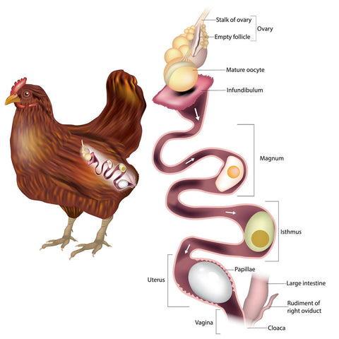
1. Egg formation
The egg is formed in the reproductive tract of the hen. The yolk is produced in the ovary and released when mature. It is captured by the infundibulum, where fertilization occurs if sperm is present.
The egg then passes through:
magnum – deposition of albumen
isthmus – formation of shell membranes
uterus (shell gland) – formation of shell and pigmentation
vagina – oviposition
2. Veterinary control of hatching
The goal of veterinary control is to ensure high fertility, high hatchability, and production of healthy chicks. This includes control of hygiene, incubation conditions, and prevention of microbial contamination.
Fertility = fertilization success
Hatchability = ability of fertile eggs to produce chicks
Hatchability is influenced by egg quality, storage duration, ventilation, and breeder nutrition, especially vitamin E and selenium levels.
Deficiency of vitamin E and selenium in breeder diets can impair embryo development and reduce hatchability due to muscular weakness (myodystrophy).
3. Handling and storage of hatching eggs
Hatching eggs are easily contaminated and should be collected frequently and kept clean. Dirty eggs should not be used for hatching due to high risk of contamination. Very large or very small eggs reduce hatchability — large eggs may result in navel defects, while small eggs provide insufficient nutrients for embryo development.
eggs are collected several times daily
fumigated with formaldehyde gas to reduce microbial load (reduces bacterial contamination by Salmonella, E. coli and improves hatchability and chick quality)
stored at 10–12°C and 70–75% humidity
before incubation, eggs are equilibrated at room temperature (~18–22°C)
Long storage reduces hatchability due to embryo aging and quality deterioration. (too long storage → reduced hatchability. embryo aging before incubation → mortality increases)
Hatchability decreases if eggs are stored for more than 7 days or if temperature changes too rapidly, leading to condensation and increased microbial contamination
4. Incubation conditions
Optimal incubation conditions are critical:
Eggs are incubated with the blunt end up to ensure correct air cell positioning, which is essential for the chick’s first breath during internal pipping.
temperature: 37.8°C
relative humidity: 50–60%
CO₂ and oxygen exchange must be ensured
Proper ventilation is essential to supply oxygen and remove CO₂; inadequate ventilation can lead to embryo suffocation and mortality.
eggs are turned every 2–3 hours to prevent embryo adhesion and ensure normal development
Turning is essential especially during early embryogenesis. prevents embryo adhesion to membranes and supports normal development of extraembryonic structures.
Critical incubation phases:
early phase = organogenesis (very sensitive)
late phase = respiratory transition (air cell use)
5. Candling and monitoring
Eggs are examined by candling:
day 5–6: (white eggs): fertility check (viability of embryo)
day 7-8 (brown eggs)
day 18: embryo development and viability (check if it moves)
This allows removal of infertile or dead embryos.
6. Hatching phase
On day 19, eggs are transferred to the hatcher:
temperature slightly reduced to ~37°C
humidity increased to 70–75%
chicks hatch around day 21
After hatching, chick quality is evaluated, and weak chicks are removed.
Very important to evaluate:
hatchability %
chick quality scoring
culling weak chicks
7. Sexing and management
Chicks may be sexed, especially in commercial layer production. Cloacal sexing (Japanese method) is commonly used. In broiler systems, sex separation may also be performed for management efficiency.
Conclusion
Successful hatching depends on proper egg handling, strict environmental control during incubation, and effective veterinary hygiene measures to ensure high fertility and hatchability and production of healthy chicks.
29. The causes of embryonal mortality & growth disorders in poultry & water fowl.
Embryonal mortality is an important economic factor in poultry production because it directly affects hatchability. It typically occurs in two critical phases during incubation:
Early mortality (around day 4) – associated with metabolic changes, especially peak lactic acid production and CO₂ exchange
Late mortality (around day 19) – when oxygen demand increases significantly before hatching
Two mortality peaks: day 4 & day 19
Embryo development and mortality are monitored by candling, usually at days 5–7 and again around day 16, where we can observe blood vessels and embryo viability.
Causes of embryonal mortality
The causes can be grouped into four main categories:
1. Nutritional factors
Deficiencies of vitamins (A, B-complex, E) and minerals (Ca, P, Mn, Zn)
Protein and lipid deficiencies
Vitamin E and selenium deficiency → muscle weakness and poor hatchability
2. Management factors
Incorrect temperature, humidity, and ventilation
Poor egg handling or storage (e.g. long storage, contamination)
Inadequate turning of eggs
Cracked shells or improper incubation conditions
3. Infectious diseases
Bacterial: Salmonella, Streptococcus
Viral: Newcastle disease, infectious bronchitis, Marek’s disease, IBD
Fungal: Aspergillus
4. Genetic factors
Lethal genes causing embryonic death
Age-related causes
Early (0–7 days): poor hygiene, trauma, vitamin deficiencies, toxicosis
Mid (7–14 days): malnutrition, infectious diseases
Late (14–21 days): malposition, oxygen deficiency, improper incubation
Growth disorders in poultry
These are mainly divided into metabolic and infectious causes, especially in fast-growing broilers.
1. Metabolic diseases
Metabolic growth disorders mainly affect the cardiovascular system, causing mortality, and the musculoskeletal system, causing lameness and poor growth.
a) Sudden death syndrome
Seen in fast-growing broilers
Due to metabolic imbalance and cardiac arrhythmias
Birds appear healthy → sudden collapse and death
b) Muscular dystrophy
Caused by selenium and vitamin E deficiency
Leads to muscle degeneration
Clinical signs:
Skeletal muscle → weakness, inability to stand
Cardiac muscle → sudden death
c) Rickets
Caused by imbalance of Ca, P, and vitamin D₃
Leads to soft, deformed bones and poor growth
2. Infectious growth disorders
Malabsorption syndrome
Multifactorial disease (viruses like reovirus, enterovirus + toxins)
Affects intestinal absorption
Clinical signs:
Poor growth and uneven flock
Diarrhea, undigested feed
Poor feathering (“helicopter wings”)
Short conclusion
So overall, embryonal mortality and growth disorders are multifactorial problems involving nutrition, management, infections, and genetics, and proper control requires good biosecurity, balanced nutrition, and optimal incubation conditions.
30. Metabolic diseases of bones - etiology, symptoms, diagnostics, dif. dg, therapy and prevention.
Metabolic bone diseases are mainly caused by disturbances in calcium, phosphorus, and vitamin D₃ metabolism, leading to defective bone formation, mineralisation, or maintenance.
1. Rickets (young birds)
Rickets is a disease of growing animals, characterized by defective mineralisation of bone and cartilage. There is a disorder of endochondral ossification in growing bones
Etiology
Deficiency of calcium, phosphorus, or vitamin D₃
Incorrect Ca:P ratio
Lack of sunlight (↓ vitamin D synthesis)
Renal or gastrointestinal disorders
Clinical signs
Enlarged joints and epiphyses, Soft, pliable beak and claws, Bone deformities, lameness, stiff gait, Poor growth and weakness
Diagnosis
Clinical signs + history (nutrition), Radiology: poor mineralisation
Post-mortem shows poorly mineralised, soft and deformed bones
Biochemically, we may see decreased calcium and phosphorus and increased alkaline phosphatase
Differential diagnosis: perosis, Marek’s disease, and vitamin deficiencies
Treatment & prevention
Correct diet (balanced Ca:P ratio)
Vitamin D₃ supplementation
Proper management and sunlight exposure
2. Osteomalacia (adult birds)
Osteomalacia is the adult form of rickets, affecting bone remodelling.
Etiology
Vitamin D₃, Ca, or P deficiency
Renal disease → phosphorus imbalance
Clinical signs
Fragile bones, fractures, Spinal deformities, Thin or soft eggshells
Key point
→ Same mechanism as rickets, but in mature skeleton
3. Osteoporosis (cage layer fatigue)
Osteoporosis is a reduction of total bone mass (both organic and inorganic components).
Etiology
High calcium demand during egg production
Inadequate calcium or vitamin D₃ intake
Protein deficiency (matrix formation)
Clinical signs
Lameness, reluctance to move, Fragile bones, fractures, Drop in egg production, Soft-shelled eggs
4. Cage layer fatigue (important clinical form)
This is a severe form of osteoporosis seen in high-producing layers.
Pathogenesis
Calcium is mobilized from bones for eggshell production
→ depletion of medullary bone → cortical bone thinning
Clinical signs
Birds unable to stand (“cage paralysis”), Very fragile bones (tibia, femur fractures), Reduced feed intake
Differential diagnosis
Infectious causes of lameness (e.g. arthritis, osteomyelitis)
Trauma
Mycotoxicosis
Treatment & prevention (general principle)
Balanced nutrition (Ca, P, vitamin D₃)
Proper Ca:P ratio
Adequate protein supply
Good management (exercise, housing)
Supplementation in high-risk periods (laying phase)
Osteopetrosis
“Osteopetrosis is the opposite condition, with thickened bones, often associated with viral diseases like leukosis.”
Strong closing sentence
So overall, metabolic bone diseases in poultry are primarily nutritional disorders, and prevention through correct diet and management is far more effective than treatment.
Clear distinction: rickets vs osteomalacia vs osteoporosis
Mention of cage layer fatigue = clinical form
Good inclusion of Ca:P imbalance + vitamin D₃
31. Infectious diseases of bones - etiology, symptoms, diagnostics, dif. dg, therapy and prevention.
Infectious bone diseases in poultry are mainly caused by viral and bacterial agents, leading to deformities, lameness, and reduced productivity.
1. Osteopetrosis (“marble bone disease”)
Osteopetrosis is a viral neoplastic disease caused by a strain of the avian leukosis virus. (retrovirus)
Pathogenesis
The virus alters osteoblast activity → excessive bone formation → thickened, dense bones → exostosis and often anemia (bone marrow compression)
Lesions
Bilateral, symmetrical thickening of long bones
Especially tibiotarsus and tarsometatarsus
Clinical signs
Lameness, difficulty moving, Emaciation (can’t reach feed/water), Weakness, recumbency
Key point
→ Bone is thick but abnormal and non-functional
2. Osteomyelitis
Osteomyelitis is a bacterial infection of bone (epiphyseal region common), often secondary to septicaemia.
Etiology
Staphylococcus aureus (most common)
E. coli, Salmonella, Streptococcus, Mycoplasma synoviae
Pathology
Bone lysis, necrosis
Replacement by caseous exudate
Chronic → sclerosis and deformity
Clinical signs
Lameness, Swollen joints, Reduced mobility
Treatment
Antibiotics, but the response is often poor
3. Femoral head necrosis (FHN)
Also called femoral head separation in fast-growing broilers.
Pathogenesis
Mechanical stress + bacterial infection → Separation of growth plate → necrosis
vascular damage → thrombosis → necrosis
Primary event is usually:
Mechanical stress + rapid growth in broilers
→ this leads to weakness at the growth plate (epiphysis)
→ separation of the growth plate = femoral head separation (FHS)
After separation:
Blood supply is compromised
The area becomes susceptible to bacterial colonization (e.g. E. coli, Staph, Strep)
Then:
Thrombosis can occur in damaged vessels
This contributes to ischemia → necrosis of the femoral head
Etiology
E. coli, Staphylococcus, Streptococcus
Clinical signs
Severe lameness, Birds may use wings for support
Prevention
Good hygiene, Proper management of fast-growing birds. ATB not recommended.
4. Avian tuberculosis
A chronic granulomatous infection caused by Mycobacterium avium.
Pathology
Granulomas in:
Liver, Spleen, Bone marrow
Clinical signs
Chronic weight loss, Lethargy, Sometimes lameness
Diagnosis
Tuberculin test, PCR, histopathology
Treatment & prevention
No treatment → culling
Prevention: hygiene, flock turnover
Differential diagnosis
Metabolic bone diseases (rickets, osteoporosis), Trauma, Nutritional deficiencies
General prevention
Biosecurity and hygiene
Good management
Control of systemic infections
Strong closing sentence
So overall, infectious bone diseases in poultry are mainly secondary to systemic infections or viral tumors, and prevention relies primarily on biosecurity, hygiene, and proper flock management.
32. Muscle disorders in poultry - etiology, symptoms, diagnostics, dif. dg, therapy and prevention.
Muscle disorders in poultry include atrophy, muscular dystrophy, and myopathies, caused by nutritional deficiencies, metabolic disturbances, mechanical damage, stress, or toxins.
1. Atrophy
Definition: ↓ size and function of the muscle after normal development
Etiology: malnutrition, decreased body weight, chronic systemic diseases (e.g. Marek’s disease)
CS: muscle wasting
Dx: clinical + history
Tx: treat underlying disease, improve nutrition
2. Muscular dystrophy (nutritional myopathy)
Etiology: deficiency of vitamin E and selenium
Pathogenesis:
Se (glutathione peroxidase) + Vit E protect membranes from oxidative damage
Deficiency → lipoperoxidation → degeneration + calcification of muscle cells
CS:
Skeletal muscle → weakness, stiffness, trembling, inability to stand
Cardiac muscle → acute heart failure, sudden death
Dx: history + lesions
Tx/Prevention: supplementation of Vit E + Se
3. Myopathies (non-inflammatory muscle degeneration)
a) Deep pectoral myopathy (“green muscle disease”)
Seen in heavy broilers/turkeys
Etiology: excessive wing flapping → muscle overuse
compartment-like syndrome due to rigid fascia
Pathogenesis:
muscle swelling inside tight fascia → vascular compression → ↓ blood flow → ischemia → hypoxia → necrosis
PM: early pale, swollen muscle → later green → fibrous capsule
Prevention: careful handling, genetic selection
b) Capture myopathy
Rare in poultry
Etiology: stress, struggle, transport. predisposed by stress + selenium deficiency
Pathogenesis: anaerobic glycolysis → lactic acidosis + hyperthermia
Lesions: pale skeletal muscle (especially legs), sometimes cardiac muscle
Peracute cases: no visible lesions
Prevention: minimize stress and handling
c) Toxic myopathy (ionophore toxicity)
Agents: monensin, salinomycin, narasin
they are ionophore anticoccidial drugs that disrupt ion transport in parasites, but in overdose they increase intracellular calcium in muscle cells, causing toxic myopathy.
Pathogenesis:
↑ cation transport → ↑ intracellular Ca²⁺ → disruption of ionic balance → muscle cell death
CS: incoordination, leg weakness, diarrhea, dyspnea, ↓ feed intake, weight loss
Ionophore toxicity is increased by ATB (tiamulin, macrolides (erythromycin), chloramphenicol)
Prevention: correct dosing, avoid drug interactions
d) Pododermatitis
Even though it is skin/joint-related, it often appears in locomotor questions:
Multi-factorial disease in broilers/breeders
Predisposing factors:
wet litter, obesity
Lesion: plantar skin erosion → ulcer → abscess → fibrosis
Leads to:
lameness + reduced fertility
Prevention:
dry litter, ammonia control, feed restriction in breeders
e) Aflatoxins → ONLY mention if examiner expands to systemic myopathies
Not necessary for muscle disorders, but if you want extra:
Aflatoxins cause neuromuscular signs (torticollis, paralysis, seizures) via liver damage → toxin effect
🔑Key exam sentence:
“Muscle disorders in poultry result from nutritional deficiencies (Vit E/Se), ischemic damage due to muscle overuse, stress-induced metabolic acidosis, or ionophore toxicity, leading to degeneration, necrosis, and impaired locomotion or sudden death.”
33. Diseases of the joints - etiology, symptoms, diagnostics, dif. dg, therapy and prevention.
Joint diseases in poultry include developmental, metabolic, and infectious disorders, affecting cartilage, growth plates, and synovial structures, leading to lameness and reduced performance.
1. Chondrodystrophy
Etiology: genetic disorder of cartilage development
Types:
Lethal chondrodystrophy → inherited → shortened cartilaginous bones
Embryonal form → skull deformity (brachycephaly), parrot beak
Pathogenesis: abnormal cartilage development → skeletal malformation
CS: deformities, non-viable chicks (severe cases)
Dx: post-mortem, congenital abnormalities
Tx: none
Prevention: breeding selection
2. Dyschondroplasia (Tibial dyschondroplasia)
Etiology:
rapid growth (broilers)
genetic predisposition
Vit D metabolism disorders
Ca:P imbalance
mycotoxins
Pathogenesis:
failure of chondrocyte maturation → avascular cartilage plug in growth plate
Most affected: proximal tibiotarsus
CS: lameness, reluctance to move, fractures
Dx: necropsy (cartilage plug in growth plate)
DDx: rickets
Tx: none specific
Prevention: balanced nutrition, genetic selection
3. Gout
Etiology:
hyperuricemia due to renal dysfunction
high protein diet
dehydration
Vit A deficiency
Pathogenesis: uric acid deposition in tissues → crystal formation
Types:
Visceral gout → organs (heart, liver, kidneys)
Articular gout → joints (swelling, inflammation)
CS:
anorexia, weight loss, diarrhea, ↓ egg production
lameness (articular form)
Dx: necropsy (urate deposits)
Tx: supportive (e.g. alkalization of water with sodium bicarbonate)
Prevention: proper nutrition, kidney protection, hydration
4. Infectious synovitis
Agent: Mycoplasma synoviae infection
Pathogenesis: respiratory infection → systemic spread → synovitis, tendinitis
CS:
lameness
swollen hocks, footpads
depression, recumbency
sternal bursitis
Dx:
ELISA, PCR, serology
clinical signs + lesions
Tx: antibiotics (variable response)
Prevention: biosecurity, vaccination, monitoring
5. Leg deformities (valgus/varus)
Etiology:
rickets (poor mineralization)
chondrodystrophy
rapid growth
Pathogenesis: weak bones → mechanical stress → angular deformities
CS: valgus (lateral), varus (medial) deviation, lameness
Dx: clinical observation
Tx: none once developed
Prevention: nutrition (Ca, P, Vit D3), growth control
6. Spondylolisthesis (“kinky back”)
Etiology:
rapid growth
conformation defects
possible bacterial involvement in breeders
Pathogenesis: vertebral displacement → spinal cord compression
CS:
posterior paralysis
inability to stand
abnormal posture
Dx: clinical signs + post-mortem
Tx: none
Prevention: genetic selection, growth control
🔑 Key exam summary
“Joint diseases in poultry are mainly caused by developmental cartilage defects (chondrodystrophy, dyschondroplasia), metabolic disorders (gout), and infectious agents (Mycoplasma synoviae), leading to lameness, deformities, and reduced locomotion performance.”