BIOL 241 Exam 4 Condensed
1/48
There's no tags or description
Looks like no tags are added yet.
Name | Mastery | Learn | Test | Matching | Spaced | Call with Kai |
|---|
No analytics yet
Send a link to your students to track their progress
49 Terms
Peripheral Nervous System Organized into:
Afferent
Going to CNS (Sensory)
Efferent
Going away from CNS (Motor)
CNS Glial Cells (Assholes Owe Everyone Money)
Astrocytes
Maintain blood-brain barrier
Oligodendrocytes
Myelination of CNS axons
Ependymal cells
Help with CSF flow
Microglia
Phagocytes
PNS Glial Cells
Satellite cells
Regulate environment around neurons
Schwann cells
Myelination of PNS axons
Anterograde vs Retrograde
Anterograde
Movement AWAY from cell body (Kinesin)
Retrograde
Movement TOWARDS cell body (Dynein)
Categorization of neurons
Structure (relationships of dendrites to the cell body)
Anaxonic neuron (no axon) (only in Brain)
Bipolar neuron (Two axons)
Unipolar neuron (One continuous axon)
Multipolar neuron (>2 dendrite clusters)
Function (S.A.M.E principle)(Sensory Afferent, Motor Efferent)
Unipolar neurons
Sensory (afferent)
Bipolar neurons
Sensory (afferent)
Multipolar
Motor (efferent)
Myelin Sheath
Protein lipoid (70% fat)
Insulates fibers = myelinated -> rapid impulses
If non-insulated = non-myelinated -> slow impulses
Myelin sheath covered areas = white matter
Gated Channels
Chemically Gated Channel (does not open until a chemical binds to it)(Ach)
Voltage-gated channel (rely on voltage difference to open)
Mechanically gated channel (rely on a force being applied to open)
Resting potential
-70mV
Neurons have more potassium channels than sodium channels
Potassium flows out more easily than sodium flows in (creates a larger negative charge inside the cell and positive outside the cell)
Cell has a resting "polarity"
Changes in membrane permeability cause:
Graded potentials
Action potentials
Graded Potential
Type of potential that can lead to an action potential (if stimulating)
Vary in size
Use chemically-gated channels
Hyperpolarization
If permeability change sends charge BELOW -70mv (cell get more negative)
Can happen if positively charged ions leave for a long time
Point: inhibits an action potential
Depolarization
If charge goes ABOVE -70mV
i.e. cell get more positive (lose polarity)
How does this happen?
Let positively charged sodium ions enter fast
Point: facilitates an action potential
Two type of graded potentials
Receptor potential
A 'receptor' is affected (NMJ)
Postsynaptic potential
Neurotransmitter -> synapse -> postsynaptic neuron
Graded potentials can be 'additive' to generate AP
Action Potential
Do not vary in size (all-or-none)
Use voltage gated channels
Action potentials do not weaken over distance
Move down axon in one direction
Main way neurons send signals
Main long-distance neural communication
Main players:
Voltage-gated sodium channels (two types)
Activation gate (opened to allow ions through)
Inactivation gate (close up channel to ions)
Voltage-gated potassium channels (one type)
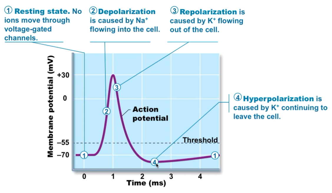
Events leading to an Action Potential
Resting state (no ion movement = -70mV)
Depolarization (sodium rushes in, potassium channels remain closed)
Becomes more positive
Repolarization (sodium channel closes, potassium channel opens)
Becomes more negative
Hyperpolarization (continued outflow of potassium, below -70mV))
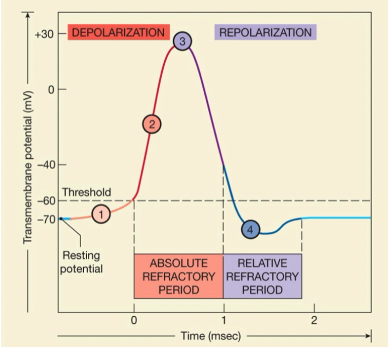
Refractory Periods
Absolute Refractory Period, sodium channels are fully open thus cannot respond to another stimulus.
Relative Refractory Period, most sodium channels are resting thus a 2nd stimulus can cause another action potential.
Propagation of APs
Recap: APs happen in one place then spread
Spread = propagation
Two ways to propagate
Continuous (non-myelinated cells) (SLOW)
After the Axon Hillock is depolarized, the AP moves down axon by opening the voltage gated sodium channels. As AP moves down each sodium channel is going from -70mV to -55mV, which lets in more sodium each time.
Saltatory (jumping) (Myelin covered cells)
Myelin sheath covered areas do not have channels under them (nodes)
AP jumps from one node to another node (FAST)
Speed of Action Potentials
Effected by:
Myelination
Axon diameter
Large area = fast flow
Large diameter = low resistance
Group A
-large diameter, lots of myelin = FAST
Position, balance, delicate touch
Group B
Medium diameter, little myelin = Medium
Temperature, touch, pain
Group C
Smallest diameter, non-myelinated = SLOW
Where do Action Potential go?
Synapses
Presynaptic neuron
AP arrives at axon terminal
Voltage gated calcium channels open and enter axon terminal
Calcium entry causes synaptic vesicles to release neurotransmitter
Postsynaptic neuron
Neurotransmitter diffuses across the synaptic cleft and binds to receptors on postsynaptic membrane
Ion movement on postsynaptic neuron causes graded potential
Neurotransmitter effects are terminated by reuptake through transport protein, enzymatic degradation, or diffusion away from the synapse.
In axosomatic synapses
Axon terminal to the body of a neuron
In axodendritic synapses
Axon terminal connects to the dendrites of the postsynaptic neuron
In axoaxonal synapses
Axon connects to another axon
Postsynaptic Potentials
Signals leaving the synapse are graded potentials
Can be
Excitatory
Called Excitatory postsynaptic potential (EPSP)
If enough build up (reach threshold -55mV) can lead to depolarization and an AP.
Inhibitory
Called inhibitory postsynaptic potential (IPSP)
Leads to hyperpolarization (away from an AP)
EPSP and IPSP can be 'additive'
Summate
By time
By area
Spatial summation: 2 simultaneous stimuli at different locations cause EPSPs that add together
Can also cause changes in membrane potential that cancel each other out. (Excitatory +, Inhibitory -)
Temporal summation: 2 excitatory stimuli close in time cause EPSPs that add together
Neurotransmitters
Acetylcholine
Type of receptor: Cholinergic
Sometimes excitatory (generates an AP)
Sometimes inhibitory (blocks an AP)
Norepinephrine
Type of receptor: adrenergic
Typically excitatory
Frontal lobe
Personality traits
Precentral gyrus
Primary Motor Cortex
Postcentral gyrus
Primary somatosensory cortex
Parietal lobe
Size and texture differentiation
Occipital lobe
Visual Cortex
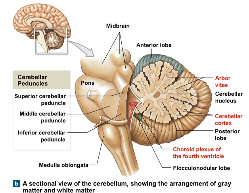
Cerebellum
Fine motor movements
Purkinje Cells
Wernicke’s Area
Speech comprehension
Broca’s Area
Speech muscle control
Thalamus
Sensory relay station
Hypothalamus
Temperature regulation
Hormone regulation (regulate pituitary)
H2O balance
Pituitary Gland
Controls glands via hormones
Controlled by Hypthalamus
Pons
Respiratory centers
Midbrain
Superior colliculus (visual reflex)
Inferior colliculus (auditory reflex)
Medulla Oblongata
Respiratory Centers
Cardiovascular centers
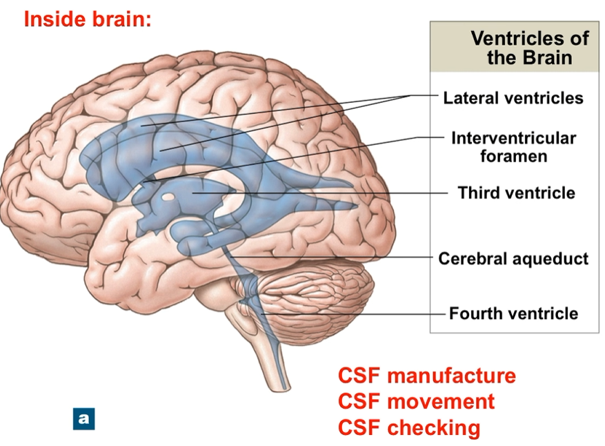
Ventricles of the brain
CSF moves through ventricles
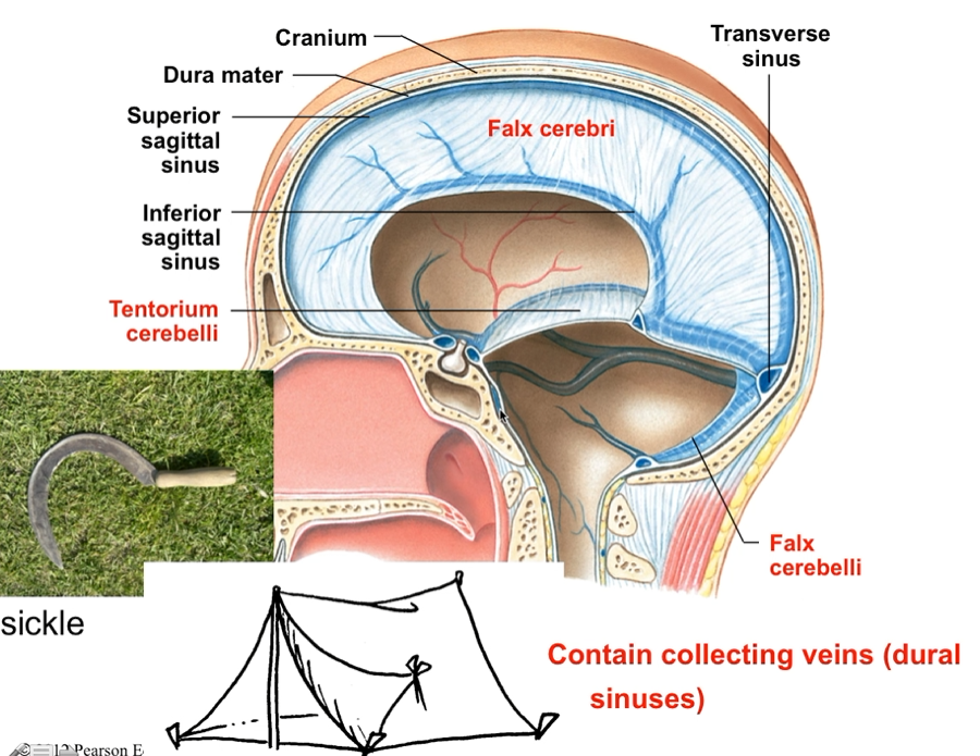
Cranial Meninges
Protect the brain from cranial trauma
Three layers
Dura mater (Outermost)
Arachnoid mater (middle)
Pia mater (innermost, lines cerebral cortex)
Dura folds
Folded inner layer of dura mater
Stabilize and support the brain
Three largest dura folds:
Falx cerebri (separates the hemispheres of the cerebrum)
Tentorium cerebelli (separates the cerebellum from cerebrum)
Falx cerebelli (separates the two hemispheres of the cerebellum)
Cerebrospinal Fluid (CSF)
Surround CNS
Functions:
Cushions delicate neural structures
Supports brain
Transports nutrients, chemical messengers, waste
Is made by the choroid plexus (circulated by ependymal cells)
Flow through ventricles
To central canal of spinal cord
Into subarachnoid granulations
Arachnoid granulation absorb CSF into venus circulation
Brainstem checks CSF for CO2 and pH
Diencephalon
Integrated sensory information and motor commands
Thalamus, Hypothalamus, Epithalamus
Basal Nuclei
Are masses of gray matter encapsulated by white matter
Are responsible for subconscious activities
Found in the cerebrum
Cranial nerves (orange orangutans on tree trunk are feeling very good vibrations AH!)
Orange - Olfactory (1): Smell
Orangutans - Optic (2): Vision
On - Oculomotor (3): Eye movements
Tree - Trochlear (4): Superior oblique eye muscle
Trunks - Trigeminal (5): Face sensation and chewing
Are - Abducens (6): lateral rectus
Feeling - Facial (7): Motor face, saliva, taste
Very - Vestibulocochlear (8): sound, rotation, gravity
Good - Glossopharyngeal (9): taste, parotid gland
Vibrations - Vagus (10): larynx/pharynx muscles, parasympathetic to thoracic and abdominal viscera
AH! - Accessory (11): Sternocleidomastoid and trapezius
Hypoglossal nerve (12): tongue, swallowing
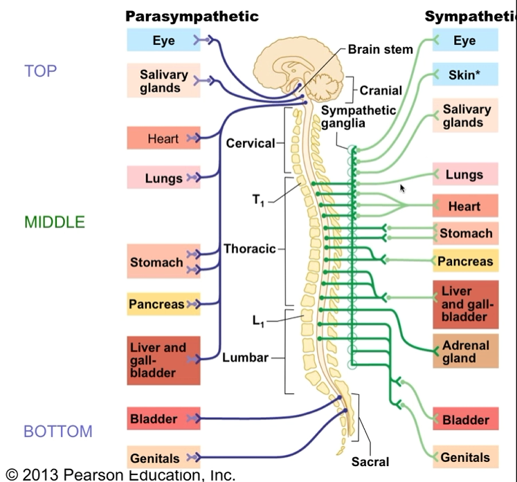
Autonomic Nervous System
Motor neurons innervate organs
Can be stimulatory or inhibitory
Subconscious control
Sympathetic (fight or flight)
Norepinephrine (NE) neurotransmitter
Parasympathetic (sit and digest)
ACh neurotransmitter
Receptor for Neurotransmitters
Cholinergic (binds ACh)
Nicotinic
All postganglionic neurons (sympathetic and parasympathetic) + NMJ
Stimulatory (depolarizing)
Muscarinic (binds ACh)
Organ cells (heart, intestine)
Inhibitory or excitatory
Speeds up heart
Slows down intestines
Adrenergic Receptors (binds NE)
Alpha
Alpha 1: blood vessel constriction
Alpha 2: pupil dilation
Beta:
Beta 1: heart (1 heart)
Beta 2: lungs (2 lungs)
Position of ganglia differs
Parasympathetic
Close to target organ
Sympathetic
Close to spinal cord
Dual Innervation
Both division at once: Autonomic plexuses (nerve network)
Sympathetic postganglionic fibers
AND
parasympathetic preganglionic fibers
Ex. Cardiac Plexus, Celiac Plexus
You have more control by having both branches innervate a structure.
Ex. Heart Dual Innervation
Opposing effects on HR
Parasympathetic
ACh -> increases hyperpolarization -> slows heart rate
Sympathetic
NE -> increases depolarization -> increase heart rate
Typically both neurotransmitters are dumped out continuously
Purkinje cell
In cerebellar cortex
Output neurons
Pyramidal cells
Cerebral cortex
Motor output
Schwann cells
Schwann cells wrap around axon to form myelin
Nerve CT Layers
Epineurium (Entire nerve)
Perineurium (Each fascicle)
Endoneurium (individual axon)