MRI
1/29
Earn XP
Description and Tags
Magnetic Resonance Imaging
Name | Mastery | Learn | Test | Matching | Spaced | Call with Kai |
|---|
No analytics yet
Send a link to your students to track their progress
30 Terms
Electromagnetism key points
electric and magnetic fields
faradays law of induction
static charges generate an electric field
moving charges generate a magnetic field
faradays law of induction: changing magnetic field induces a current in a conducting wire ( right hand rule )
loop of current generates ‘dipole’ magnetic field (e.g. bar magnet)
features of MRI ( an introduction) (5 things)
shows brain structure and function
non invasive and safe ( uses non-ionising radiation)
whole brain 3d imaging technique ( with ~mm resolution)
excellent contrast between different tissues
very versatile ( different contrasts )
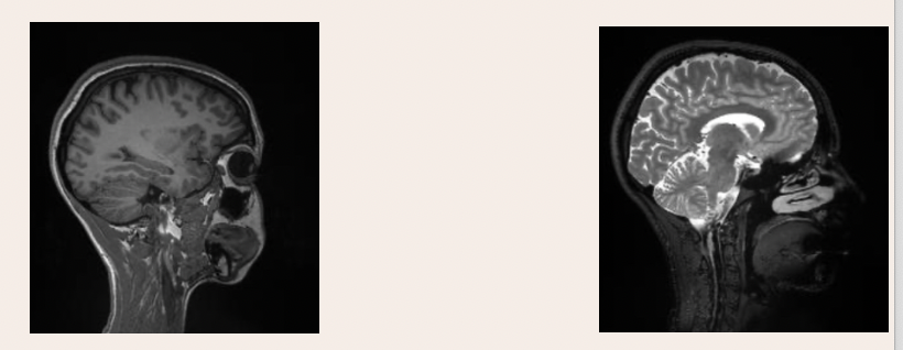
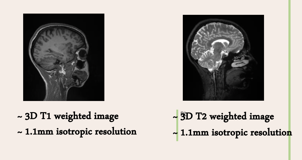
which image is T1 weighted and which image is T2 weighted
what are each respectively better for
T1 - better for anatomical detail and normal brain structure
T2 - better for tumours, inflammation, trauma as sensitive to water ( so tumour would appear as a light liquid)
what is ‘spin’
also known as ‘ intrinsic angular momentum’ of particle/atoms
creates a magnetic dipole
what are 3 fundamental properties of a particle
mass
charge
spin
what is B0
strong, uniform magnetic field you lay in when in an MRI scanner
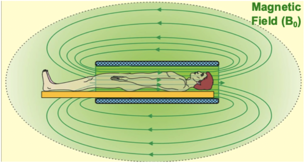
what happens when you go in an MRI scanner
refer to magnetic moments
define bulk magnetism
magnetic moments ( define strength and orientation of magnetic source) of hydrogen atoms in the water in our body are randomly oriented
when go into B0 they either align with or against B0
slightly more of our H magnetic moments align with B0 - this creates small bulk magnetism ( this is the M in MRI)
bulk magnetism (M) is the vector sum of all spin ‘ups’ and spin ‘ downs’
what is precession
when an object is spinning ( angular momentum) , it rotates around direction of applied force
same thing happens to M if tipped away from B0 which induces a current ( faraday) which can then be detected in RF( Radio frequency) coil conductors around persons head
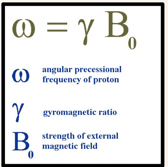
what is lamour equation
resonance
resonance - strong response of an oscillating system to a driving force that matches its own natural frequency
contexyual understanding:
Resonance is when something vibrates a lot because you’re adding energy at exactly the right timing for how it naturally wants to move. ( like pushing a swing at the right time to make it go higher)
state simply how we tip M away from B0 to cause it to precess
we apply another magnetic field (B1) perpendicular to B0
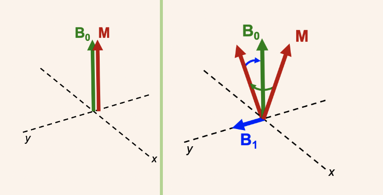
what is the most efficient way to apply B1 to achieve resonance ( 2 things)
keep B1 on continuously AND rotate it with M so it always pushes in the right direction
(concept of resonance is need to match frequency of of B1 rotation with frequency of M’s precession( lamour frequency) to consistently tip it away from B0 and generate measurable signal ) - dont memorise this bit
what is the radiofrequency (RF) pulse
RF pulse - the B1 field applied t M away from B0
Rf because rotates at lamour frequency to stay synchronised with M as it precedes
pulse because applied for short time just enough to rotate M away from B0
what 2 features does frequency depend on
properties of nucleus ( 𝛾 in lamour equation different for different nuclei)
magnetic field strength B0
( this is basically lamour equation)
describe how gradients are used to work out the position of signals
changing the gradients means different positions have different frequencies which gives them different signals
( imagine you have a field of bells and you hear a ‘ding’ dont know which bell it came from - gradients would give each bell a different ‘ pitch’ aka signal so we know which bell made which ‘ding’)
how is the fourier transform used in MRI
it mathematically separates all of the frequencies from different positions and their amplitudes
( imagine it like separating the notes out from hearing a piano chord)
what is contrast and what is it used for
contras= difference in appearance of different tissues in an image
used to characterise brain structure and anatomy
separate healthy and diseased tissue
2 things measured signal depends on
water concentration ( proton density)
timings of acquisition ( relaxation property of M)
why does brain tissue disrupt B0
inhomogenous - some areas have iron which increases local B0, some are dimagnetic (oppose main field) which reduces local B0
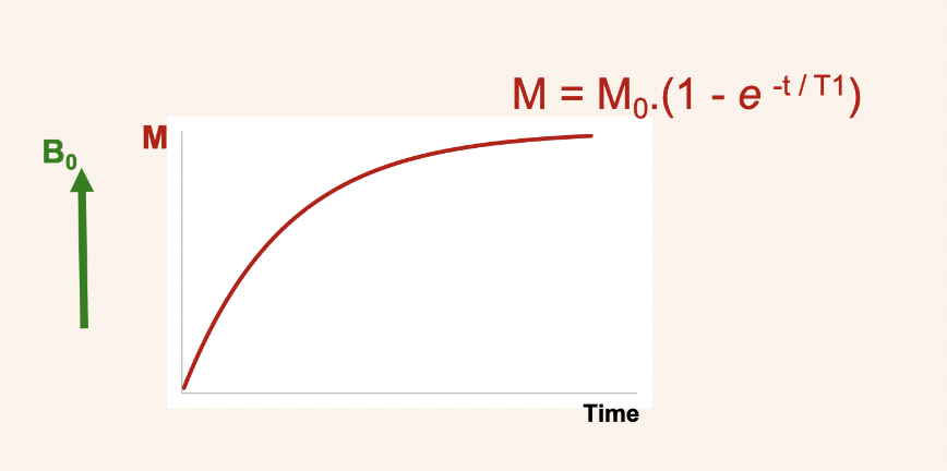
recovery of M = t1 relaxation
after M is tipped away from B0
M naturally wants to realign with B0 but this is a gradual process which follows an exponential recovery curve with time constant T1
T1 exponential function
for t1 low values mean quick recovery so M returns back to original value sooner
long T1 will be dark in T1 weighted image
describe what happens in spin echoes (T2 relaxation)
90 degree rf protons spread out (dephase) because of magnetic field inhomogeneities (T2*) and T2.
to reverse this, 180 degree rf pulse is applied → this flips spins and brings protons back together (rephasing). This fixes T2* so isolates T2
once rephased this = spin echo = pure T2
T2 and T2* exponential function
T2*: M(t) = M0 . e-t/t2*
T2:M(t) = M0 . e-t/t2
T2>T2*
T2* relaxation
what is it
what does it define
2 things it depends on
T2* is time constant for signal decay
defines how quickly for signal disappears
depends on tissues types (grey matter,white matter, cerebrospinal fluid)
depends aswell on overall uniformity of magnetic field
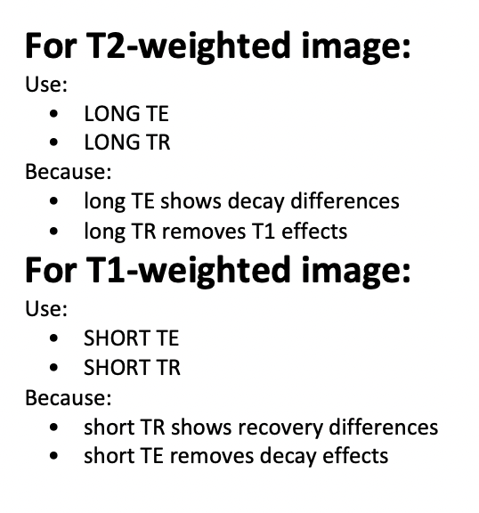
how to you pick echo time (TE) / repetition time (TR)
include what te and tr actually means
As time goes on, signals decay (T2 decay).
If we wait longer before taking the photo (long TE), some tissues lose more signal than others, so we see decay differences. This gives a T2-weighted image.long TR removes T1 effects(recovery)
For T1, instead of focusing on decay, we focus on recovery.
If we restart the experiment quickly (short TR), some tissues have recovered more than others. This creates signal differences based on recovery speed, giving a T1-weighted image.short TE removes decay effects (T2 weighting).
TE = deciding how long to wait as decay over time , until take the picture
TR = how long to wait before repeating the pulse
which type of haemoglobin is more paramagnetic( weakly attracted to poles but no permanent magnetism)
deoxy
what are the effects of the presence of deoxyHb in a magnetic field
causes inhomogeneities ( irregularities) = dephasing
( makes spins precess at different frequencies (magnetisation partially cancels out))
what weighting does blood oxygenation level affect and how
higher blood oxygenation = higher T2* values
how does blood oxygenation level affect the contrast
increasing blood oxygenation decreases the contrast
what happens to the concentration of deoxyHb in veins on neuronal activation
Cerebral blood flow (CBF) increases because need more exygen and glucose
Efficiency of oxygen extracted into brain decreases because blood flows through capillaries quicker
Therefore conc of deoxyHb in veins decreases