patho lab final
1/85
There's no tags or description
Looks like no tags are added yet.
Name | Mastery | Learn | Test | Matching | Spaced | Call with Kai |
|---|
No analytics yet
Send a link to your students to track their progress
86 Terms
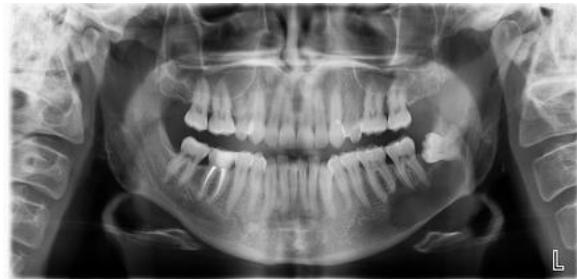
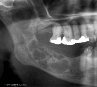
A 19 year old patient present with mild swelling of lower jaw. Intra orally a small swelling was visible over left third molar region. A radiograph taken. Provide Differential diagnosis
Dentigerous cyst
OKC
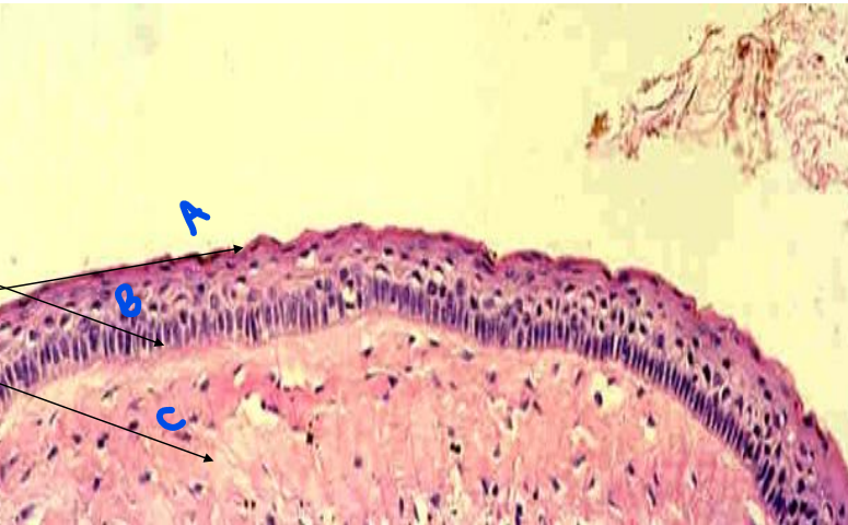
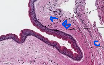
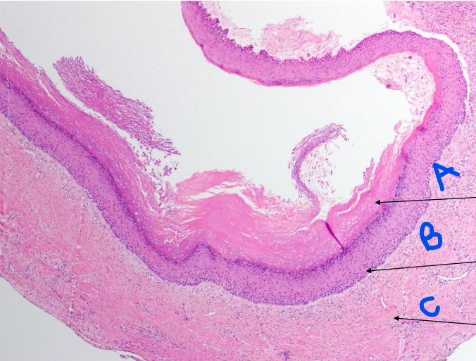
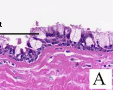
name and identify
OKC
a- parakeratinized lining epithelium
b- tombstone appearance of basal cells
c- connective tissue capsule
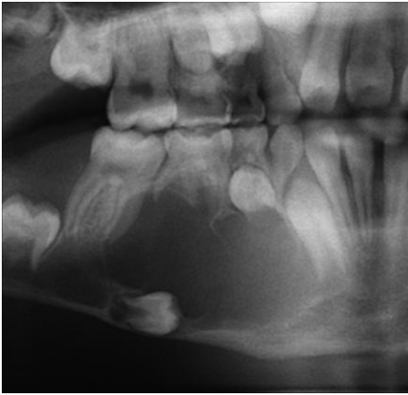
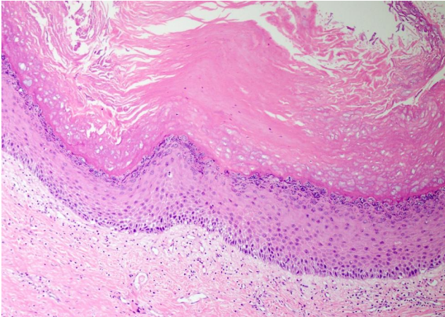
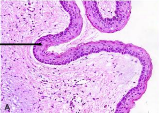
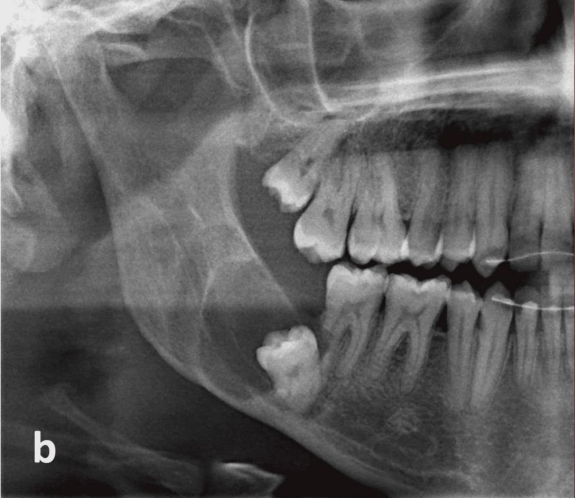
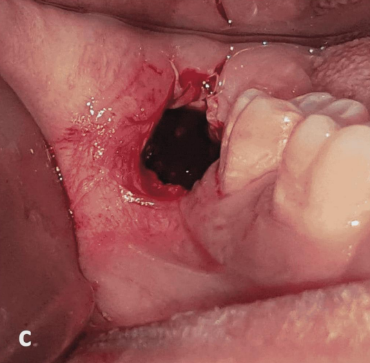
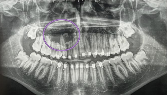
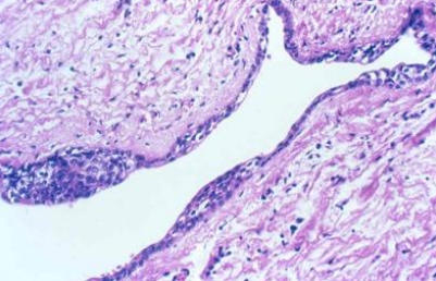
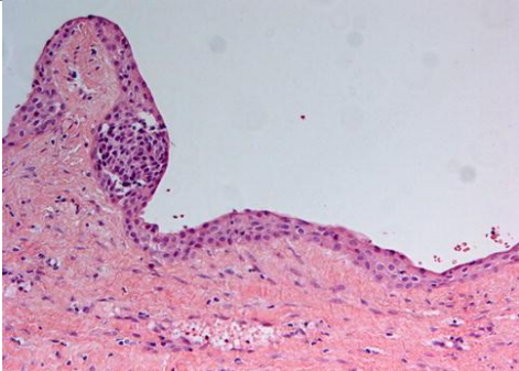
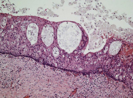
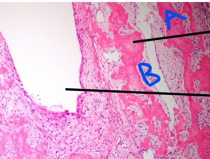
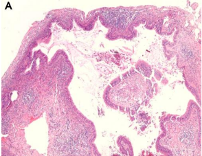
A 35 year old patient present with mild swelling of lower jaw. Intra orally a small swelling was visible over left third molar region. Radiographic and histologic images are shown below
OKC
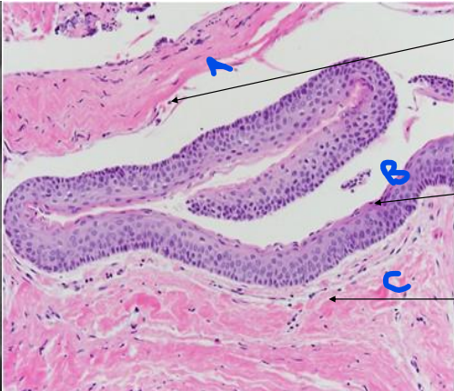
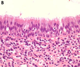
A 35 year old patient present with mild swelling of lower jaw. Intra orally a small swelling was visible over left third molar region. Radiographic and histologic images are shown below
OKC
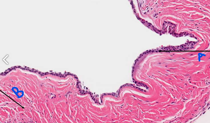
a- separation of epithelium from connective tissue
b- parakeratinized stratified squamous epithelium
c- connective tissue capsule
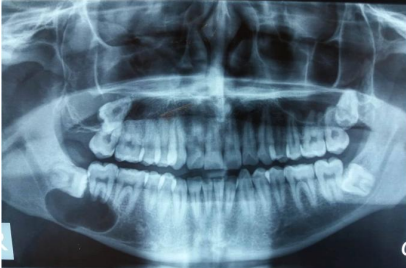
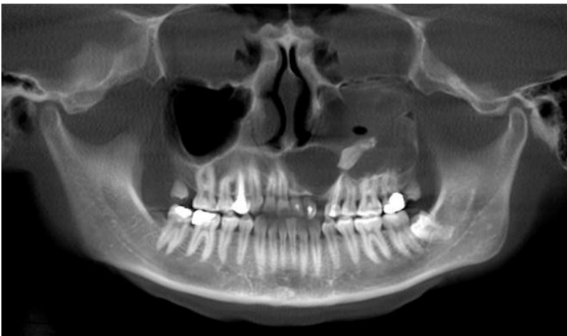
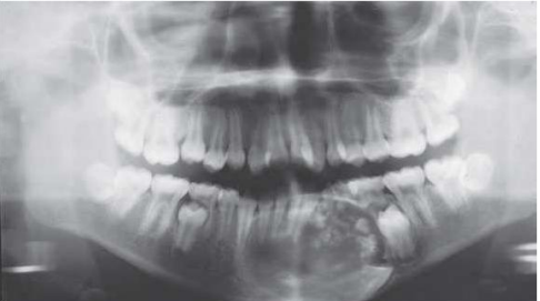
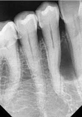
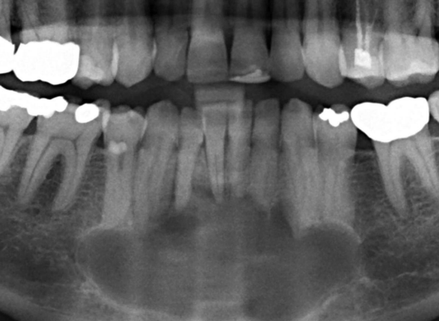
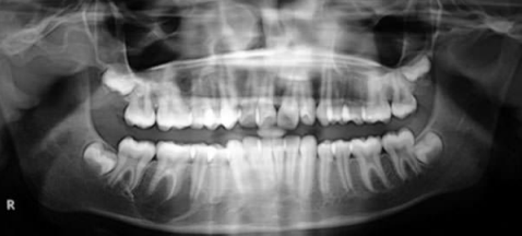
A 25 year old patient present with mild swelling of lower jaw. Intra orally a small swelling was visible over right third molar region. A radiograph taken. Provide Differential diagnosis and justify
OKC
Ameloblastoma
Multilocular radiolucency
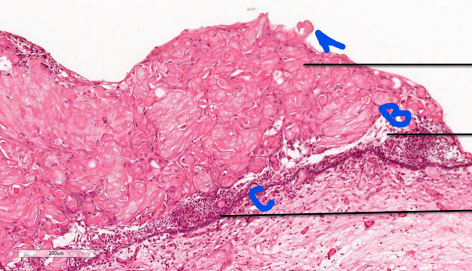
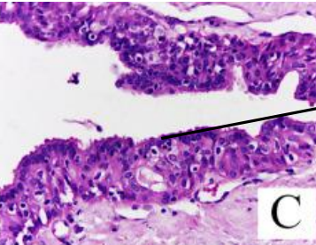
OKC
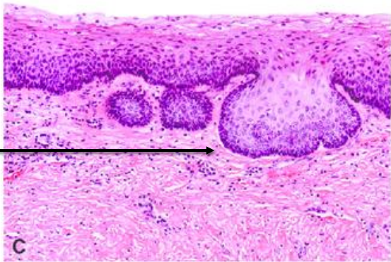
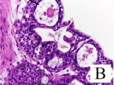
a- palisading row of basal layer
b- parakeratinized stratified squamous epithelium with corrugated surface
c- connective tissue capsule
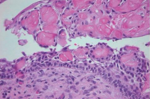
A 25 year old patient present with mild swelling of lower jaw. Intra orally a small swelling was visible over right molar region. A radiograph taken. Provide diagnosis and justify
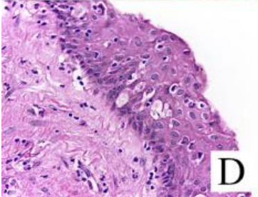
Orthokeratinised odontgenic cyst
Orthokeratinised odontgenic cyst
OOC
a- orthokeratinized stratified squamous epithelium
b- cuboidal basal cell layer
c- connective tissue capsule
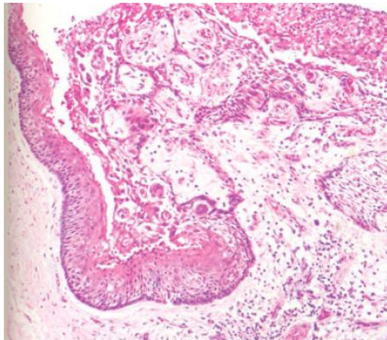
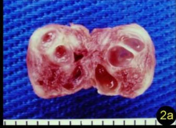
A 55 year old patient present with mild swelling of upper jaw. A biopsy was taken and the image is provided below.
OKC (Inflamed towards one side)
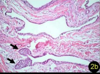
OKC
-lining epithelium
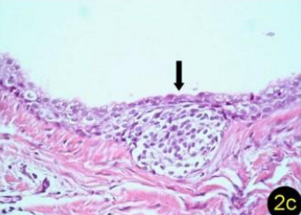
OKC
- daughter cyst
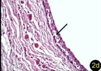
OKC
- basal cell budding
OKC
- odontogenic epithelial rests
A 25 year old patient present with mild swelling of lower jaw. Intra orally a small swelling was visible over right third molar region. A radiograph taken. Provide Differential diagnosis
Dentigerous cyst
OKC
A 12 year old with cyst of jaw
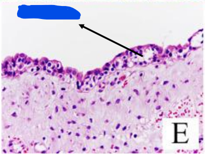
Dentigerous cyst
a- Non keratinized stratified squamous Epithelial lining (2-3 layer thickness)
b- connective tissue capsule
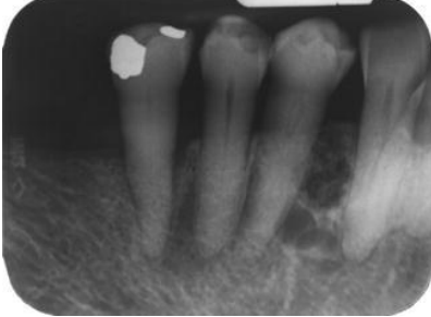
Marsupialization and the cyst have shrunken
Marsupialization and the cyst have shrunken
Dentigerous cyst
a- Non keratinized stratified squamous Epithelial lining (2-3 layer thickness)
b- Connective tissue capsule
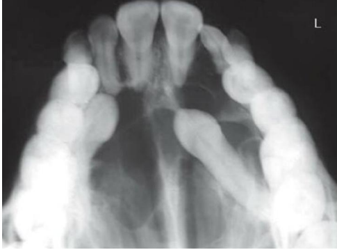
A 25 year old patient present with mild swelling of front jaw. Intra orally a small swelling was visible and 23 was missing. A radiograph taken. Provide Differential diagnosis
Dentigerous cyst
Adenomatoid
odontogenic tumor
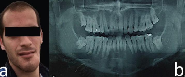
A swelling of palate for 3 years duration showing progressive enlargement. Provide DD. Justify your DD
OKC
Ameloblastoma
Multilocular appearnace
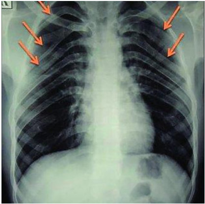
Gorlin - Goltz Syndrome
- bifid ribs
Gorlin Goltz syndrome
Multiple radiolucencies in jaw suggestive of OKC
Complain of missing teeth in 5 year old boy. Provide DD
Dentigerous cyst
Adenomatoid odontogenic tumor
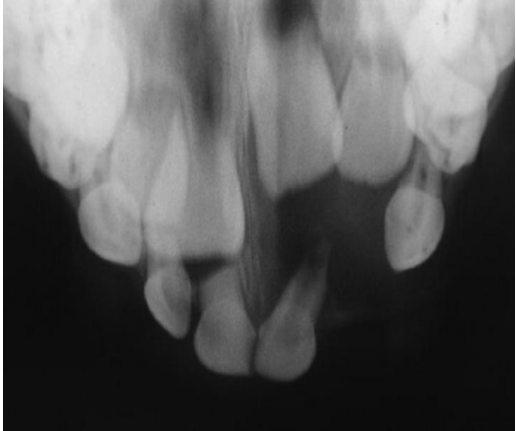
Dentigerous cyst
Adenomatoid odontogenic tumor
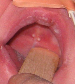
The mother of a 2 month old baby has a concern of white nodules in the mouth of her kid. The clinical image is shown below. There is no pain or any other symptoms. Provide diagnosis
Epstein pearls
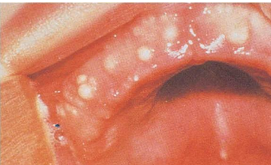
The mother of a 1 month old baby has a concern of white nodules in the mouth of her kid. The clinical image is shown below. There is no pain or any other symptoms. Provide diagnosis
Bohn nodules
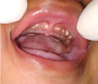
A full term newborn boy, weighing 3 kg, born out of an uncomplicated pregnancy, was brought to us for evaluation of a few small, white and round bumps on the gingival surface. Examination of the oral cavity showed multiple, firm, pearly-white papules measuring 2 to 4 mm in diameter, grouped over the vestibular aspect of the alveolar ridge of the maxillary arch
Bohn nodules
The mother of a 1 month old baby has a concern of white nodules in the mouth of her kid. The clinical image is shown below. There is no pain or any other symptoms. Provide diagnosis and managent
Bohn nodules
No treatment
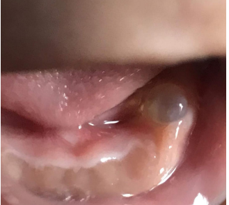
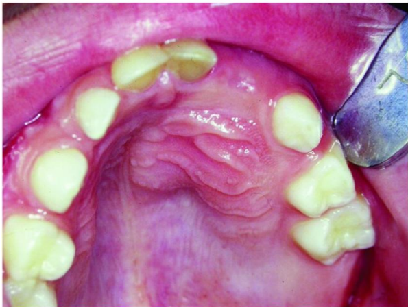
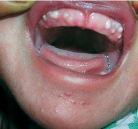
The mother of a 1 month old baby has a concern of a bubble in the mouth of her kid. The clinical image is shown below. There is no pain or any other symptoms. Provide diagnosis and synonym
Gingival cyst of newborn/infant
Dental lamina cyst of infants
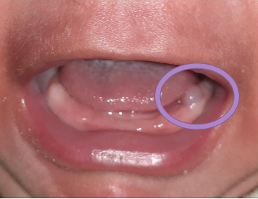
Spot the cyst and provide diagnosis
Gingival cyst of newborn/infant
Dental lamina cyst of infants
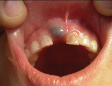
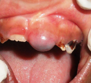
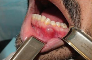
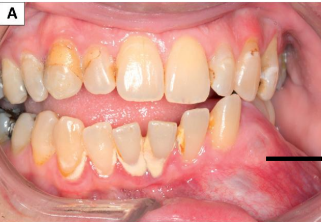
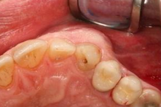
12-year old male patient, which presented as a swelling involving the gingiva in the region of 11 of 5 year duration
Eruption cyst
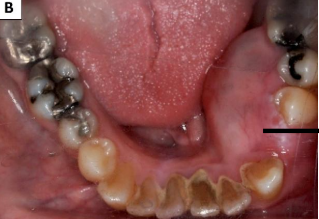
7-year old male patient, which presented as a swelling involving the gingiva overlying the crown of 21
Eruption cyst
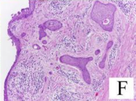
A 30 year old female presented with complaint of swelling in the lower jaw. Intraorally a swelling was not over the left vestibule spanning over 43 to 35. Provide differential diagnosis.
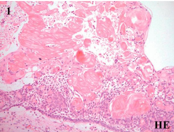
Calcifying odontogenic cyst
Calcifying epithelial odontogenic tumor
calcifying odontogenic cyst
a- ghost cells
b- odontogenic epithelial lining with stellate reticulum like cells
c- ameloblasts like basal cells
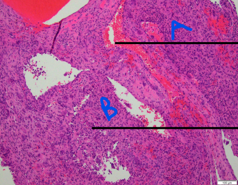
Calcifying odontogenic cyst
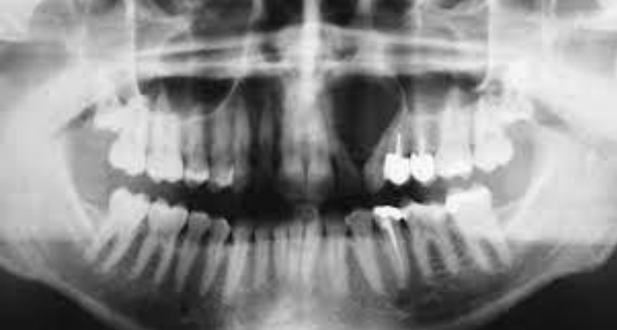
A 17-year-old patient referred by his orthodontist following the fortuitous discovery of a mixed radiolucent/radiopaque image in the right jaw. Provide DD
Calcifying odontogenic cyst
Calcifying epithelial odontogenic tumor
Calcifying odontogenic cyst
Calcifying epithelial odontogenic tumor
Calcifying odontogenic cyst
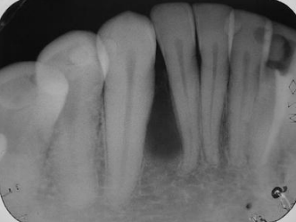
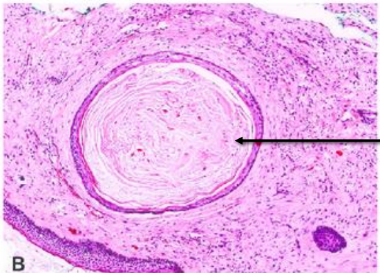
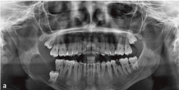
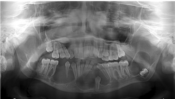
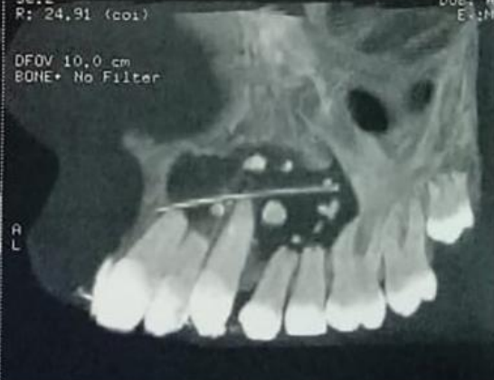
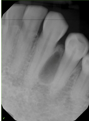
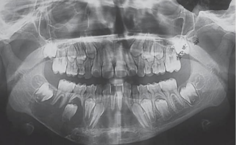
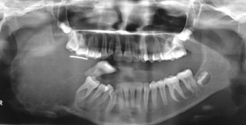
A 51-year-old male patient presented with a well-delimited, radiolucent, mandibular lesion, located between the roots of the right lower lateral incisor and canine and evidenced during routine radiographic examination. Provide 2 differential diagnosis
Lateral periodontal cyst
OKC
Lateral periodontal cyst
A 43-year-old male patient with a complaint of painless swelling in the left mandibular premolar region. Although the history revealed a presence of the swelling for more than one year without any associated symptoms, the patient preferred to obtain clinical consultation as he was concerned about the swelling. The patient had no previous history of dental treatment except for periodic oral prophylaxis. Provide two differential diagnosis
Lateral periodontal cyst
OKC
Provide two differential diagnosis
Lateral periodontal cyst
OKC
Provide two differential diagnosis
Lateral periodontal cyst
OKC
Lateral periodontal cyst
Provide 1 provisional diagnosis
Botroid odontogenic cyst
Botroid odontogenic cyst
Botroid odontogenic cyst
Botroid odontogenic cyst
Botroid odontogenic cyst
A 35-year-old woman presented with the chief complain of asymptomatic swelling in the lower front region of jaw since 4 months. On clinical examination, hard swelling was present on body of mandible in relation to 33 to 43, obliterating the buccal vestibule and measuring about 1.5 cm in diameter. On palpation, the lesion was nontender, with no discharge, and the overlying mucosa was normal. Provide DD
OKC
Glandular odontogenic cyst
Odontogenic tumor
A cyst of jaw
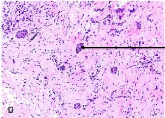
Glandular odontogenic cyst
glandular odontogenic cyst
- mucous cell/goblet cell
glandular odontogenic cyst
- microcyst
glandular odontogenic cyst
- cilia
glandular odontogenic cyst
glandular odontogenic cyst
- clear cells
glandular odontogenic cyst
Clinical presentation of cysts and tumors of jaw
Buccal expansion
Clinical presentation of cysts and tumors of jaw
Lingual expansion
buccal expansion
palatal expansion
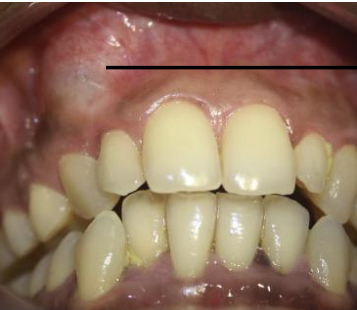
32 year old male patient with complaint of swelling in midline of palate of 3 months duration. There is no pain or any other symptoms. Provide differential diagnosis and justify
Periapical cyst
ncisive canal cyst
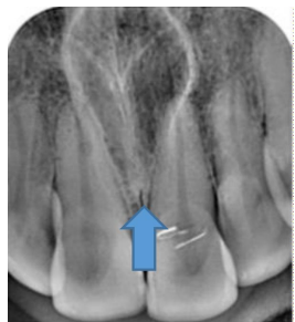
62 year old male patient with complaint of swelling in midline of palate. Provide differential diagnosis and justify
Incisive canal cyst
OKC
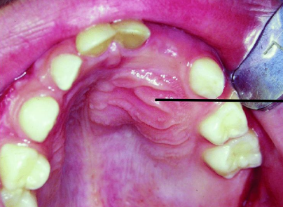
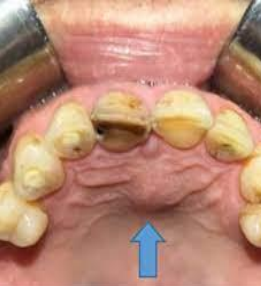
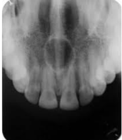
A 23 year old presenting with a cystic swelling in midline in the anterior region of palate. Provide differential diagnosis
Incisive canal cyst
OKC
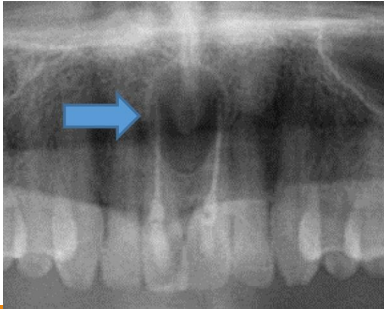
A 52 year old presenting with a cystic swelling in midline in the anterior region of palate. Provide differential diagnosis
Periapical cyst
Incisive canal cyst
Incisive canal cyst
a- pseudostratified ciliated columnar epithelium
b- goblet cells/ mucous cells
A 31 year old presenting with a cystic swelling in lateral aspect between lateral incisors and canine. Provide differential diagnosis
OKC
Lateral periodontal cyst
A 31 year old presenting with a cystic swelling in lateral aspect between lateral incisors and canine. Provide differential diagnosis
OKC
Lateral periodontal cyst
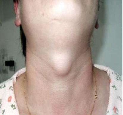
A 45 year old presented with a cystic swelling in midline of the neck. Provide DD
Thyroglossal duct cyst
Dermoid/epidermoid cyst
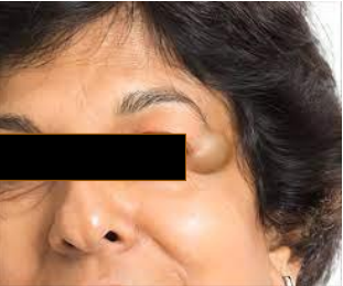
A 35 year presented with cystic swelling on lateral part of forehead of 2 cm diameter. Provide DD
Dermoid cyst
Epidermoid cyst
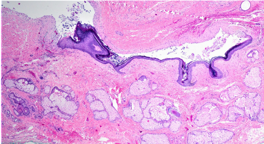
A 35 year presented with cystic swelling on lateral part of forehead of 2 cm diameter. The microscopic image is shown below.
Dermoid cyst
A 35 year presented with cystic swelling on left side of the neck of 2 cm diameter. Provide DD
Dermoid cyst
Epidermoid cyst
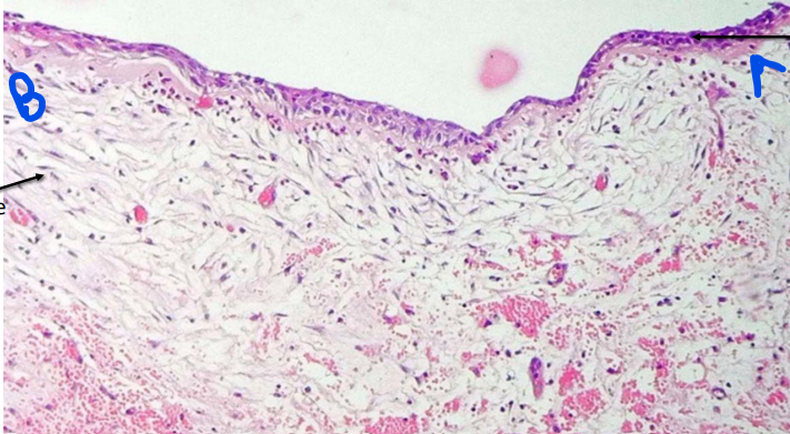
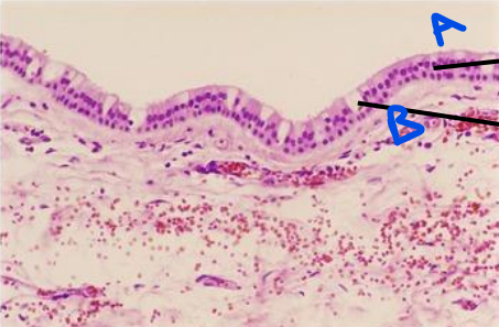
epidermoid cyst
a- keratin
b- orthokeratinized stratified squamous epithelium
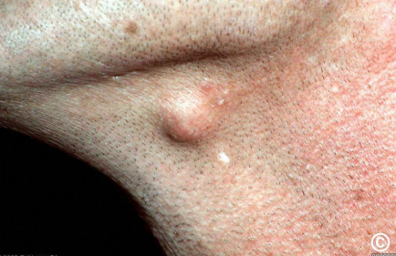
Accidental finding, no expansion of bone
OKC
Solitary bone cyst
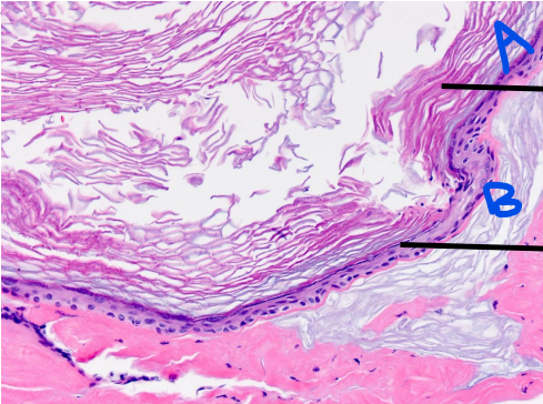
solitary bone cyst
a- bone
b- cavity without lining epithelium
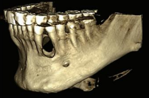
A 20 year old boy presented with swelling of the lower face. Provide DD
OKC
Ameloblastoma
Aneurysmal Bone Cyst
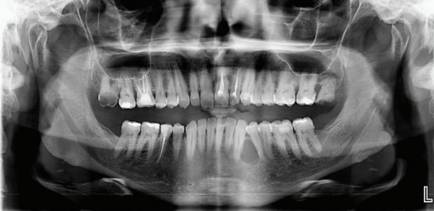
A 9 year old boy presented with swelling of the lower face. Provide DD
OKC Ameloblastoma Aneurysmal Bone Cyst
Aneurysmal Bone Cyst
a- blood filled sinuses/spaces
b- giant cells
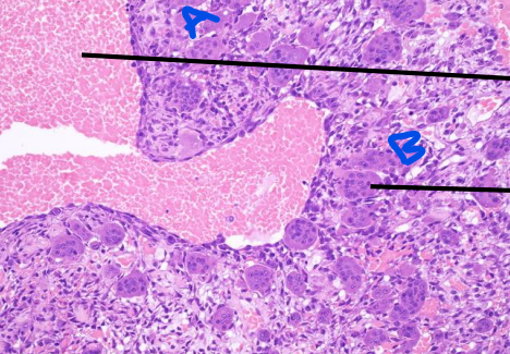
A 18 year old boy presented with cystic swelling of mandible. Provide DD and justify
Aneurysmal bone cyst
Ameloblastoma
Aneurysmal Bone Cyst
a- blood filled sinuses/spaces
b- giant cells
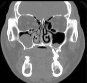
An 18-year-old woman presented with unilateral rhinorrhea for past 2 months. She had a history of road accident 12 months ago. Provide DD
Surgical cyst of maxillary antrum
Mucosal cyst
A 42-year-old woman came complaining of intermittent stinging pain in her right cheek. There were no clinically abnormal findings in physical and oral examinations. Radiograph showed a cystic swelling in maxillary antrum. The patient only had a history of orthognathic surgery on both the maxilla and the mandible performed about 21 years prior to this visit
Surgical cyst of maxillary antrum