Head anatomy SDL
 Call Kai
 Learn
Learn Practice Test
 Spaced Repetition
 Match
Match Flashcards
 Knowt Play
1/60

There's no tags or description
Looks like no tags are added yet.
Name | Mastery | Learn | Test | Matching | Spaced | Call with Kai |
|---|
No analytics yet
Send a link to your students to track their progress
61 Terms
Where is the oral mucosa
a lining of the entire oral cavity
what is protective oral mucosa?
stratified squamous epithelium
more keratinised = more wear areas
can have conified ‘spikes’
supported by the lamina propria
what is the lamina propria?
dense collagenous tissue that supports oral mucosa
how does the thickenss of the protective oral mucosa differ in herbivores to carnivores?
much thicker in herbivorous animals
which animals may have cornified ‘spikes’ on their protective oral mucosa?
cats
herbivores
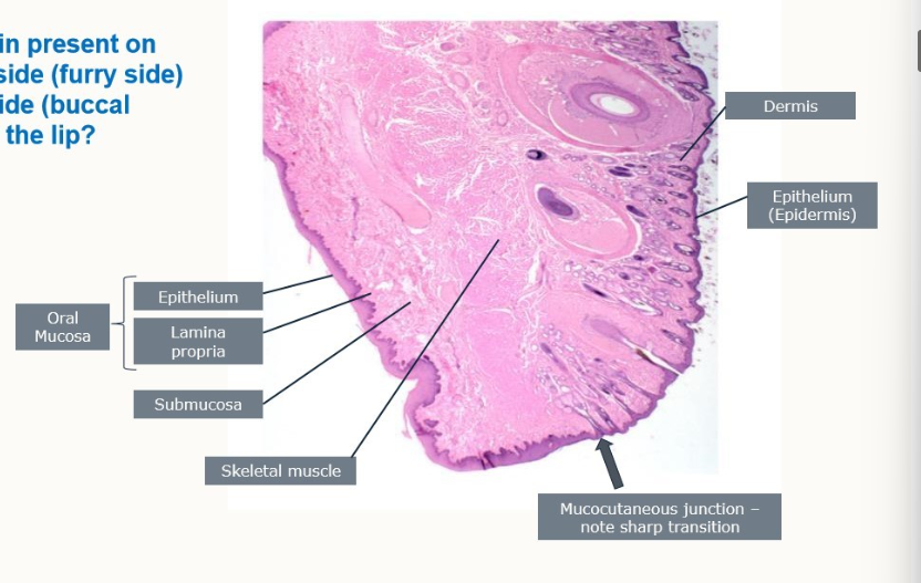
Is keratin present on the outside of inside of the lip?
buccal side is non-keratinised epithelium
keratin present on the outside
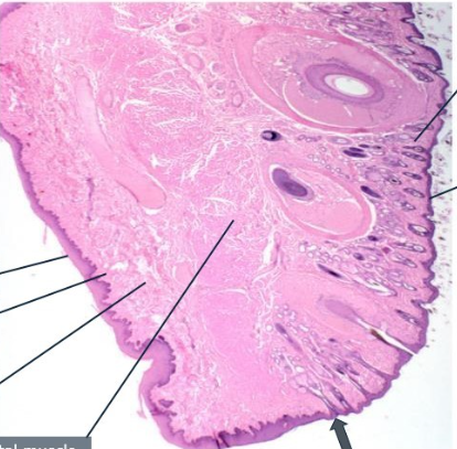
Label this image
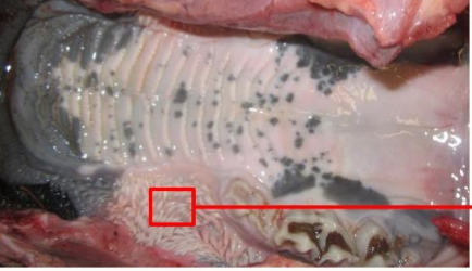
What is this showing?
keratinised papillae
How is the keratin replaced in the keratin layer of the cornified spikes?
keratinised stratified squamous epithelium
keratinocytes continually turnover which produce keratin
functions of the tongue?
prehend food
control food bolus
antagonist to cheek muscles to push food side-to-side over occlusal surfaces
catching ruminal bolus during cudding
carries taste buds
suckling in neonatal mammals
What are key parts of the tongue structurea?
lingual root
median groove
lingual apex
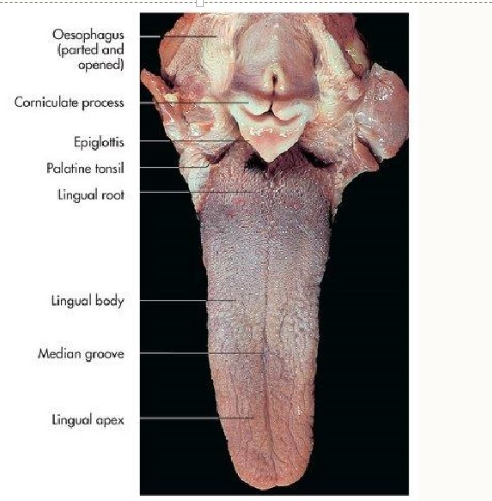
can you relate tongue shape to function?
carnivores →smoother and sharper tongue used for tearing meat
herbivores have a flatter tongue to hold and remove plant material
omnivores have versatile tongues
what are the 2 types of papillae?
mechanical
gustatory
what are mechanical papillae and what are some features of them?
protection, markedly keratinised
Features:
conical
marginal
filiform (small and numerous)
what are gustatory papillae and what are some features of them?
taste, contain taste buds
features
fungiform
civumvalate/valate
foliate
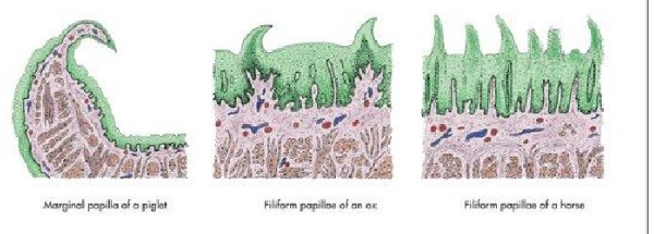
What papillae are these?
mechanical
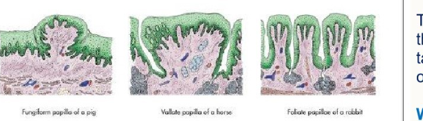
What papillae are these?
gustatory
what stem cells do we have in the oral cavity?
linguinal epithelium stem cells
why are stem cells needed in the oral cavity?
exposed to harsh substances, wear the epithelium down
therefore due to the reate of turnover of the lingual epithelium, need stem cells to constantly replace.
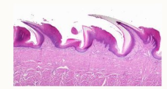
what papillae are these?
mechanical
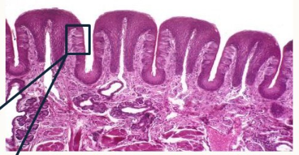
What papillae are these?
gustatory papillae
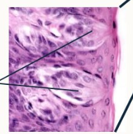
What are these?
taste buds
why do gustatory papillae need to be wet?
taste is enhanced when molecules are dissolved → water acts as a solvent
enables solutes to be transported into the clefts to the receptors on the taste buds
enables taste buds to be activated
keeps taste receptors activated
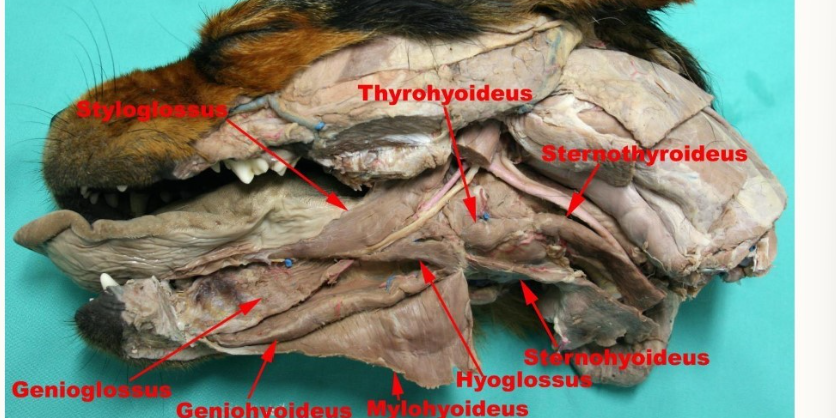
what are the extrinsic tongue muscles?
geniohyoideus
genioglossus
hyoglossus
styloglossus
mylohyoideus
What is the location of:
geniohyoideus
genioglossus
hyoglossus
styloglossus
mylohyoideus
chin to hyoids
chin to base of tongue
hyoid to base of tongue
stylohyoid to tongue
transverse fibres across jaw, supporting tongue
which nerve contains the motor supply to the intrinsic and extrinsic tongue muscles?
CNXII
what are the two types of sensation in the tongue?
gustatory
somatosensory
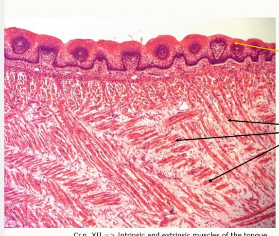
What histological features canw e identify on the tongue muscle?
Top = epithelium with papillae
bottom = intrinsic striated muscle fibres in x, y, z axes
what is wooden tongue?
disease in cattle
caused by actinobacillus lignieressi → a commensal on the URT
opportunistic infection, as it’s a commensal
sof tissue damage being affected of oral cavity and throat
see granulomatous lesions → firm/woody texture of these areas
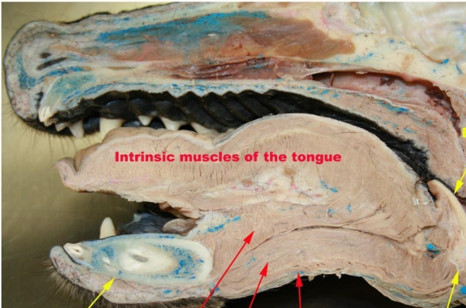
What can we identify here?
Left to right:
Mandibula (yellow)
Genioglossus
Geniohyoideus
mylohyoideus
top yellow = epiglottis
bottom yellow = hyoid
sternohyoideus
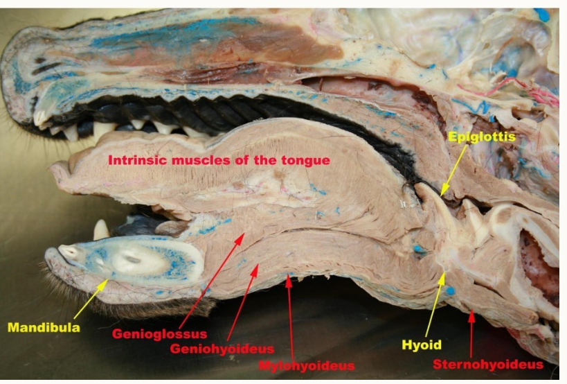
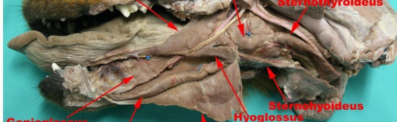
Label the muscles here
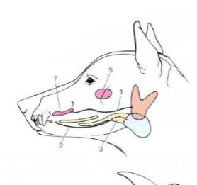
What glands can we identify here?
parotid gland
mandibular duct
monostomatic part of sublingual s.g.
zygoamtic salivary gland
buccal salivary gland
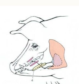
What can we identify in this image?
Orange = parotid
Light blue = mandibular
Pink linear = buccal
Yellow = sublingual
4 = ppolystomatic part of sublingual s.g.
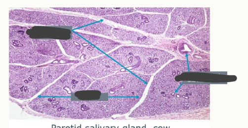
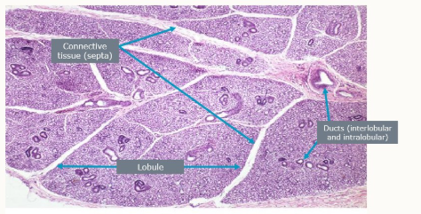
what structures can we identify in this salivary gland?
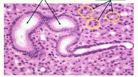
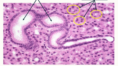
What structures can be identified in this salivary gland?
Left = ducts
yellow circles = secretory units
What are myoepithelial cells?
contractile cells found in glandular tissue
contract → aiding secretions being expelled by glandular epithelial cells
5 roles of myoepithelial cells?
support structurally
allows even distribution of secretion
respond to hormonal and neurological signs for fine-tuning glandular activity
maintain balance of fluids and substances in the body
efficient release of secretions into ducts and external environments
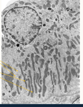
What can we identify here? what is its function?
basal striation
role in pumping Na out of duct
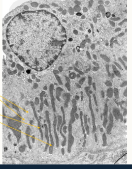
what is the purpose of the mitochondria and folded basement membrane in salivary glands?
folded to increase SA → more aerobic respiration
for K/Na active transport
what is the oesophagus?
the continuation of laryngopharynx connecting to pharynx to stomach
what are the 3 parts of the oesophagus?
cervical = upper aspect
thoracic = beginning at thoracic inlet
abdominal (oesophageal hiatus → stomach)
what is the thoracic oesophagus covered in?
serosa
where does the thoracic oesophagus run?
through inlet on left, moves back to previous dorsal to trachea
runs in mediastinum
where does the oesophagus terminate?
at cardia of the stomach
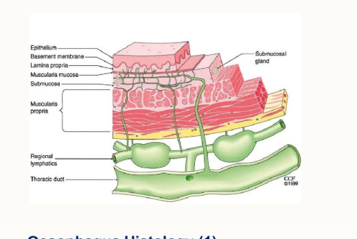
Layers of the oesophagus?
epithelium
basement membrane
lamina propria
muscularis mucosa
submucosa
musclaris propria
regional lymphatics
thoracic duct
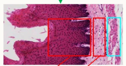
Label
left to right:
strat. sq. epithlium
lamina propria
muscularis mucosa
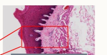
What is this red box?
the mucosa
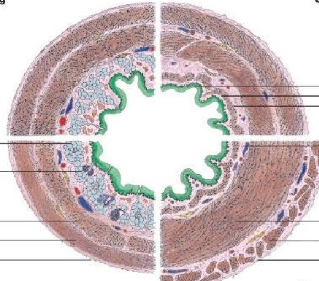
What does this iamge show?
species difference in the oesophagus
for ruminant oesophagus:
muscularis externa structure
muscularis mucosae structure
mucous glands found where
mucosal epithelium structure
skeletal muscle whole length into reticulum
isolates smooth muscle cranially, a sheet caudally
only at junction of pharynx and oesophagus
very keratinised - strat. sq. epithlium from oesophagus to stomach
for horse oesophagus
muscularis externa structure
muscularis mucosae structure
mucous glands found where
mucosal epithelium structure
cranial is skeletal → smooth more caudally
isolated bundles cranial, continuous sheet caudal
only at pharynx-oesophagus junction
some - strat.sq.epithelium continues from oesphagus to stomach
for pig oesophagus:
muscularis externa structure
muscularis mucosae structure
mucous glands found where
mucosal epithelium structure
cranial 1/3 is striated, caudal 2/3 is smooth
lamina muscularis is present caudal, absent cranially
glands abundant cranially, not caudally
some keratinised strat. sq. epithelium from oesophagus to stomach
for dog oesophagus
muscularis externa structure
muscularis mucosae structure
mucous glands found where
mucosal epithelium structure
skeletal from oesophagus to junction with stomach. layers of striated muscle
lamina mucosa present caudal, not cranially
glands throughout but more cranially
non-keratinised stratified squamous epithelium
for cat oesophagus
muscularis externa structure
muscularis mucosae structure
mucous glands found where
mucosal epithelium structure
skeletal cranially, changes to smooth in caudal third
isolated bundles cranially, continuous sheet caudally
glands only at at pharynx and oesophagus junction
non-keratinised stratified sq. epithelium - abrupt transition to columnar
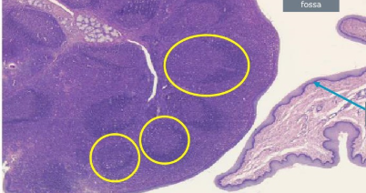
What is this a histological section of
palatine tonsil
yellow circles = lymphoid follicles
blue arrow = stratified sq. epithelium
space = tonsillar fossa
what clinical signs may occur if an animal has a blocked nasal cavity?
noisy respiration
mouth breathing
discharge from nose
facial distortion
sneezing
what clinical signs may we see with a blocked oral cavity?
inappetance
dysphagia
aphagia
distress/rubbing/pawing at mouth
sialorrhoea
ptyalism
tongue protrusion
what is sialorrhoea
dribbline
what is ptyalism?
excess saliva production
what conditions may affect prehension?
facial paralysis, botulism
what conditions may affect chewing/biting
broken jaw
TMJ pain
dental disease
what conditions may affect swallowing?
botulism
pharyngitis
foreign body