OB/GYN Davies: Extrapelvic Pathology Associated with Gynecology (Part 17)
1/9
There's no tags or description
Looks like no tags are added yet.
Name | Mastery | Learn | Test | Matching | Spaced | Call with Kai |
|---|
No analytics yet
Send a link to your students to track their progress
10 Terms
E. Hepatic adenoma
631. A patient who has taken oral contraceptives for more than 5 years is at increased risk for developing:
A. Heart disease
B. Ectopic pregnancy
C. Lung cancer
D. Renal cancer
E. Hepatic adenoma
D. Meigs syndrome
632. Pelvic ascites and right-sided pleural effusions can be associated with benign ovarian fibromas. This condition is known as:
A. Turner syndrome
B. Carcinoid syndrome
C. Stein-Leventhal syndrome
D. Meigs syndrome
E. Pseudosyndrome
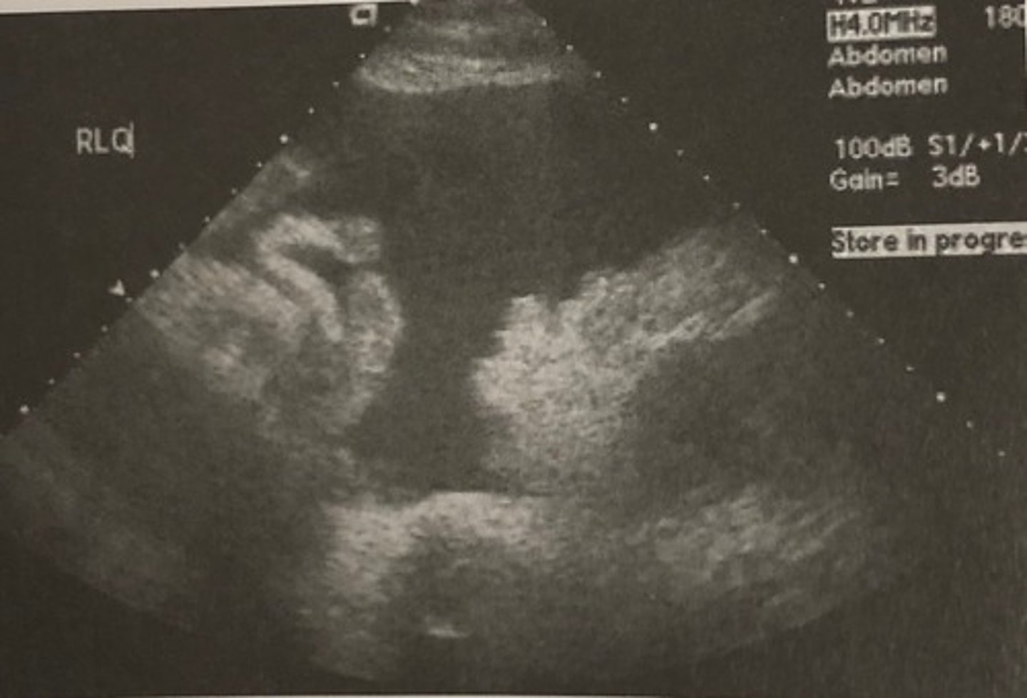
A. Hydronephrosis
633. This patient presents in her mid second trimester with right flank pain. Her obstetric sonogram is normal so the doctor orders an ultrasound exam of her right upper quadrant. The longitudinal scan below demonstrates:
A. Hydronephrosis
B. Cholecystitis
C. Gallstones
D. Pyelonephritis
E. Liver enlargement
E. Fitz-Hugh-Curtis syndrome
Perihepatitis can be associated with pelvic inflammatory disease, causing right upper quadrant tenderness and pain. This condition is known as:
A. PID
B. Stein-Leventhal syndrome
C. Indistinct uterus
D. Meigs syndrome
E. Fitz-Hugh-Curtis syndrome
B. Check Morison's pouch
635. Whenever you suspect pelvic ascites, you should:
A. Scan the liver
B. Check Morison's pouch
C. Have the patient void and then rescan
D. A and B
E. B and C
D. Malignancy
636. A patient presents with abdominal swelling, low back pain, and an extremely elevated CA-125. These clinical findings suggest:
A. Nonspecific findings
B. Infection
C. Pregnancy
D. Malignancy
E. Hemorrhage
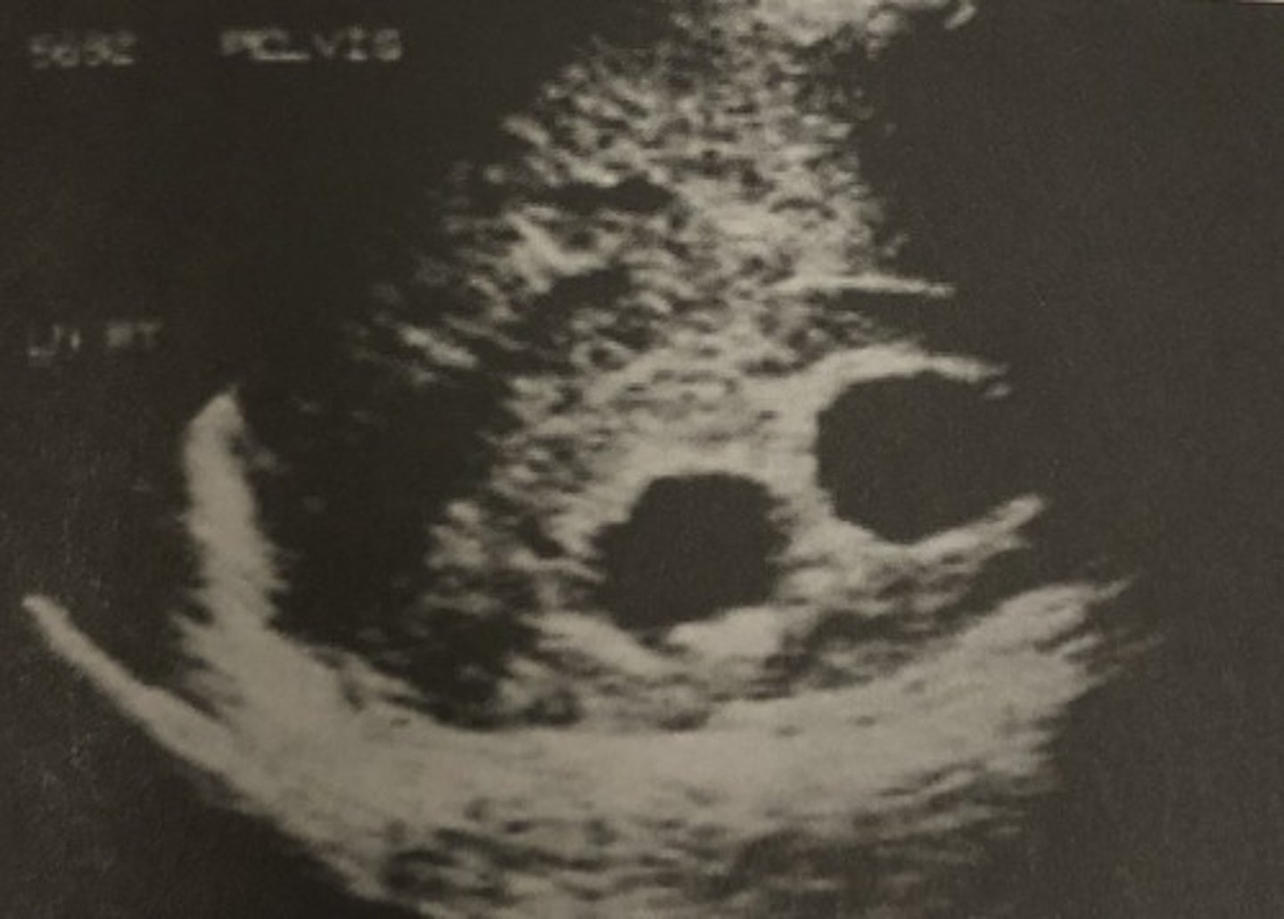
C. Pelvic ascites.
637. This longitudinal sonogram through the right lower quadrant of the patient described in question 636 suggests:
A. Ruptured appendix
B. Endometriosis
C. Pelvic ascites
D. Ruptured ectopic pregnancy
E. Pelvic inflammatory disease
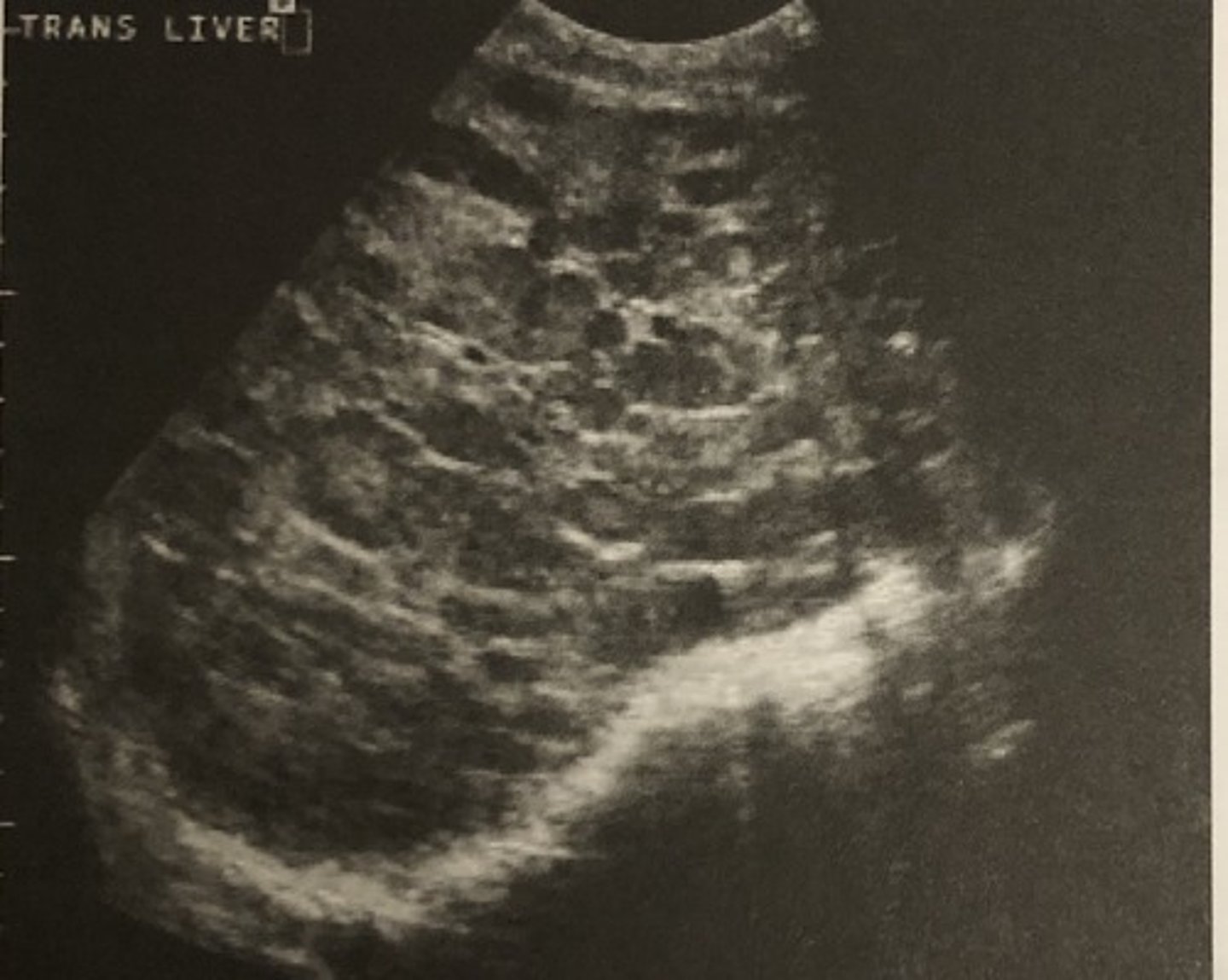
A. Metastatic liver disease
638. In the patient described in question 636, evaluation of her right upper quadrant indicates:
A. Metastatic liver disease
B. Liver abscess
C. Normal liver
D. Abdominal ascites
E. Perihepatitis
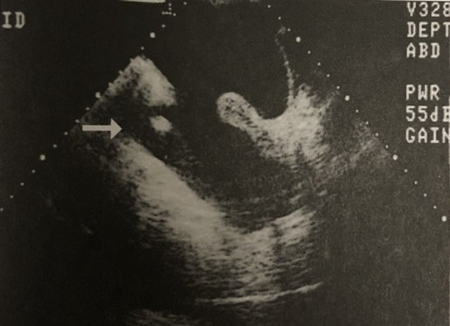
A. Pelvic ascites
639. On bimanual pelvic examination, the clinician cannot palpate the uterus or ovaries. This image suggests:
A. Pelvic ascites
B. Atrophied organs
C. Bowel obstruction
D. Patient obesity
E. Constipation

A. Slice-thickness artifact
640. The arrow in the image in question 639 is pointing to:
A. Slice-thickness artifact
B. Bowel
C. Side lobe artifact
D. Debris
E. Septation