2.2: Tests and Measures
1/75
There's no tags or description
Looks like no tags are added yet.
Name | Mastery | Learn | Test | Matching | Spaced | Call with Kai |
|---|
No analytics yet
Send a link to your students to track their progress
76 Terms
What provides the first key to differential diagnosis and clinical decision-making?
Location
What do you need to use for reference of wound location?
anatomical landmarks
What periwound does this describe: Red, irritated skin due to chemical irritation?
Excoriation
What periwound does this describe: White, moist/mushy skin due to excessive moisture on the skin?
Maceration
What periwound does this describe: Excessively dry skin?
Desiccation or anhydrous
What periwound does this describe: Brown/red-brown discoloration of periwound skin?
Hemosiderin deposits
What periwound does this describe: redness?
ErythemaWhat
What is hemosiderin deposition often associated with?
Long-standing venous insufficiency
What are red flags indicating infection?
Edema, induration (hard swelling), fever, and erythema (deep "angry" red)
How is erythema in inflammation described?
The redness is typically well-defined, localized immediately around the wound, and proportional to the extent of the tissue damage. It is often described as a healthy "pink" tone.
How is erythema in infection described?
The redness is often a deep, "angry" red. It frequently has poorly defined borders and is significantly out of proportion to the size of the wound, potentially spreading far into the extrinsic tissue.
How is edema in inflammation described?
Swelling is usually localized and proportional to the injury.
How is edema in infection described?
Swelling becomes more generalized and may be accompanied by induration, which is a hardening of the periwound skin.
How is temperature (calor) in inflammation described?
There is a localized increase in temperature around the wound site.
How is the temperature in an infection described?
While localized heat is present, a systemic fever (increased body temperature) is a major "red flag" for infection.
How is wound drainage and odor in inflammation described?
Produces serous (clear/straw-colored) or serosanguinous (clear/tinged with red) exudate, which is thin and watery in consistency.
How is wound drainage and odor in infection described?
Produces purulent drainage, which is thick, opaque, and creamy, ranging in color from yellow to green or brown. This is often accompanied by a foul odor, which is a strong subjective indicator of infection.
What are wound bed characteristics during inflammation?
Typically shows healthy, beefy red granulation tissue.
What are wound bed characteristics during infection?
The granulation tissue may become pale, dusky, or friable (bleeds easily), indicating poor blood supply or the presence of pathogens. You may also see new areas of tissue breakdown or a "stalled" healing process.
What does a grade of 1+ on the pitting edema scale mean?
Barely perceptible depression
What does a grade of 2+ on the pitting edema scale mean?
easily identifiable depression, rebounds in <15 seconds
What does a grade of 3+ on the pitting edema scale mean?
Depression rebounds in 15-30 seconds
What does a grade of 4+ on the pitting edema scale mean?
Depression lasts for >30 seconds
How do you test circulation?
Peripheral pulses or capillary refill
What does a grade 0 mean on a peripheral pulse scale?
Absent pulse
What does a grade 1+ mean on a peripheral pulse scale?
Diminished pulse
What does a grade 2+ mean on a peripheral pulse scale?
Normal pulse/easy to palpate
What does a grade 3+ mean on a peripheral pulse scale?
Bounding or accentuated pulse
Why might you have difficulty finding a peripheral pulse?
Presence of peripheral vascular disease
What does capillary refill test?
Indicates surface arterial blood flow
What is a normal time for color return during capillary refill?
3 seconds
How do you test protective sensation?
5.07 monofilament that assesses 10 g of pressure
What is wound drainage?
A breach in the epidermis leaking of intra- and extracellular fluids
What should be noted about wound drainage?
- Type and color: indicating components present like RBCs, WBCs, and pathogens
- Consistency: provides information about hydration and possible infection
- Amount: indicates the wound/systemic hydration and perfusion, and possible presence of infection
What is the description of serous exudate or transudate drainage?
Clear, yellow (straw colored)
What is serous exudate composed of?
A mixture of water, electrolytes, and protein (serum)
What is the description of serosanguinous (normal) drainage?
Clear, tinged with red or brown
What is serosanguineous composed of?
Serum and red blood cells; if the tinge is dark brown, it indicates old or dried blood
What is the description of exudate?
Translucent/opaque, creamy yellow
What is exudate composed of?
Dead white blood cells (WBCs) and denatured proteins
What does exudate indicate?
inflammatory response
What is the description of purlulent/purlence drainage?
Thick, opaque, varied colors (yellow, brown, green) and odors
What is purlulent/purlence drainage composed of?
Dead pathogens, white blood cells, and proteins
What does purlent/perlence drainage indicate?
infection
What is the direct measurement of wound dimension?
This method involves measuring the longest length and the widest width of the wound using a consistent scale, typically in centimeters.
How is the length dimension oriented?
cephalocaudally, meaning it follows a "head-to-toe" direction. When using the clock method, the 12-o’clock position is assigned to the part of the wound located closest to the patient’s head, and the 6-o’clock position is closest to the feet.
How is the width dimension oriented?
This is the longest dimension perpendicular to the length. On a clock face, this would generally be measured from the 3 o'clock to the 9 o'clock position
How can you keep the wound dimension measuring consistent?
The patient should be in the same position for every measurement, and ideally, the same clinician should perform the measurements
How can you calculate the surface area of the wound?
Length X Width
When would wound tracing or planimetry be more reliable?
for representing the actual wound area and reflecting changes in irregularly shaped or circular wounds
What is tunneling?
A narrow passageway created by the separation or destruction of fascial planes .
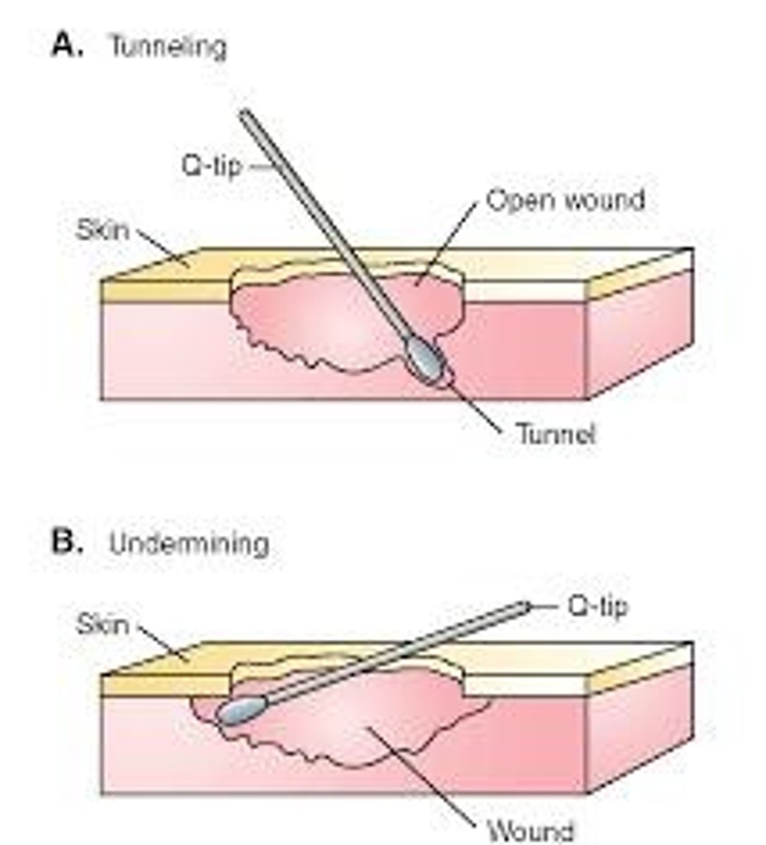
How is tunneling measured?
It is measured by inserting a probe into the passageway until resistance is felt. The depth is the distance from the probe tip to the point where it is level with the wound edge.
What is the typical etiology for tunneling in wounds?
Most commonly found in surgical wounds and neuropathic ulcerations.
What is undermining?
Occurs when the tissue under the wound edges becomes eroded, resulting in a large wound underneath a small surface opening.
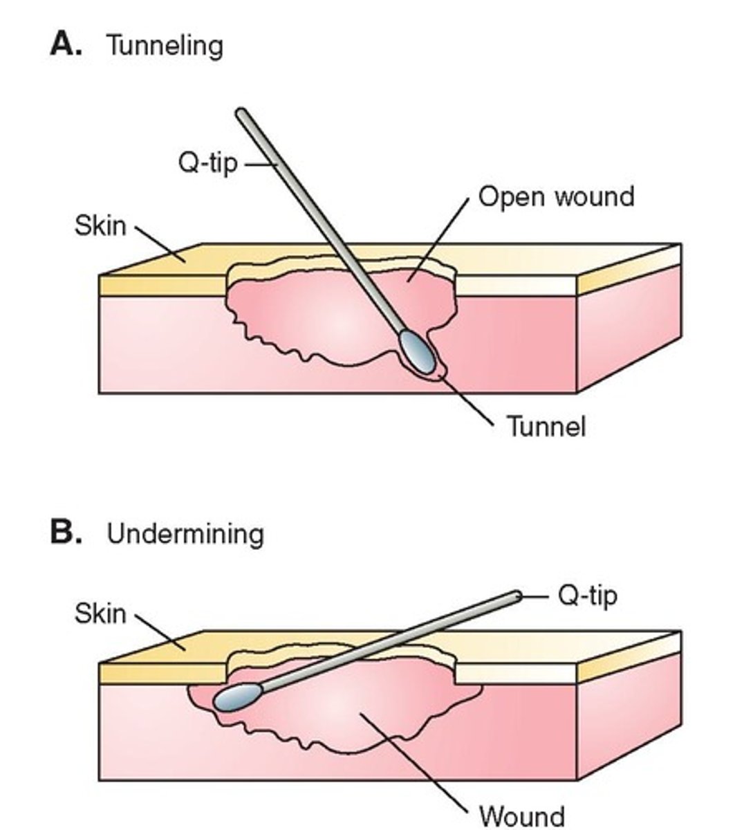
How is undermining measured?
It is measured by inserting a probe under the wound edge, directed almost parallel to the wound surface, until resistance is felt.
What is the typical etiology of undermining?
More commonly found in pressure injuries or neuropathic ulcers
What tissue(s) contribute to the characteristic wound base color of Red?
Healthy granulation tissue, healthy dermis, and healthy muscle
How does healthy granulation tissue contribute to the characteristic wound base color of red?
This is a hallmark of the proliferative phase of healing and appears "beefy red" due to the presence of oxygen-rich capillaries. In the acute proliferation stage, it is often described as shiny and bright pink to red.
How does healthy dermis tissue contribute to the characteristic wound base color of red?
Viable dermal tissue typically has a pink or rosy red tone.
How does healthy muscle tissue contribute to the characteristic wound base color of red?
Viable muscle tissue is bright or dark red and bleeds readily when traumatized
What is indicated by the characteristic wound base color of yellow?
Can indicate either healthy deep structures or various forms of necrotic debris.
What tissue(s) and their conditions contribute to the characteristic wound base color of dry yellow?
Fibrin, crust, and dehydrated adipose
How do fibrin and crust tissue contribute to the characteristic wound base color of dried yellow?
When a wound bed is allowed to air-dry, dead cells and proteins (like fibrin) dehydrate to form a yellow or tan crust
How does dehydrated adipose tissue contribute to the characteristic wound base color of dried yellow?
While healthy fat is pale yellow, it can become a deeper yellow if it becomes dehydrated
What tissue(s) and their conditions contribute to the characteristic wound base color of moist yellow?
Slough and healthy deep structures
How does slough tissue contribute to the characteristic wound base color of moist yellow?
This is necrotic (dead) tissue that is yellow or tan with a stringy or mucinous consistency. It is composed of bacteria, white blood cells, and degraded extracellular matrix
How does healthy deep-structure tissue contribute to the characteristic wound base color of moist yellow?
Certain healthy tissues are naturally yellow. The plantar fascia is described as pale yellow, and healthy adipose tissue (fat) is glistening white to pale yellow
What tissue(s) and their conditions contribute to the characteristic wound base color of black?
Eschar and nonviable deep tissues
How does eschar contribute to the characteristic wound base color of black?
necrotic tissue that can be either hard or soft. Wounds covered in this are typically full-thickness injuries.
How does nonviable deep tissue contribute to the characteristic wound base color of black?
When deeper structures lose their blood supply, their color shifts
What does a nonviable muscle look like?
gray or black
What does a nonviable tendon or ligament look like?
Dark in color and may appear leathery
What does nonviable bone look like?
dark and bruised
When there is the presence of necrotic or granulation tissue, what do you need to do?
Identify the color and estimate the area of the wound surface by percentage and or quartiles
After taking all the results from the examination, what do you do next?
Evaluation, making a clinical judgement about the patient's condition
After you make an evaluation, you make a diagnosis to...
serve as a basis to develop prognosis/goals and interventions