urinary system
 Call Kai
 Learn
Learn Practice Test
 Spaced Repetition
 Match
Match Flashcards
 Knowt Play
1/46

There's no tags or description
Looks like no tags are added yet.
Name | Mastery | Learn | Test | Matching | Spaced | Call with Kai |
|---|
No analytics yet
Send a link to your students to track their progress
47 Terms
Urinary system
Filtration of blood leading to secreting urine
Components of urinary system
kidneys (2)
Ureters (2)
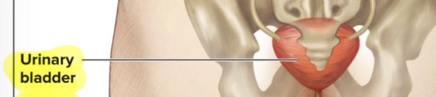
Urinary Bladder
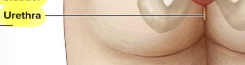
Urethra
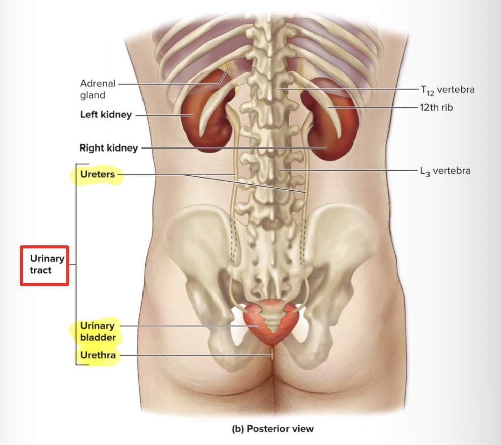
Urinary tract
Composed of:
Ureters (2)
Urinary bladder
Urethra
Local (specific) functions of urinary system
Kidneys: filter the blood by removing waste products and convert filtrate into urine
Ureters: transport urine from kidneys to urinary bladder
Bladder: expandable muscular sac that stores as much as 1L of urine
Urethra: eliminates urine from the body
Kidneys function
filter blood by removing waste products
Convert filtrate from blood into urine
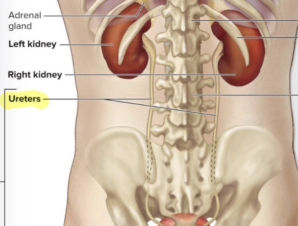
Ureters function
Transport urine from kidneys to urinary bladder
Urinary bladder function
Expandable muscular sac that stores as much as 1L of urine
Urethra function
Eliminates urine from the body
Systemic (body) functions of the urinary system
water volume regulation + solute concentration regulation
Acid base balance: help ph in blood to be regulated by kidneys
Excretion of metabolic wastes, toxins, drugs by kidneys
Activation of vitamin D
Endocrine function:
Renin: regulation of BP
Erythropoietin: regulation of RBC production
Renin hormone
Regulates blood pressure
Erythropoietin hormone
Regulates Red Blood Cell production
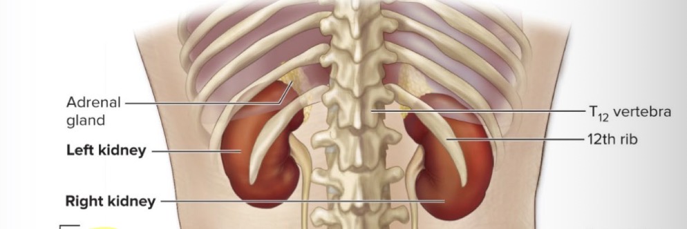
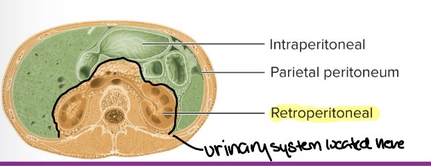
Urinary system location
Retroperitonral (behind parietal peritoneum): in the back
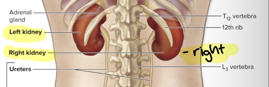
Kidneys location
Left: between T12 and L3 vertebrae
Right: 2cm inferior to left kidney, between T12 and L3
Internal Anatomy of Kidneys
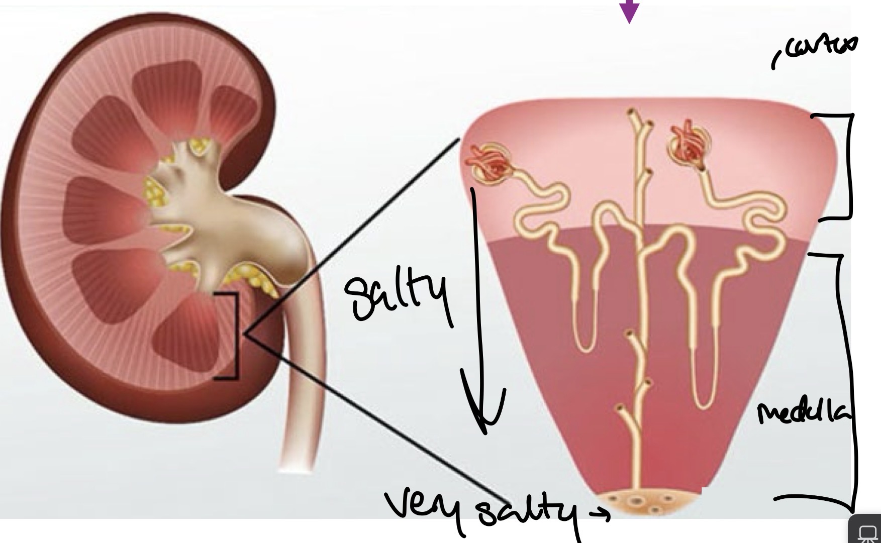
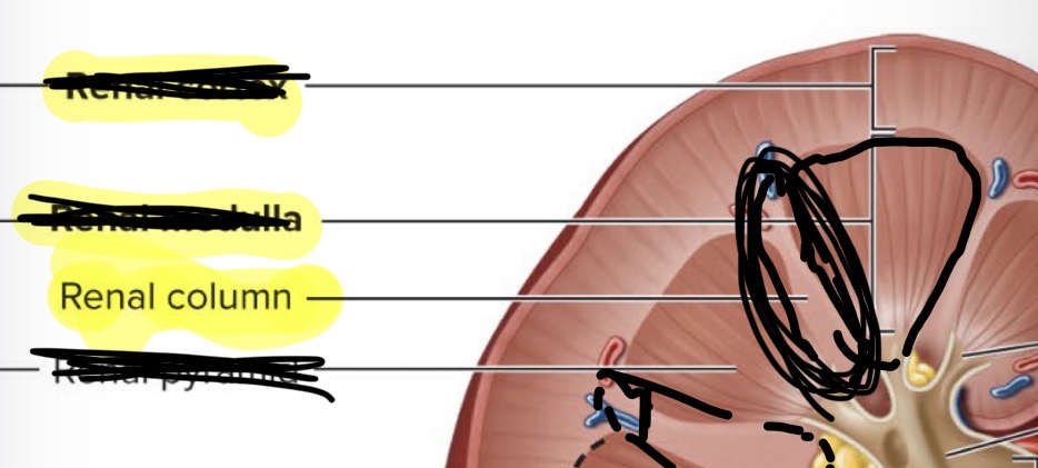
renal cortex
Renal medulla
Renal pyramid
Renal column
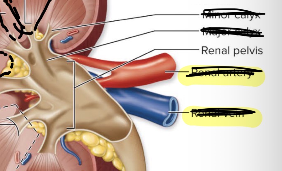
Minor calyx
Major calyx
Renal pelvis
Renal cortex
Top part of internal kidney (around whole kidney)

Renal medulla
Middle part of renal pyramid

Renal pyramid
Pyramid shaped darker sections inside of kidneys
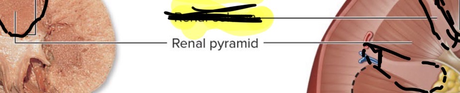
Renal column
The parts in between each renal pyramid
Minor calyx
Smallest funnel leading to bigger funnel which leads to biggest funnel
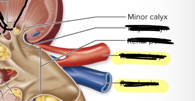
Major calyx
Big funnel leaded to biggest funnel
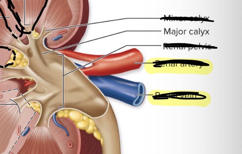
Renal pelvis
Biggest pyramid shaped funnel that leads to ureter
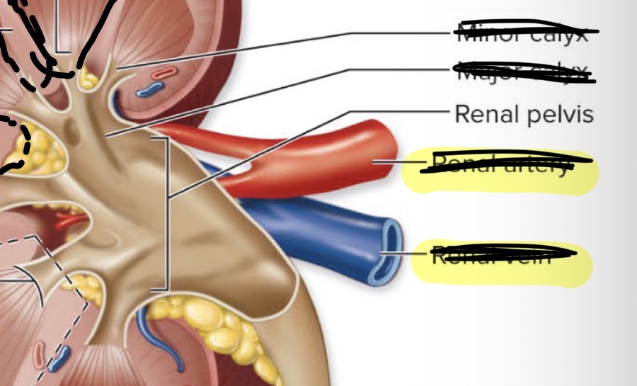
External kidney anatomy
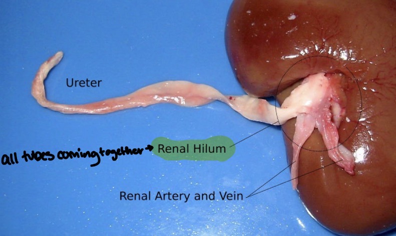
renal hilum
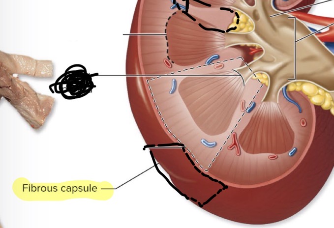
Renal/Fibrous capsule
Renal artery
Renal vein
Ureter
Renal hilum
Where all tubes come out of kidney together including the
ureter
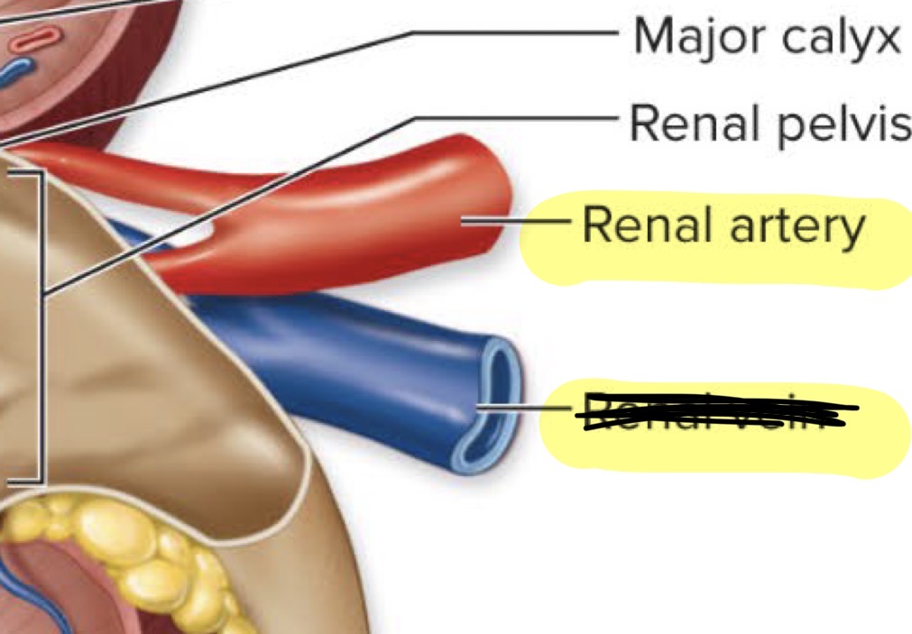
Renal artery
Renal vein
Renal/Fibrous Capsule
Protective layer surrounding kidneys
Renal vein
Carries filtered blood away from the kidney back to the heart through the IVC
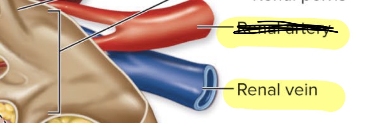
Renal artery
Supplies kidneys with oxygenated blood from aorta
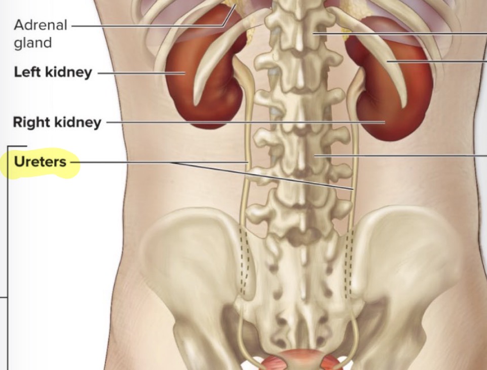
Ureter
Takes urine to bladder
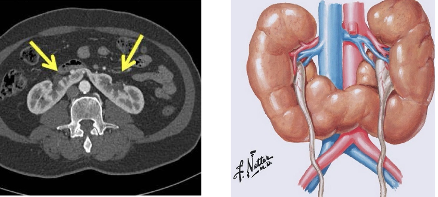
Horseshoe kidney
Very large horseshoe shaped kidneys that make a person prone to kidney stones and UTI
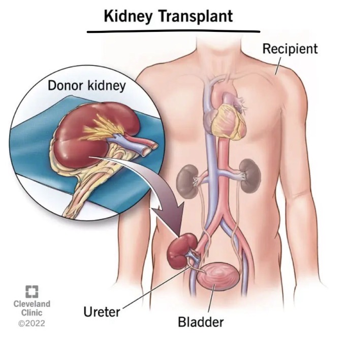
Kidney transplant
Cut open groin, add 3rd kidney to iliac veins and arteries
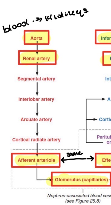
Path for renal artery
(Blood to kidneys): Aorta → renal artery → afferent arteriole → glomerulus (capillaries)
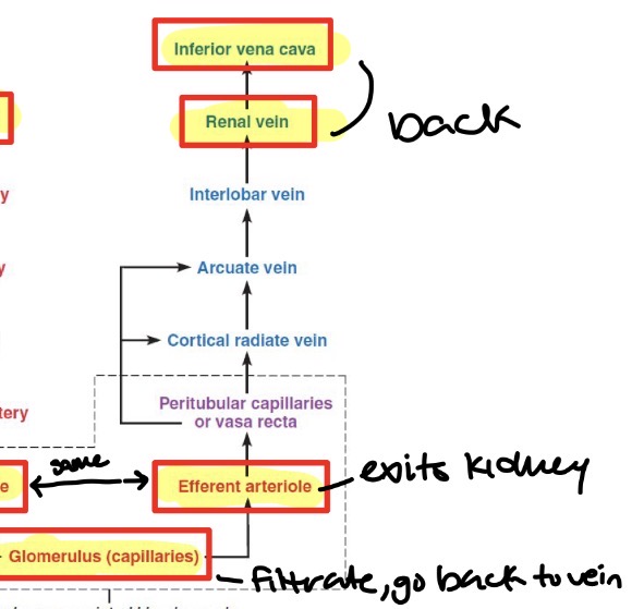
Path for renal vein
(Blood leaving kidneys): Glomerulus (capillaries) → efferent arteriole → renal vein → IVC
Nephron
Smallest unit in kidneys that form urine (start in cortex and go down into medulla until entering minor calyx)
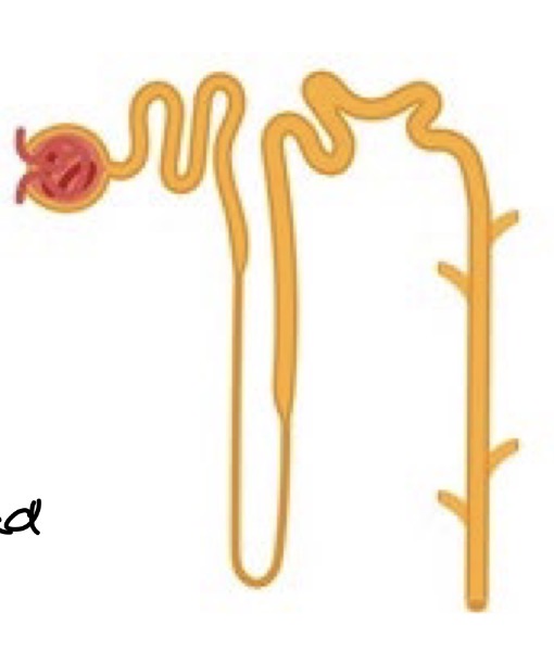
Two main parts of nephrons
renal corpuscle
Renal tubule
Renal corpuscle
Circle head at top of nephron
Contains:
glomerulus which connects to efferent and afferent arterioles
bowman’s capsule
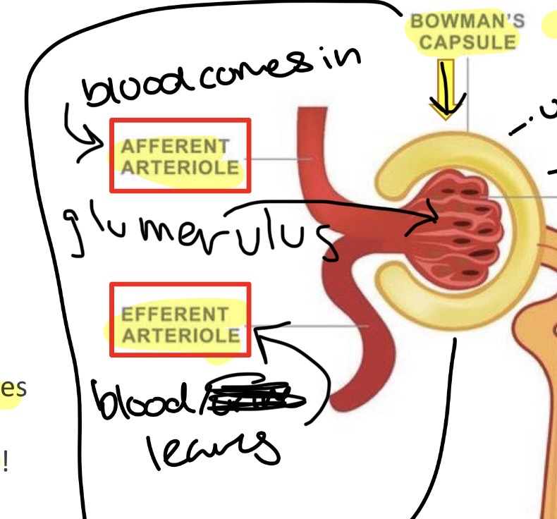
Glomerulus
Connects to efferent and afferent arterioles
Filtration of blood occurs here
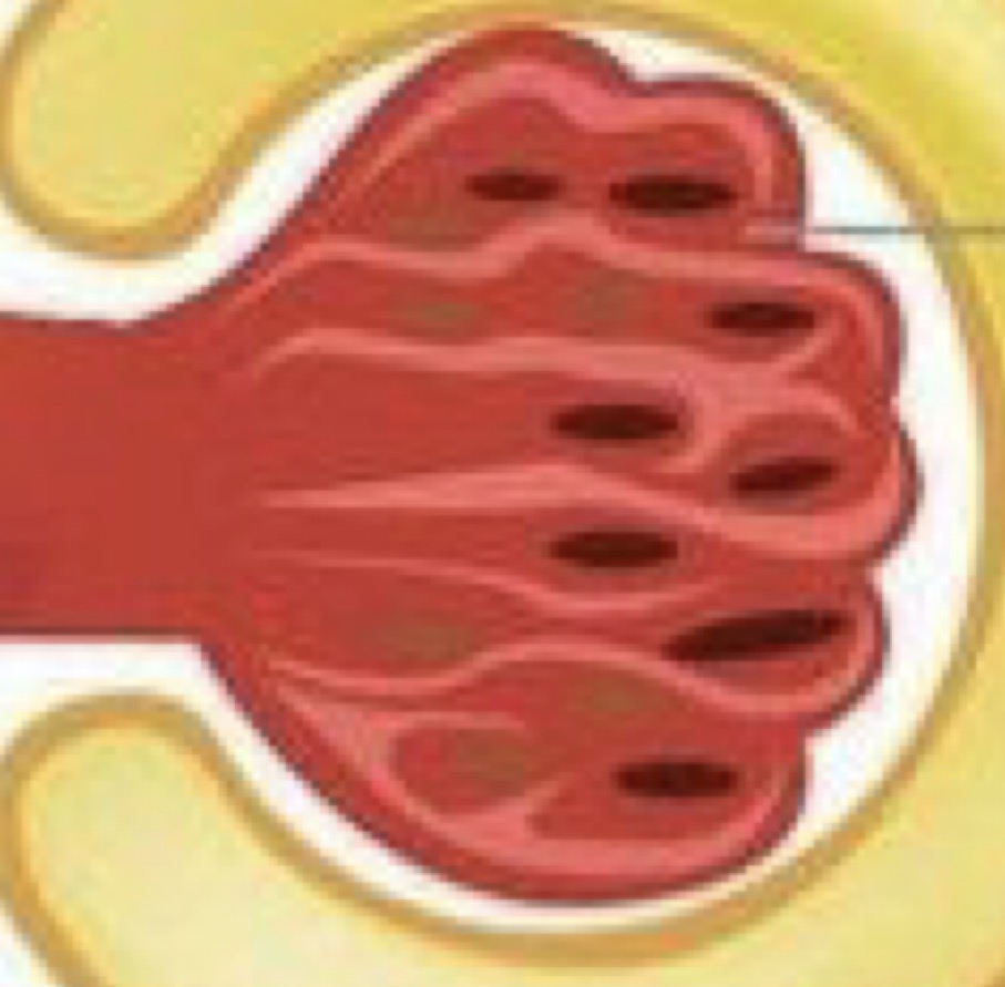
Bowman’s capsule
Surrounds glomerulus (Aka: glomerular capsule)
Where water, salts, glucose and wastes are pushed into from blood filtering
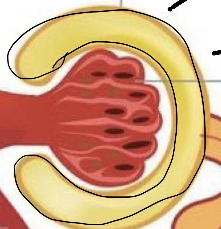
Renal Tubule
Wiggly part
Proximal Convoluted tubule (PCT
Nephron Loop (Henle’s Loop)
- descending limb
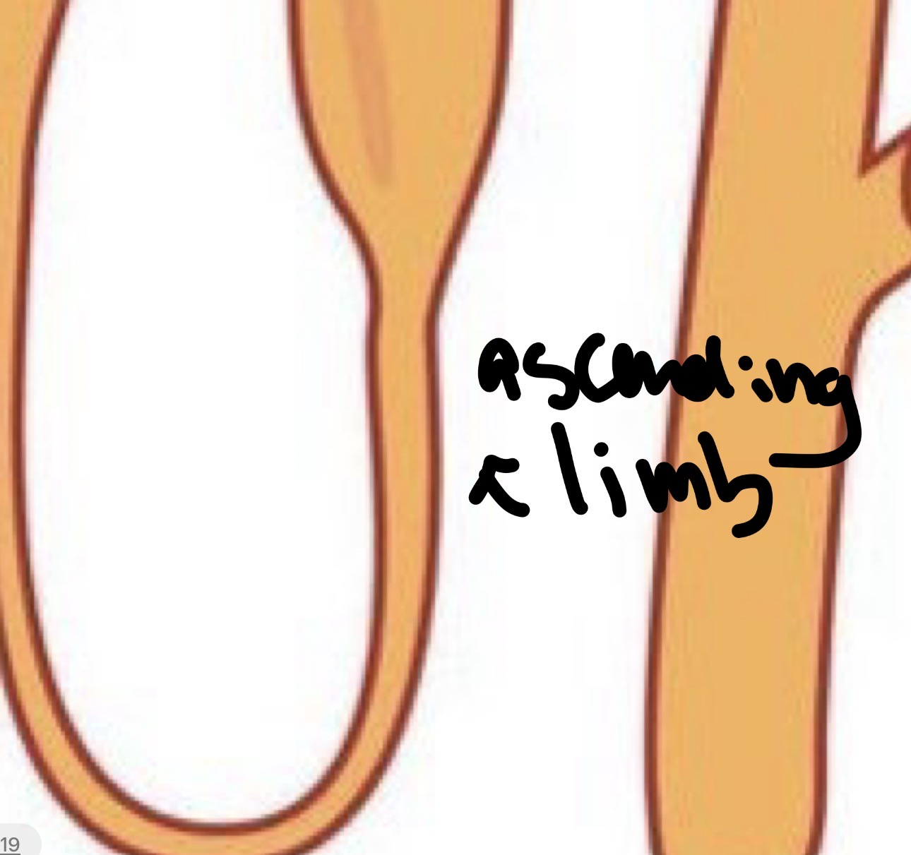
- ascending limb
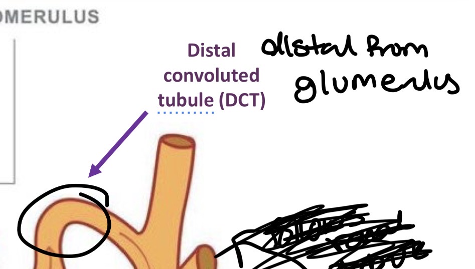
Distal Convoluted tubule (DCT)
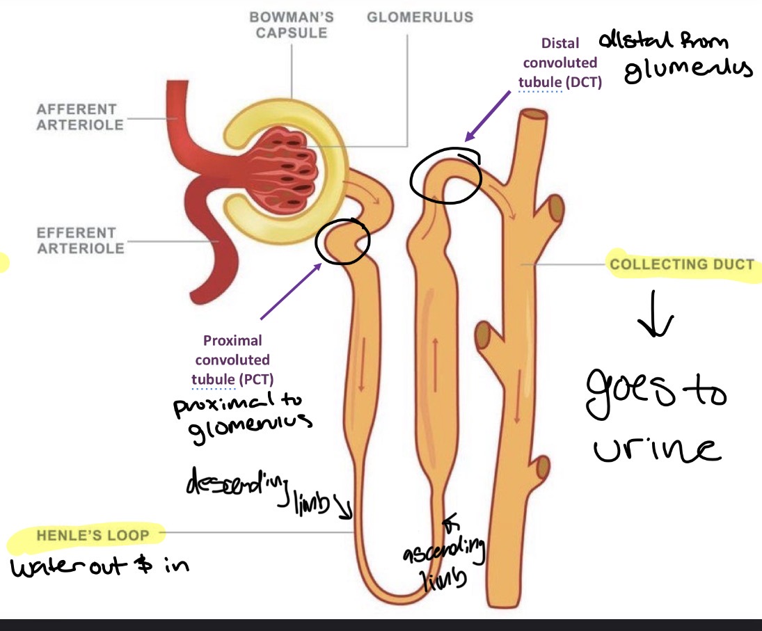
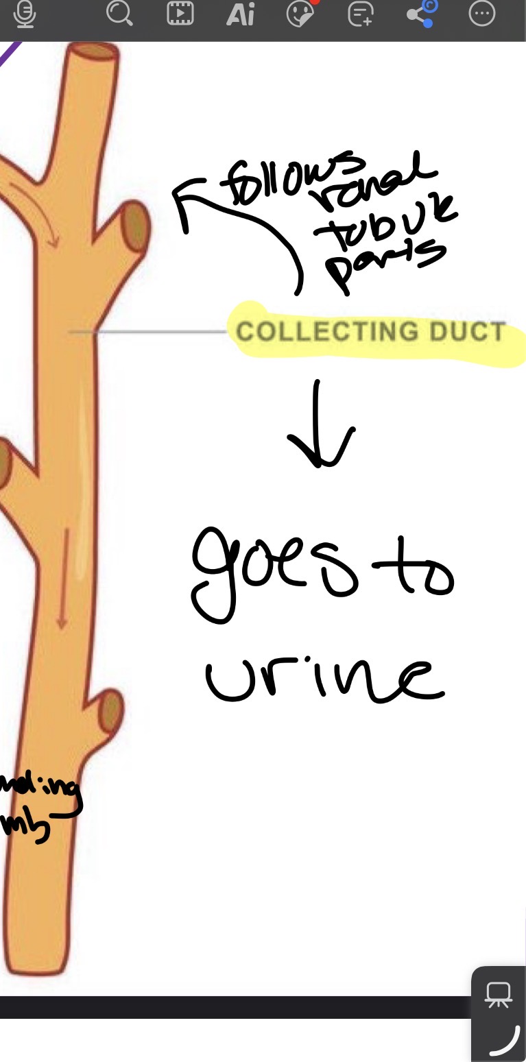
Collecting Duct in regards to renal tubule
Follows renal tubule after DCT
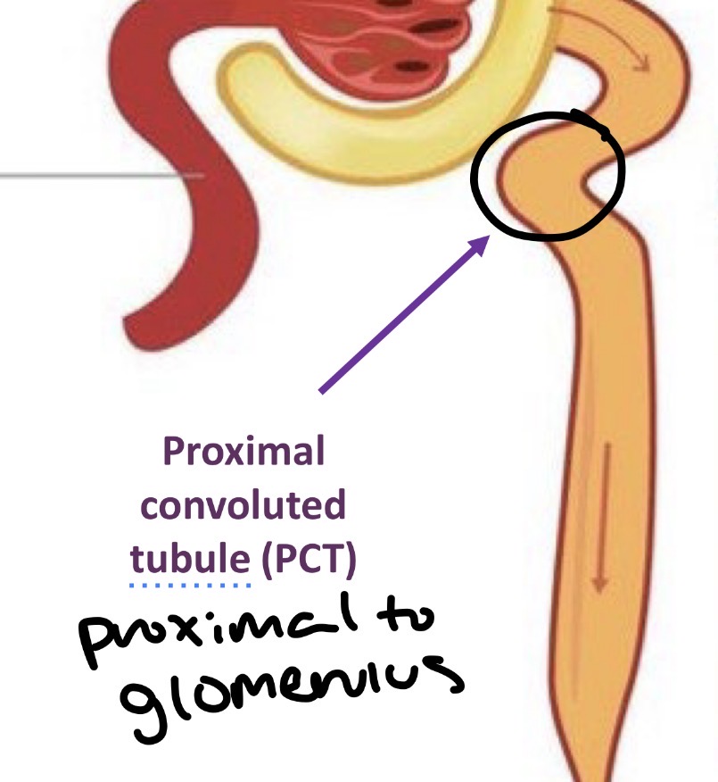
Proximal Convoluted Tubule (PCT)
Proximal (close) to glomerulus
Reabsorbs
65-70% of water
Sodium and other ions
Nearly all glucose
Amino acids
From the bowman’s capsule
Nephron Loop (Henle’s Loop)
Two parts
descending limb
Ascending limb
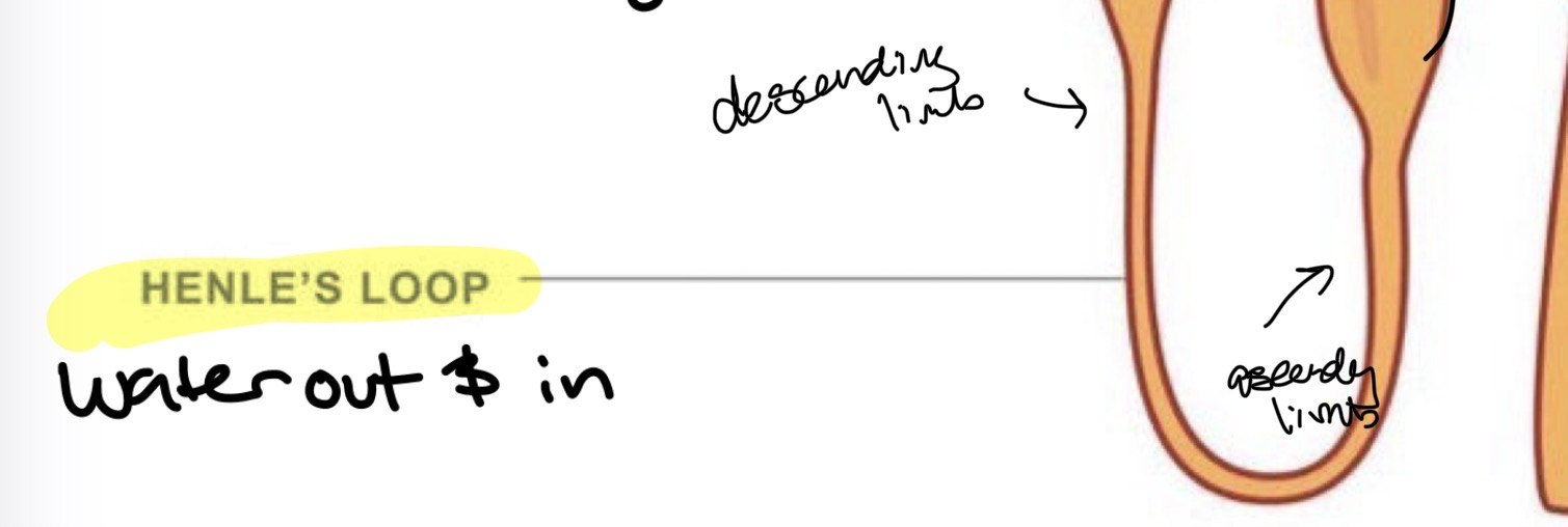
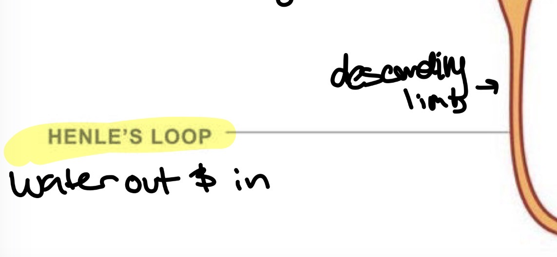
Descending limb of Henle’s Loop
Water leaves and returns to blood (permeable to water) (osmosis)
Salt stays
Happens due to osmosis of water, this limb has more water than surrounding kidney tissue which has more salt. Water goes to where there is less water which is the kidney tissue and leaves the limb. Vasa recta picks up water and takes it back into blood stream
Ascending limb of Henle’s Loop
Salt leaves
Water already gone (impermeable to water)
Thin part: salt diffuse out naturally (diffusion)
Thick part: kidney cells actively pump sodium and chloride out into surrounding tissue so water leaves in descending loop and salt can leave in ascending (active transport)
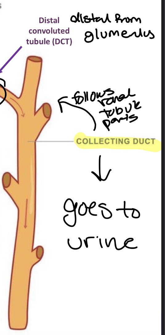
Distal Convoluted Tubule (DCT)
Distal (far) from glomerulus
adjusts sodium levels
Adjusts potassium levels
Helps regulate blood pH
Responds to hormones such as aldosterone
Collecting Duct
Last stop before urine leaves kidney
If body needs water: ADH (antidiuretic hormone) released causing more water reabsorbed by body instead of leaving with urine: darker and less urine
If body is well hydrated: less water is reabsorbed: lighter and more urine
Two types of nephrons
cortical nephrons
Juxtamedullary nephrons
Cortical nephrons
Nephrons in renal cortex only
Juxtamedullary nephrons
Nephrons close to medulla (dives all the way into medulla)
most important for urine concentration as its saltiest toward the end of the pyramid