Week 13 Part A: Cranial Nerves Part B: Spinal Nerves Part C: Physiology Lab: Nerve Conduction Velocity
1/75
There's no tags or description
Looks like no tags are added yet.
Name | Mastery | Learn | Test | Matching | Spaced | Call with Kai |
|---|
No analytics yet
Send a link to your students to track their progress
76 Terms
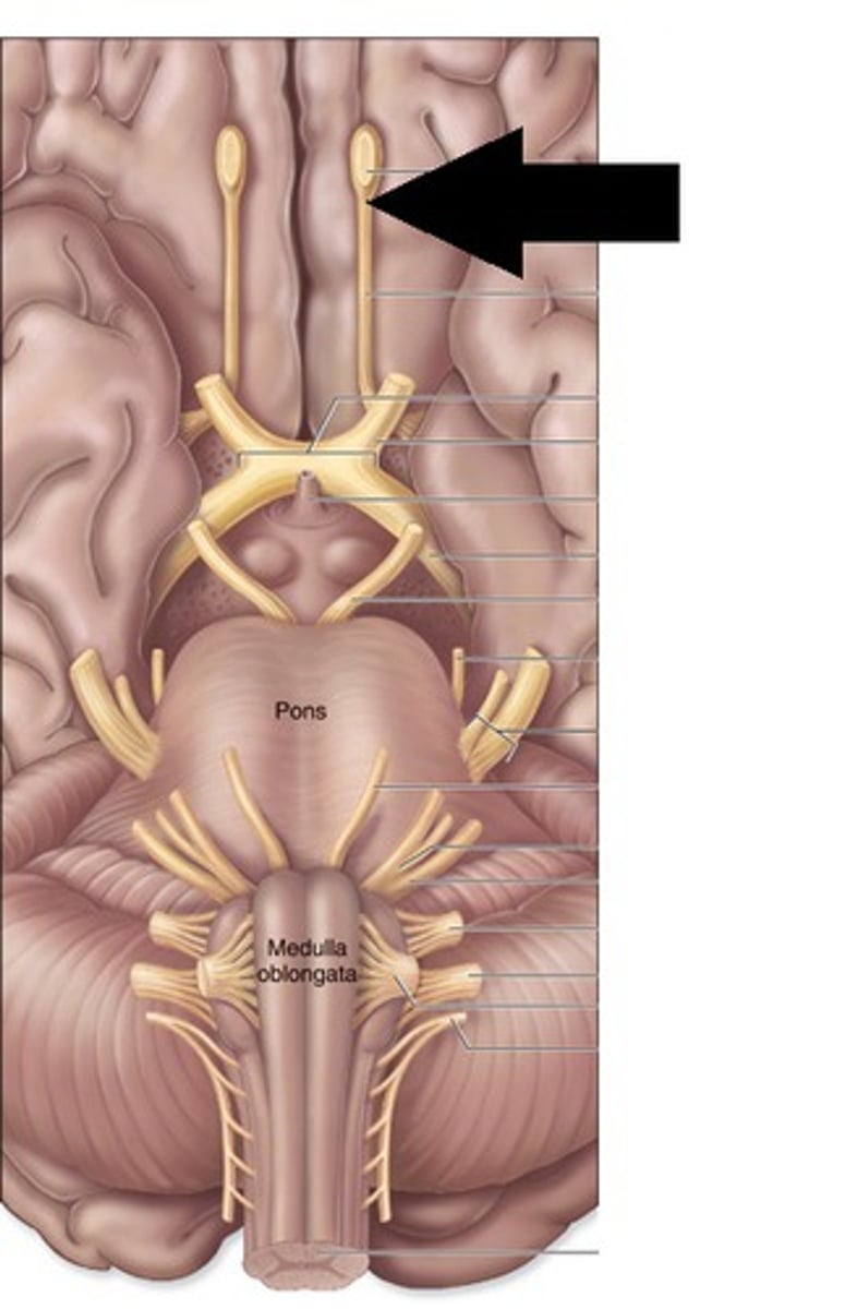
CN I Olfactory nerve
Sensory for smell; located in nasal cavity; fibers pass through cribriform foramina to olfactory bulb
Filaments of olfactory nerve
Fibers passing from nasal mucosa through cribriform foramina
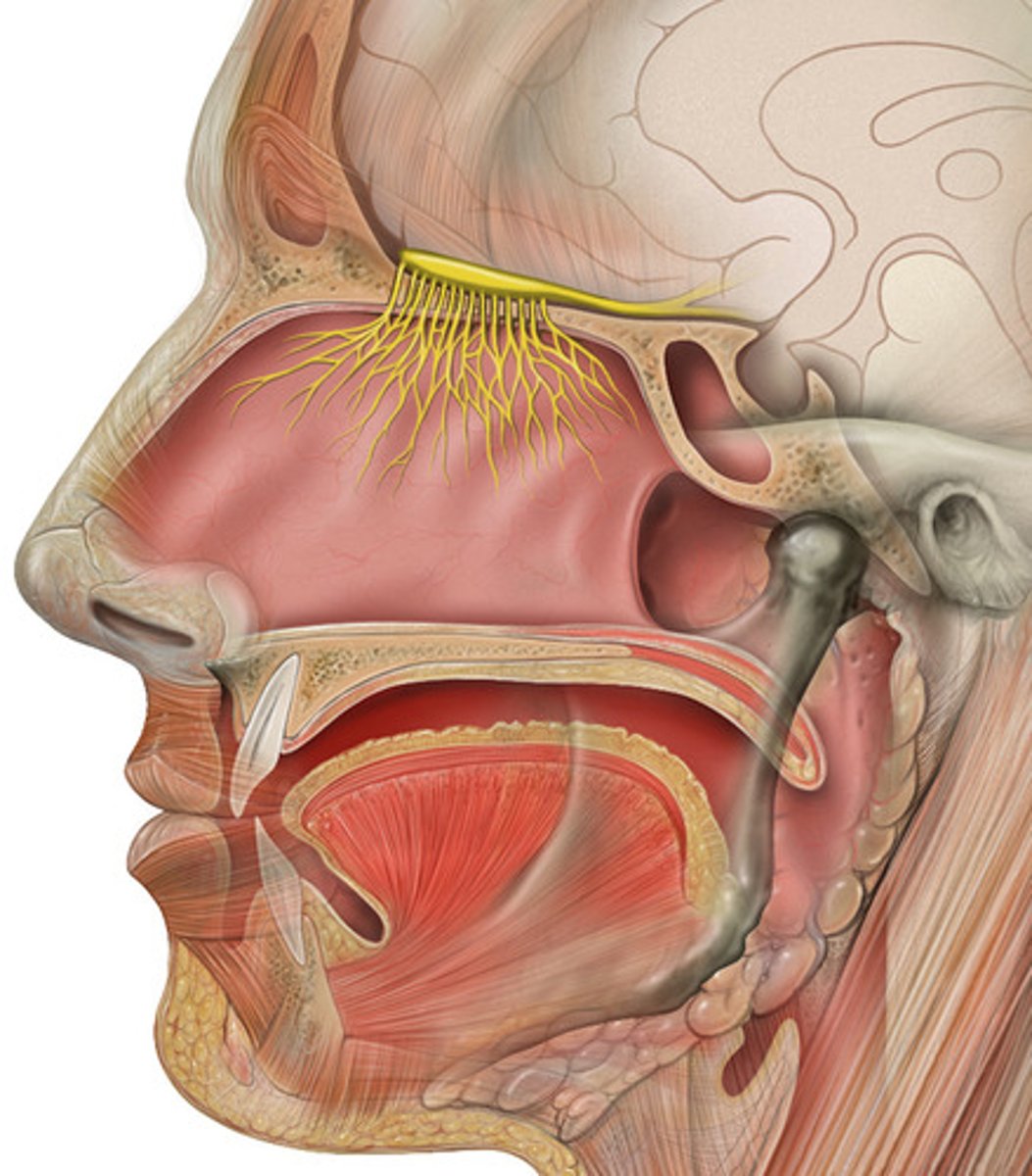
Cribriform foramina
Tiny holes in ethmoid bone for CN I filaments
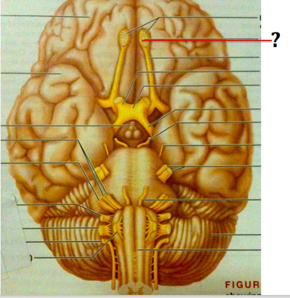
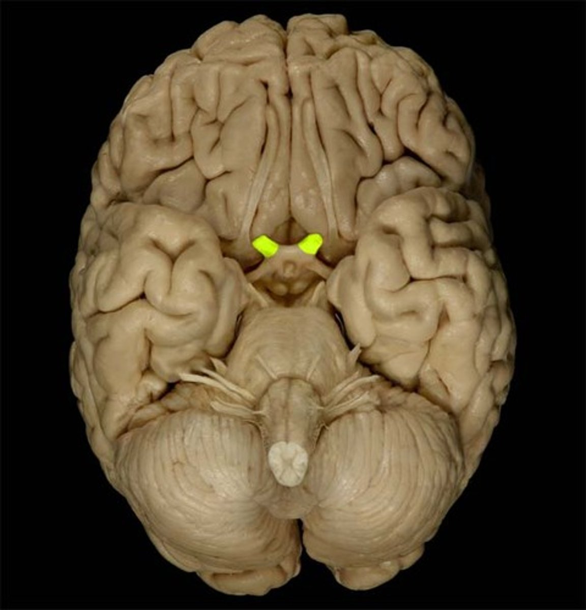
Olfactory bulb
Structure in frontal lobe receiving CN I signals
CN II Optic nerve
Sensory for vision; passes through optic canal to optic chiasm
Optic canal
Opening in sphenoid bone for CN II
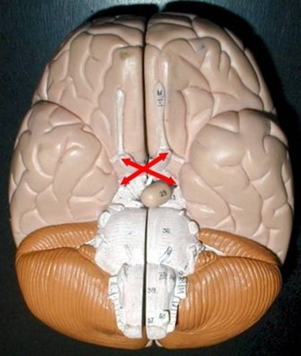
Optic chiasma/chiasm
Crossing of optic nerves anterior to pituitary gland
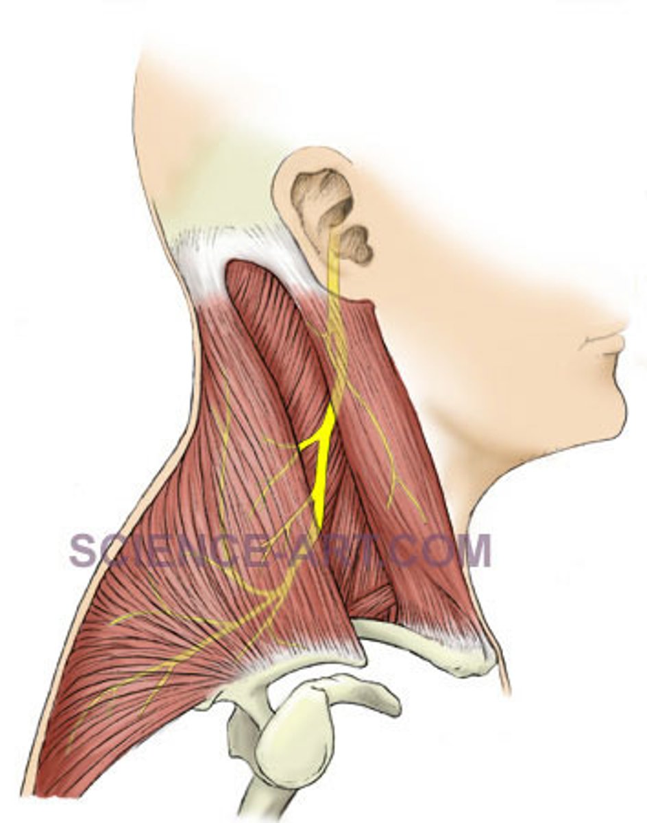
CN V Trigeminal nerve
Mixed nerve of the face; emerges from pons
Ophthalmic division (V1)
Sensory; passes through superior orbital fissure
Superior orbital fissure
Opening for CN III, IV, V1, VI
Maxillary division (V2)
Sensory; passes through foramen rotundum
Foramen rotundum
Passageway for CN V2
Mandibular division (V3)
Mixed; passes through foramen ovale
Foramen ovale
Opening for CN V3
CN VIII Vestibulocochlear nerve
Sensory for hearing and balance; enters skull through internal acoustic meatus
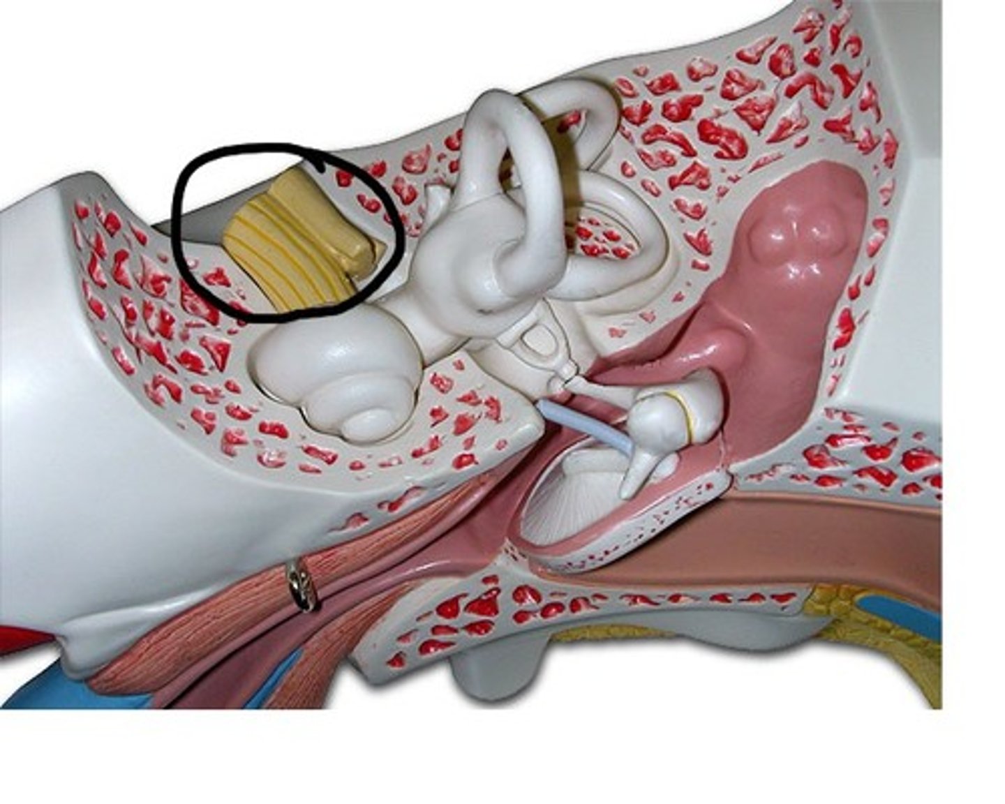
Vestibular nerve
Balance branch of CN VIII
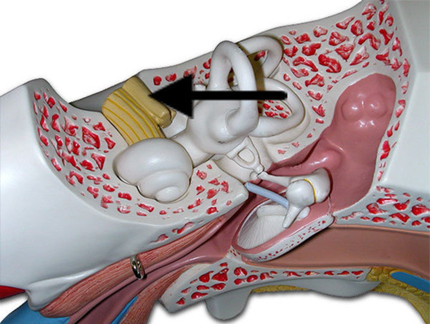
Cochlear nerve
Hearing branch of CN VIII
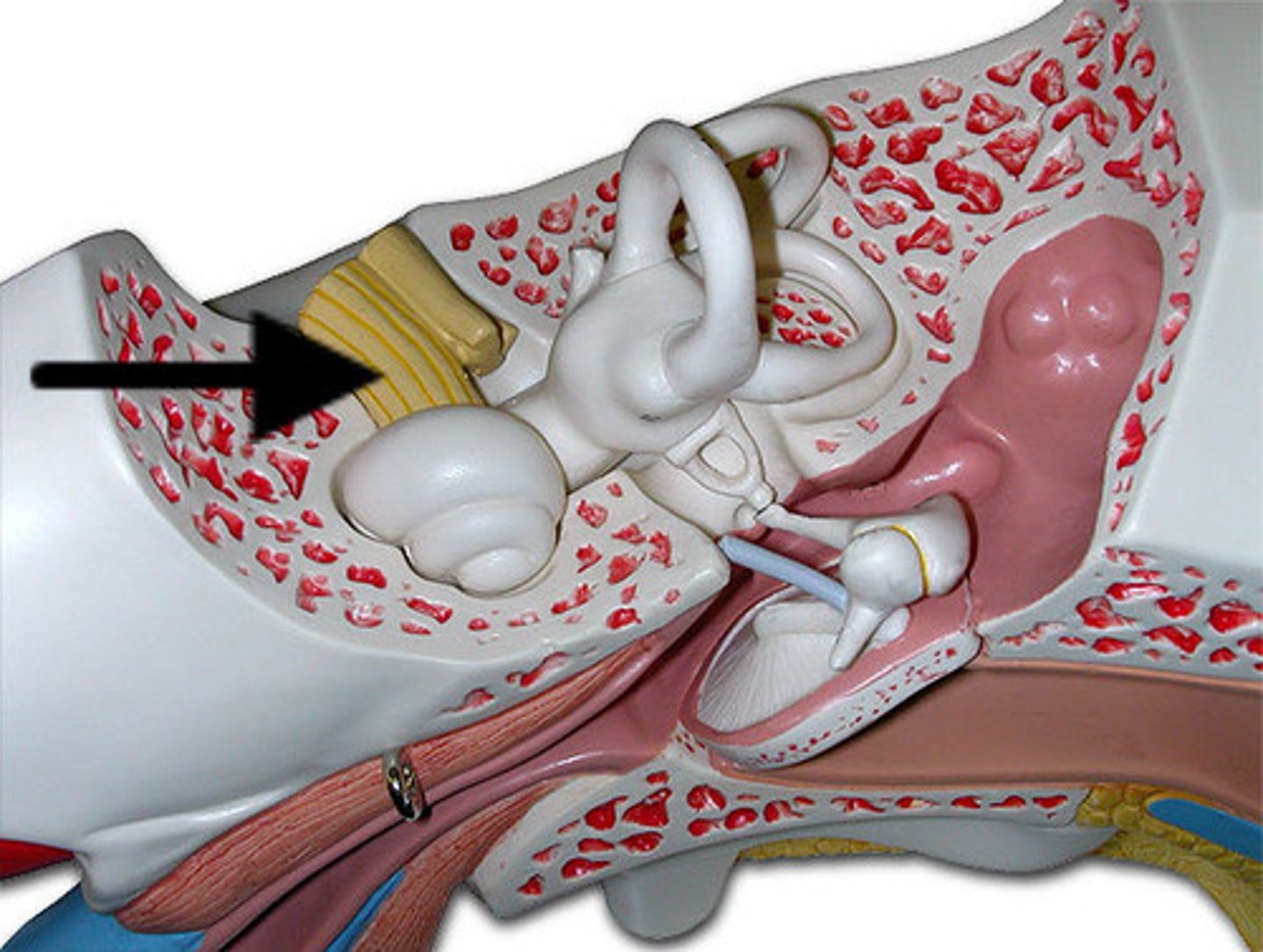
Internal acoustic meatus
Passage for CN VII and VIII
CN X Vagus nerve
Mixed; sensory & motor to viscera; exits skull via jugular foramen
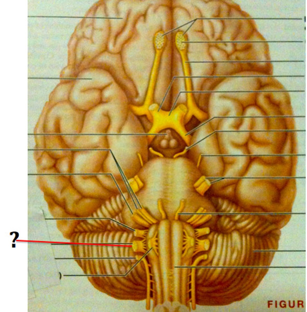
Jugular foramen
Opening for CN IX, X, XI
Sensory and motor for viscera
Function of CN X
CN XI Accessory nerve
Motor to sternocleidomastoid & trapezius; root enters via foramen magnum
Foramen magnum
Entry point for spinal root of CN XI
Sternocleidomastoid
Innervated by CN XI
Trapezius
Innervated by CN XI
Olfactory nerve
I
Optic nerve
II
Trigeminal nerve
V
Ophthalmic division of trigeminal nerve
V1
Maxillary division of trigeminal nerve
V2
Mandibular division of trigeminal nerve
V3
Vestibulocochlear nerve
VIII
Vestibular branch of vestibulocochlear nerve
VIII (Balance)
Cochlear branch of vestibulocochlear nerve
VIII (Auditory)
Vagus nerve
X
Accessory nerve
XI
Epineurium
Outer connective tissue covering of a nerve
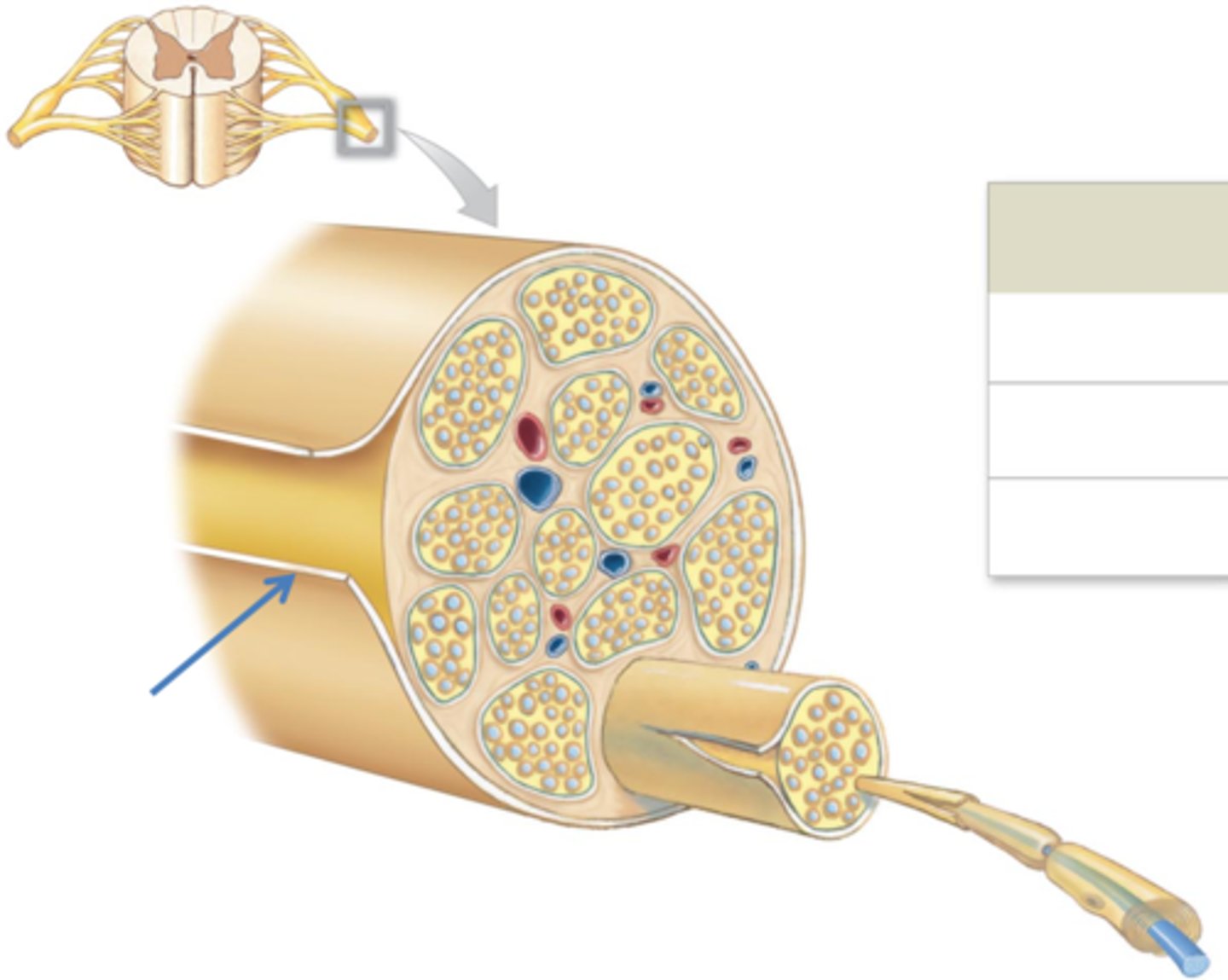
Perineurium
Surrounds each fascicle
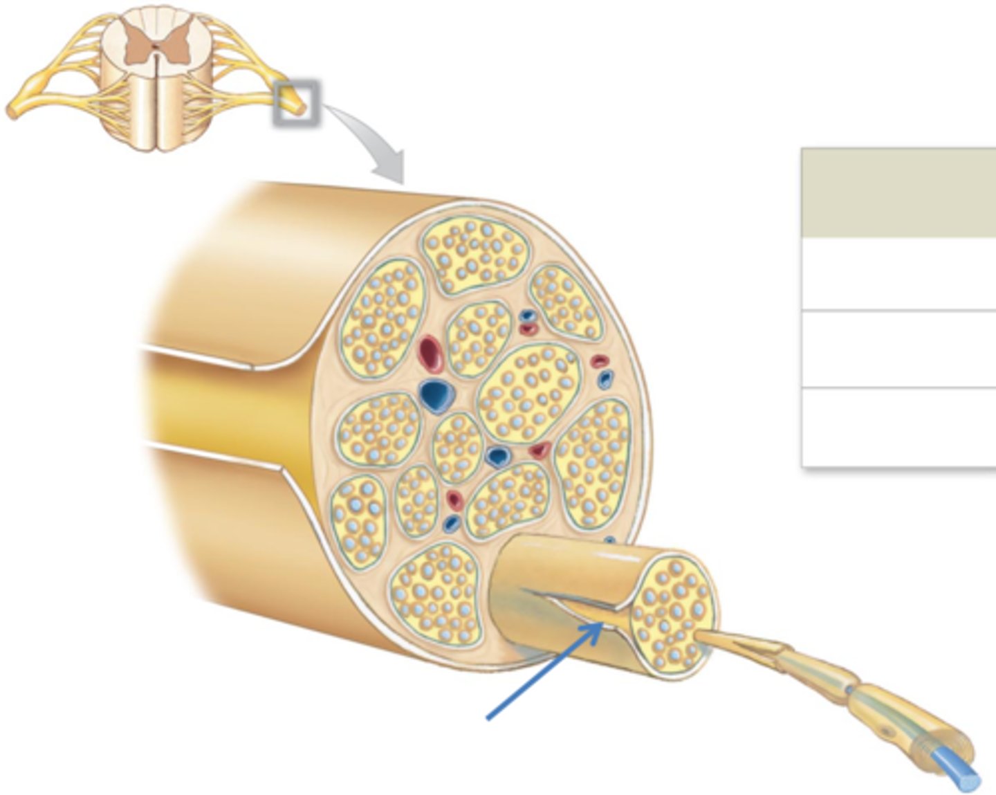
Fascicle
Bundle of axons within a nerve
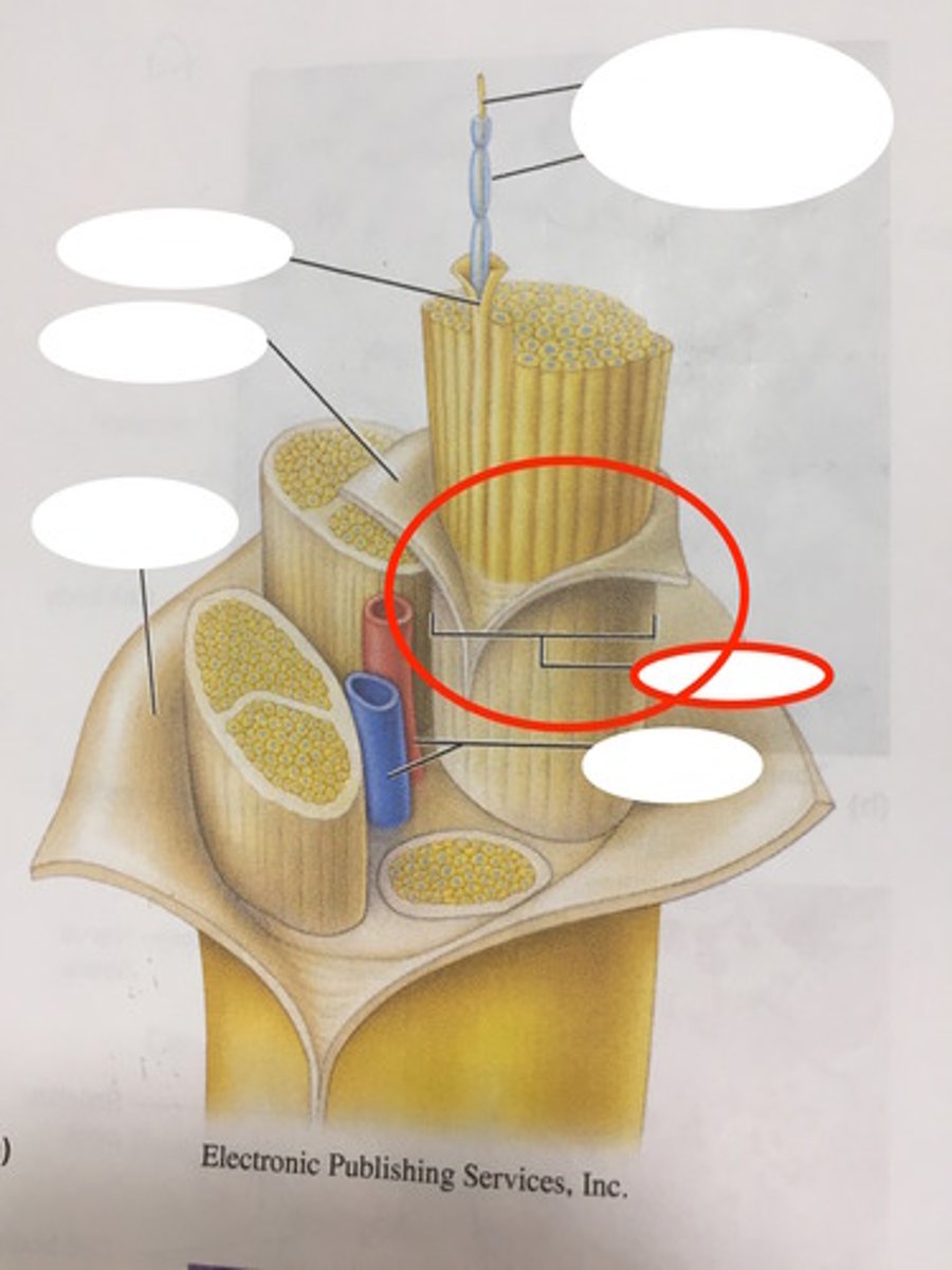
Endoneurium
Connective tissue surrounding each axon
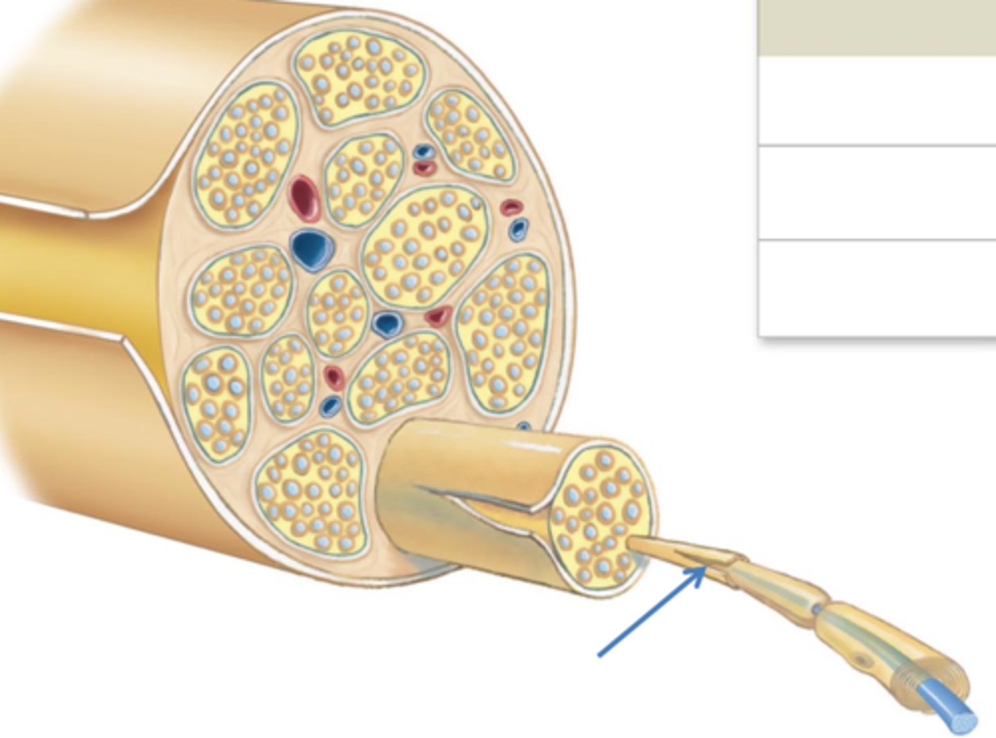
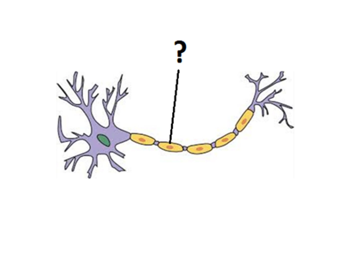
Axon
Nerve fiber that carries impulses
Myelin sheath
Fatty insulation increasing conduction speed
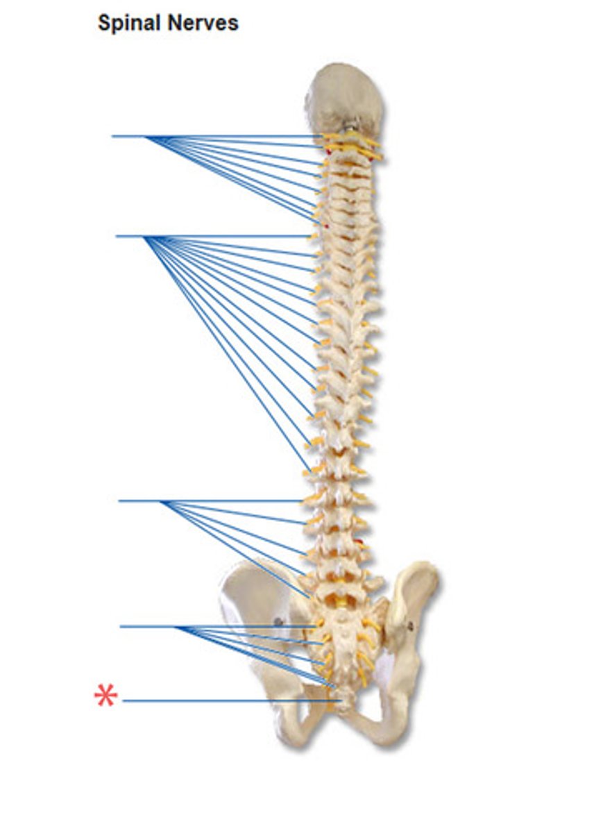
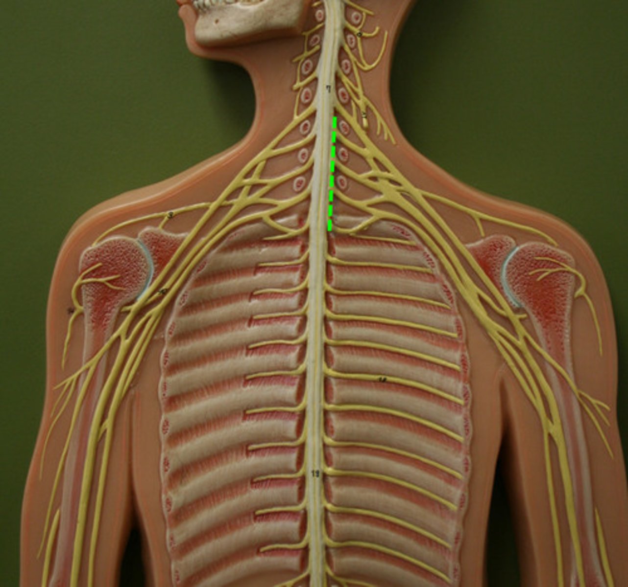
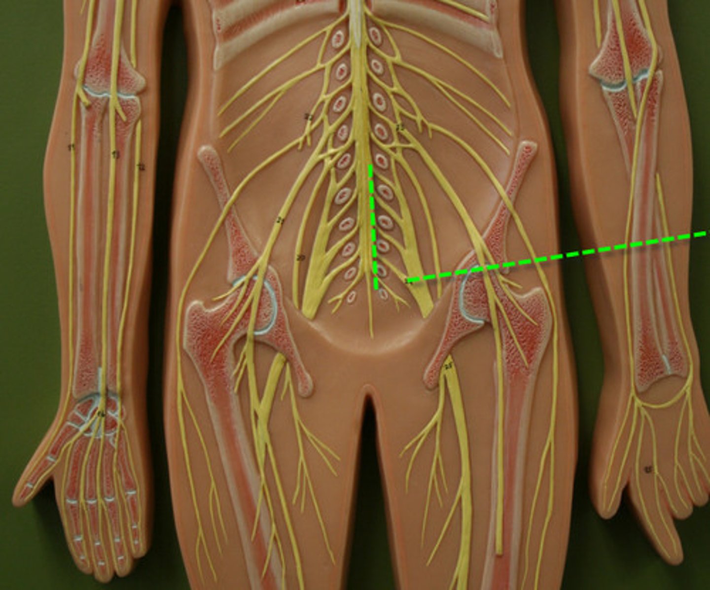
Cervical nerves (C1-C8)
First spinal nerve group

Thoracic nerves (T1-T12)
Twelve spinal nerves
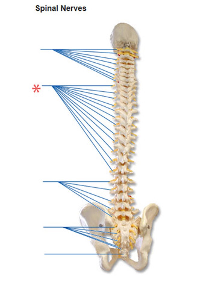
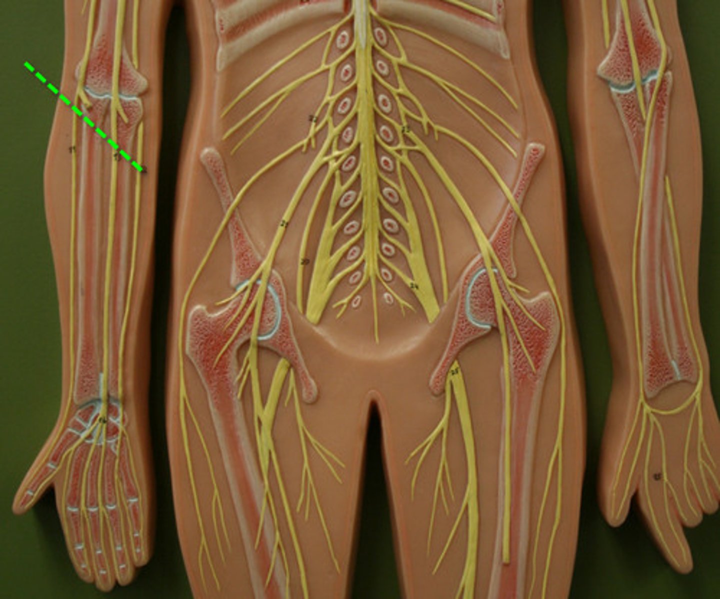
Lumbar nerves (L1-L5)
Five spinal nerves
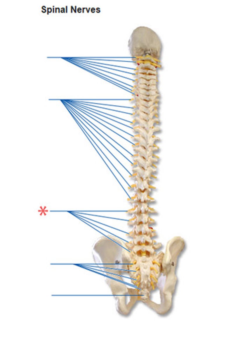
Sacral nerves (S1-S5)
Five spinal nerves
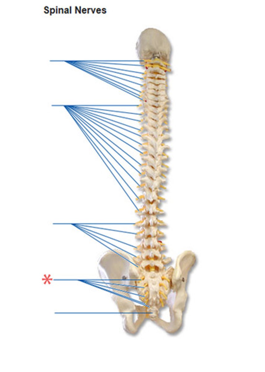
Coccygeal nerve (Co1)
Single nerve
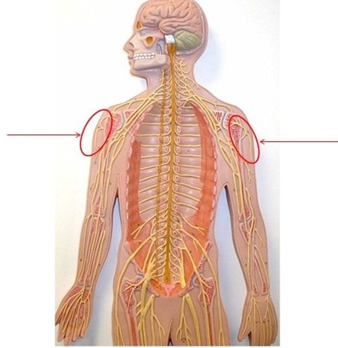
Cervical plexus
Formed by C1-C5
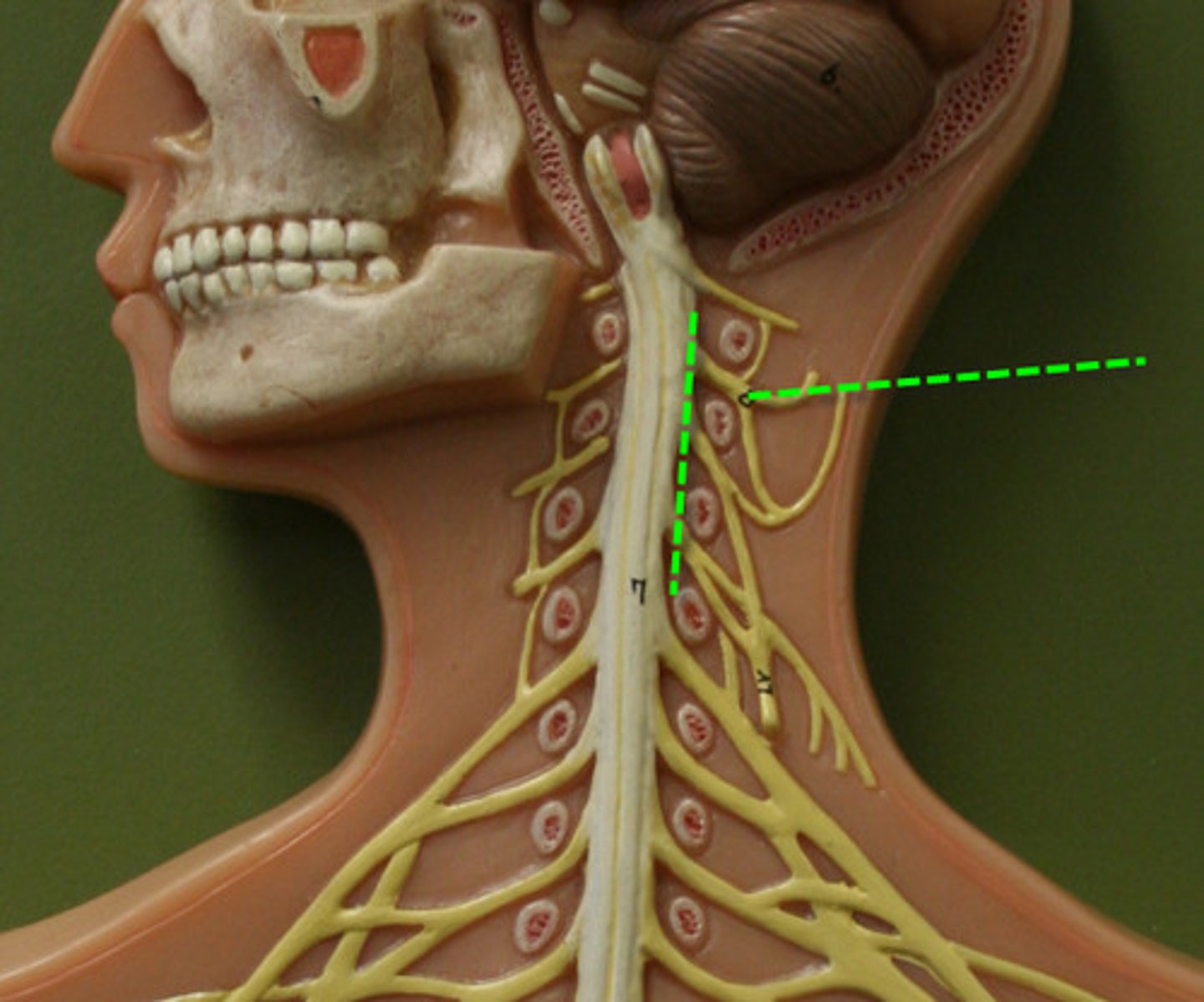
Phrenic nerve
From C3-C5; innervates diaphragm
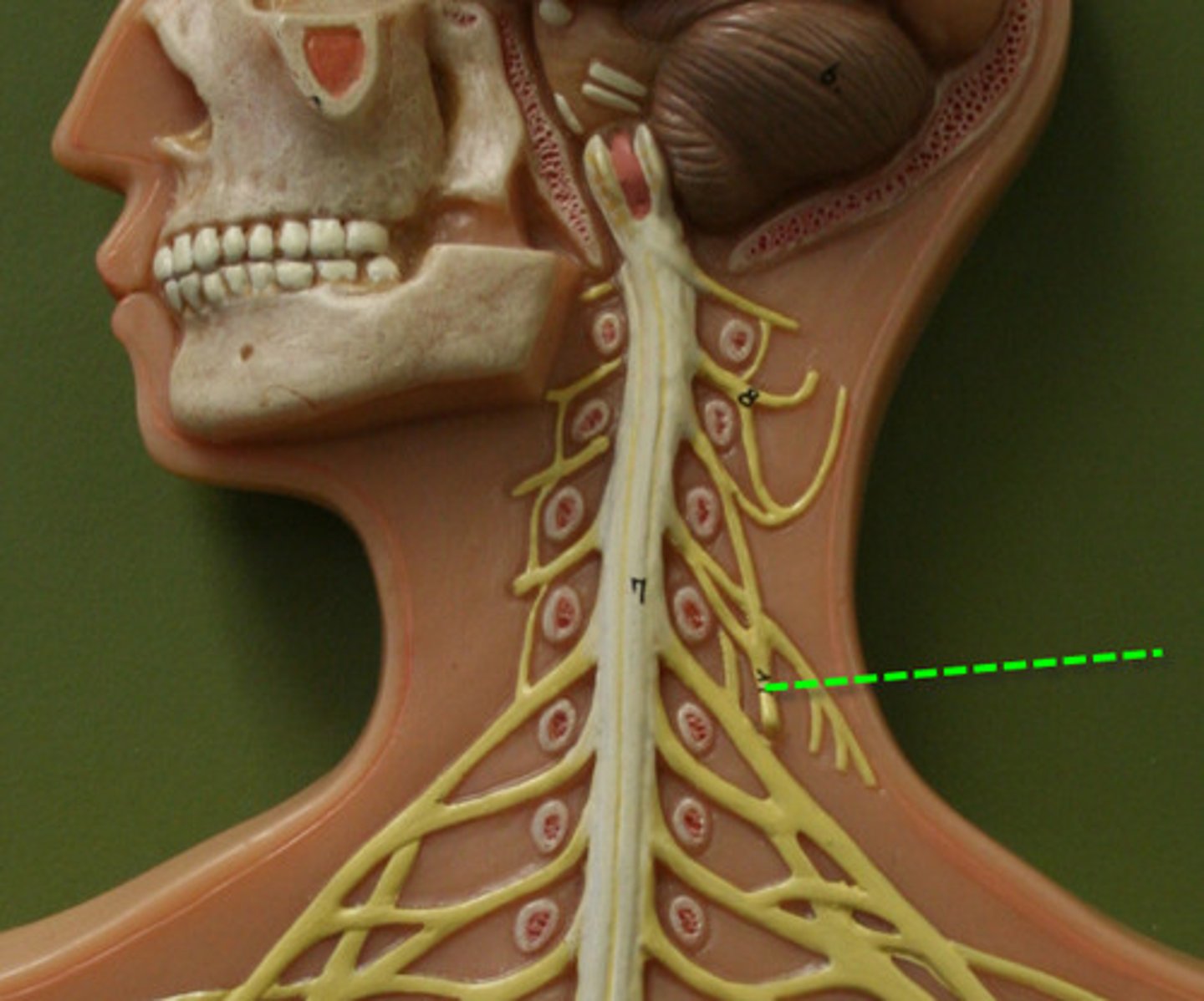
Brachial plexus
Formed by C5-T1
Musculocutaneous nerve
Flexor muscles of brachium
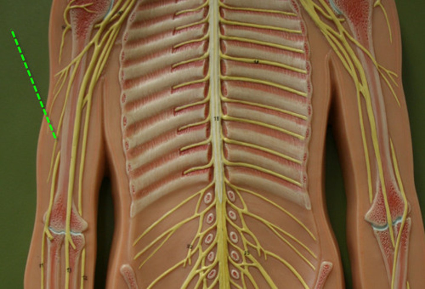
Median nerve
Flexors of antebrachium; carpal tunnel nerve

Ulnar nerve
Flexors of antebrachium & hand
Radial nerve
Extensors of brachium & antebrachium
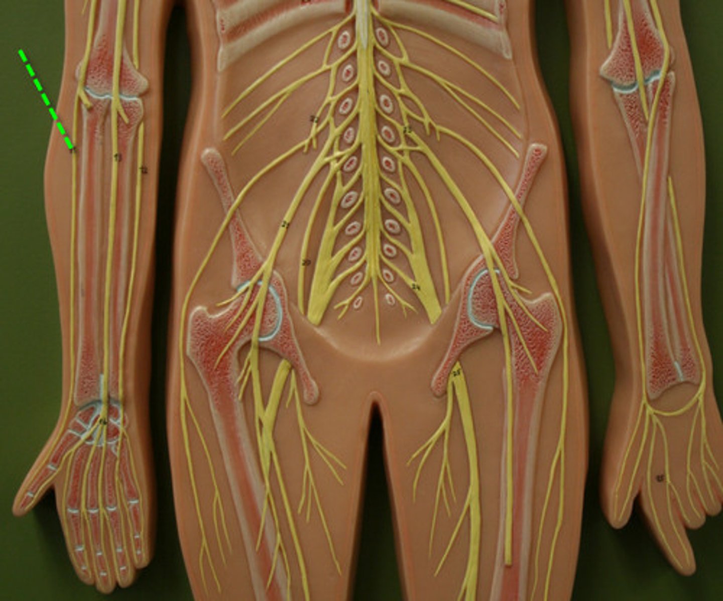
Axillary nerve
Deltoid and teres minor
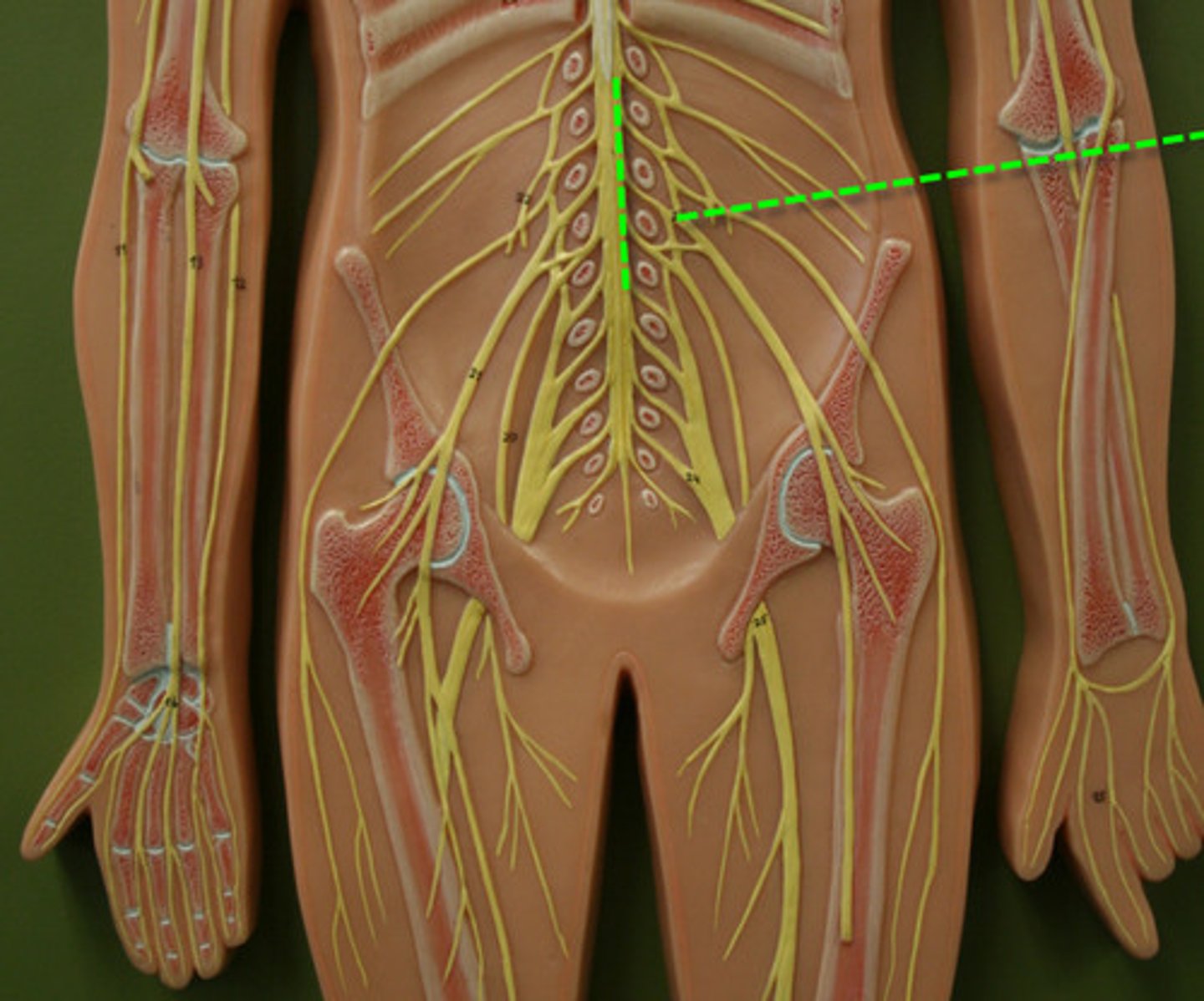
Lumbar plexus
Formed by L1-L4
Femoral nerve
Quadriceps, sartorius, pectineus, iliacus
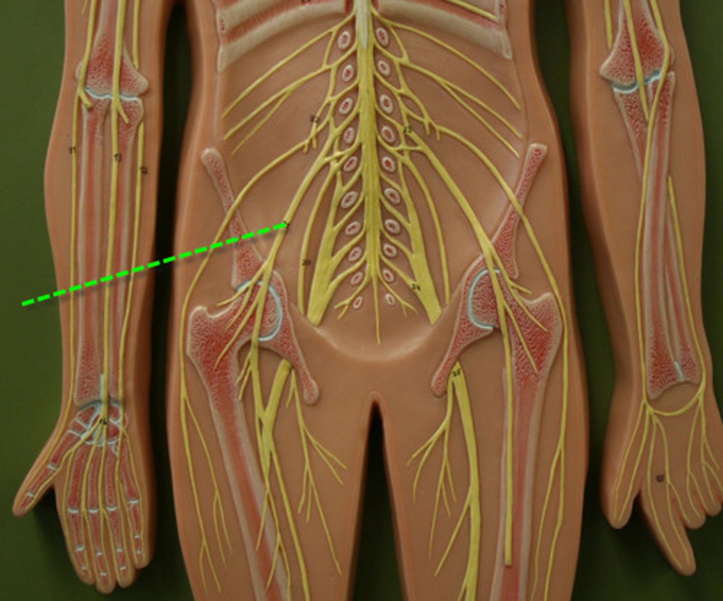
Obturator nerve
Adductors of thigh, gracilis
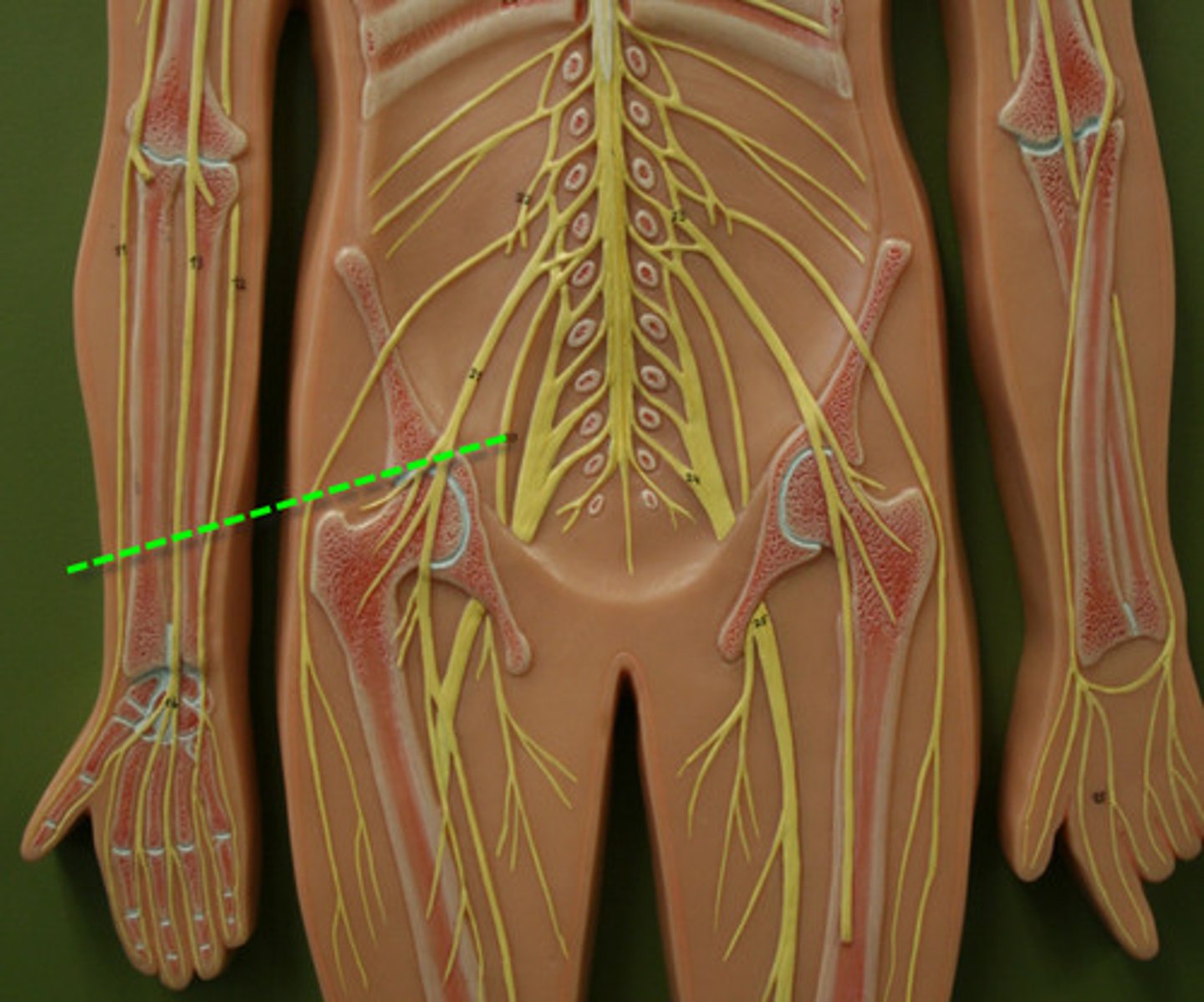
Sacral plexus
Formed by L4-S4
Sciatic nerve
Largest nerve: divides into tibial + common fibular
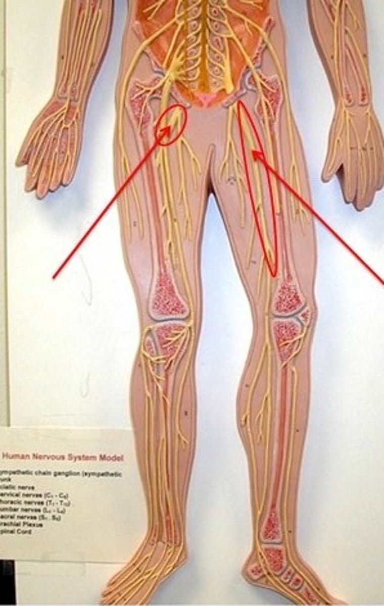
Tibial nerve
Posterior thigh (except short head of biceps femoris), leg, foot muscles
Common fibular nerve
Short head of biceps femoris, tibialis anterior, fibularis group, toe extensors
Power Lab
Device that records nerve activity and EMG signals
Stimulating bar electrode
Delivers electrical stimulus to nerve
2 electrode cables (positive and negative)
Carry electrical signals for recording
Foam electrodes
Stick-on electrodes used to record EMG
Electrode paste
Improves electrical conductivity
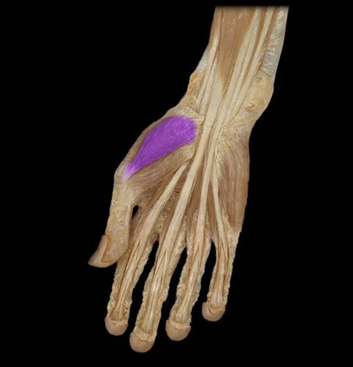
Abductor pollicis brevis
Muscle that contracts during the NCV experiment
Nerve conduction velocity (NCV)
Speed of action potential along a nerve
How NCV is calculated
Distance between stimulation points / (latency elbow - latency wrist)
Purpose of NCV
To measure conduction speed of the median nerve
Why elbow latency is longer
Greater distance for impulse to travel from elbow to muscle
Structure stimulated during lab
Axon of somatic motor neuron to abductor pollicis brevis
How a shock triggers an AP
Electrical stimulus depolarizes axon membrane directly
What EMG graph shows (x-axis)
Time in milliseconds
What EMG graph shows (y-axis)
Voltage in millivolts