Lecture 2.16 - Cadiac/Smooth, MuscIII; Ca2+, Twitch, SO/FOG, Gap Junction, Intercalated Disc, Myogenic
1/23
There's no tags or description
Looks like no tags are added yet.
Name | Mastery | Learn | Test | Matching | Spaced | Call with Kai |
|---|
No analytics yet
Send a link to your students to track their progress
24 Terms
iClicker
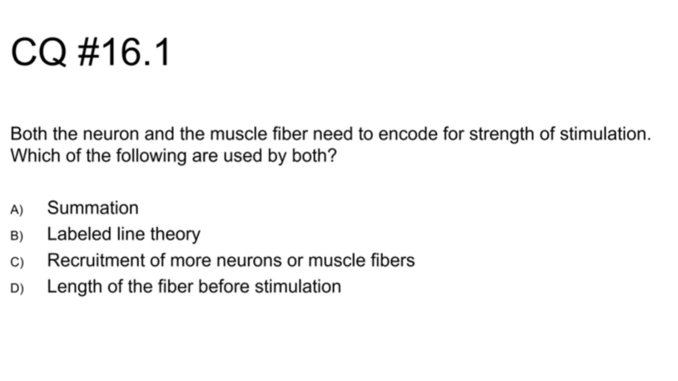
A) Neurons : spatial, temporal, or both = more likely to reach AP ; Muscles : recruiting more neurons
B) Only for neurons
C) More neurons not necessary for strength - only more frequent action potentials ; is true for neurons
D) Only for muscles
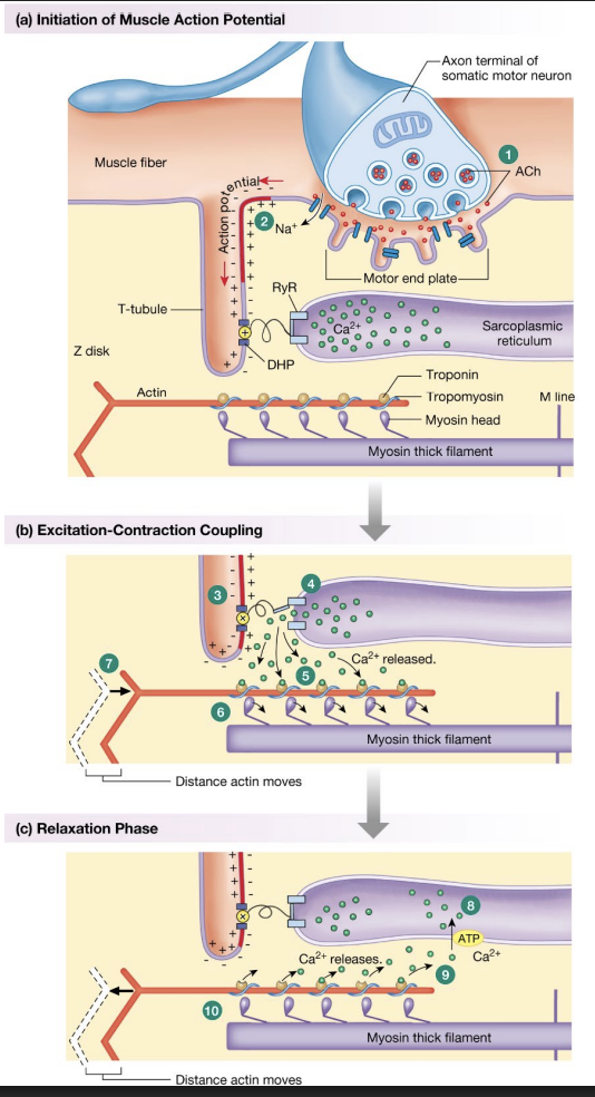
Summary of skeletal muscle contraction
NMJ - where neuron meets sarcolemma at motor end plate
ACh released from motor neuron via exocytosis
ACh binds to nicotine receptor, opens, allowing Na+ in and causing an AP if large enough
AP passes down sarcolemma into T-tubules, allowing AP to enter muscle fiber by reaching DHPR / L-type channel
DHP(voltage-gated Ca2+ channel) connected to RyR channel (in skeletal muscles), allows Ca2+ to rush into sarcoplasm and Ca2+ to be released from intracellular stores
Ca2+ binds to troponin C, pulls on I, T →moves tropomyosin
Tropomyosin reveals myosin binding sites
Cross-bridge cycling
Myosin flexes, closing distance between actin fibers, causing contraction
Ca2+ removed via ATP (AChE), goes back into sarcoplasmic reticulum via SERCA, tropomyosin cover binding sites…
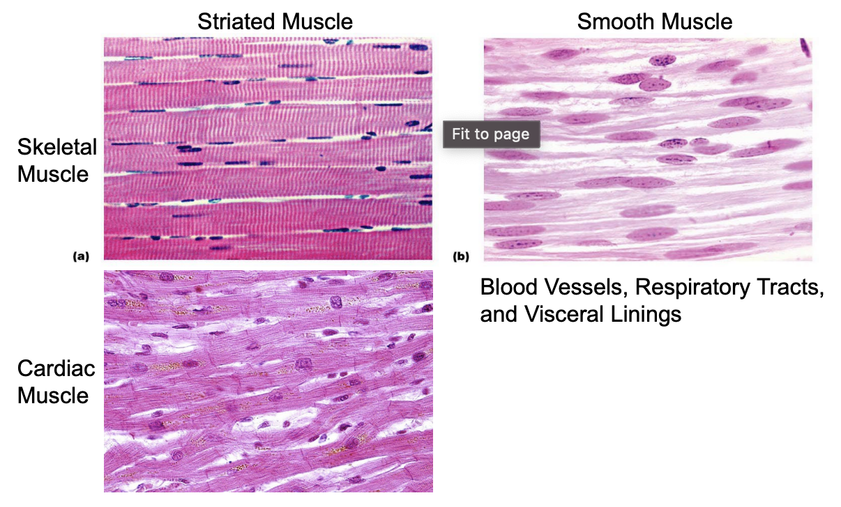
Striated muscle
Due to organization of Actin and Myosin
Dark spots - nuclei
Skeletal muscles = Linear, contracts in same direction
Cardiac muscles = branch
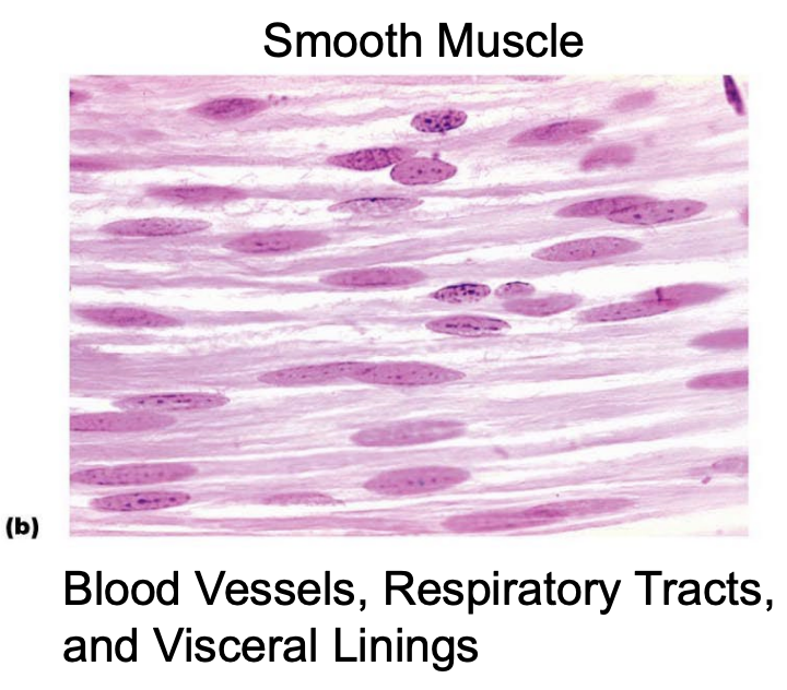
Smooth muscles - not striated
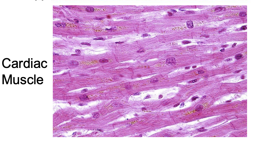
Cardiac muscle, striated
Striated, but branches
Can be multi-nucleated, sometimes binucleate nucleated, but usually mono-nucleated
Dark lines - Z
Light lines - M
Smooth muscle
NOT STRIATED
Mon-linear organization of Actin and Myosin
Mono-nucleated
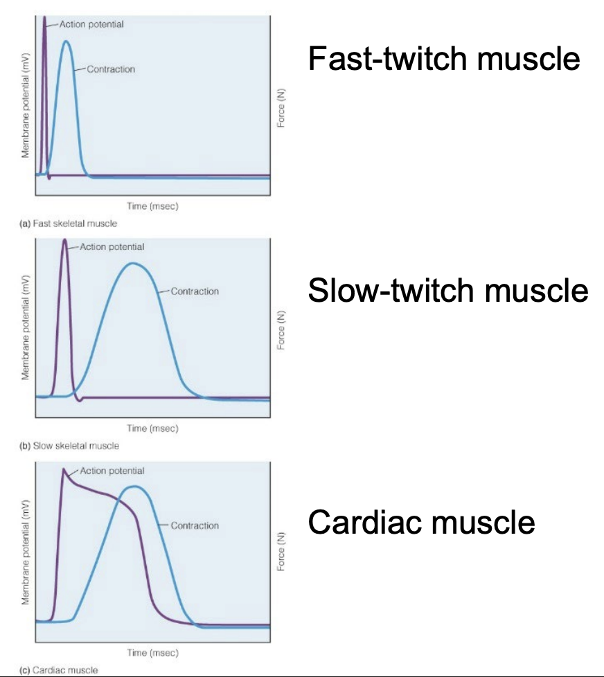
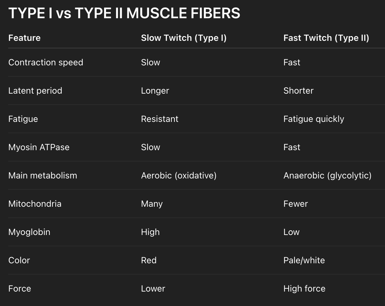
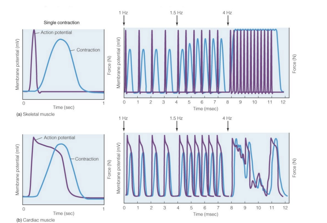
Differences - AP + Twitch speed
Not all skeletal muscle is the same - latent period between AP and Contraction can vary
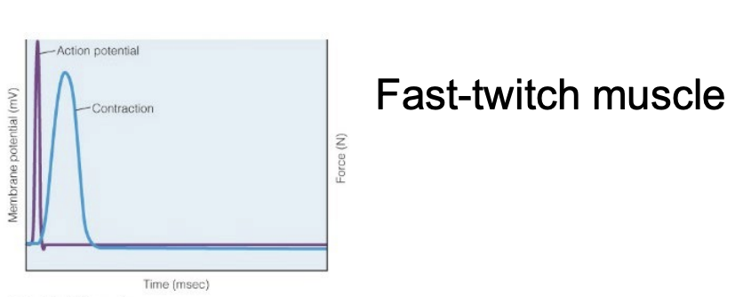
Fast Twitch / FG (Type 2)- contraction right after AP
Faster depolarization
Shorter latent period
Shorter contraction
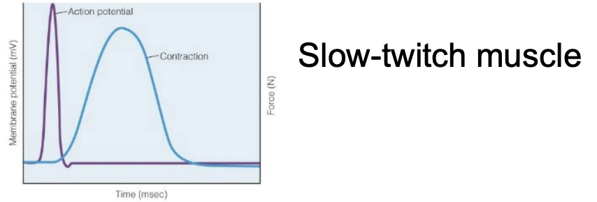
Slow Twitch / SO (Type 1)- contraction a little after AP
Slower depolarization
Longer latent period
Longer contraction
Muscles can have combinations of slow/fast twitch fibers with some predominating
Smallest force out of the 3 Twitch types
Fast-twitch muscle (Type 2) / Fast Glycolytic (FG) and Fast Oxidative Glycolytic (FOG)
PURPOSE:
quick bursts
large force output
Reaches depolarization faster
Due to number and properties of Na+/K+ channels
Short latent period
Contraction occurs right after AP
Fatigue quickly
Fewer mitochondria
Pale/While color
High force
**TWO TYPES:
FOG - Fast Oxidative/Glycolytic
Medium force
FG - Fast Glycolytic
Greatest force
EXAMPLES
Gastrocnemius (jumping calf muscle)
Biceps brachii
Finger flexors
Eye muscles (extraocular muscles — extremely fast)
Slow-twitch muscle (Type 1) / Slow Oxidative (SO)
PURPOSE:
maintain posture
repeated activity
low-force endurance
Can last all day
Slower depolarization
Longer contraction cycle
Longer latent period
Electron Transport chain - Use Oxygen, unlimited ATP
Resistant to fatigue
More mitochondria
Red color
Lower force
EXAMPLES
Soleus (calf postural muscle) → standing
Back extensor muscles (erector spinae) → posture
Neck stabilizers
Diaphragm (important!) → continuous breathing
Slow vs Fast Twitch muscle chart
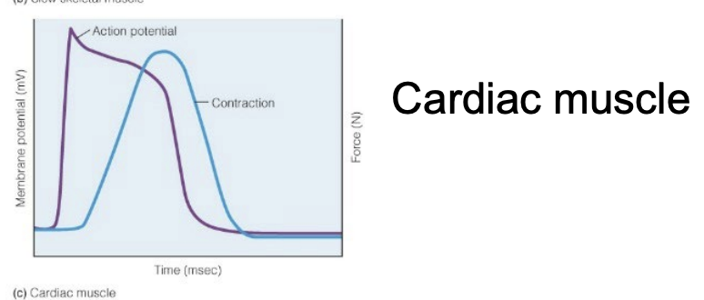
Cardiac Muscle - Twitch
Normal depolarization phase, but very long strangely shaped repolarization phase, that goes into the contraction
Contraction resembles that of the Slow Twitch
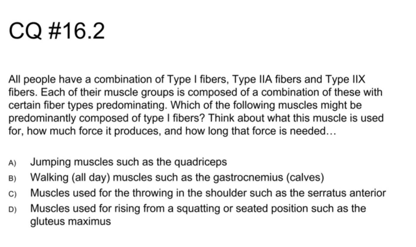
iClicker
Predominantly Type I = Slow Twitch?
→ Think of ALL DAY things
Answer = B
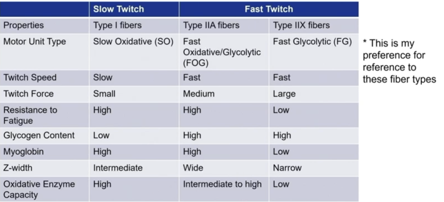
SO vs FOG/FG Chart (MEMORIZE - Glycogen, Myoglobin, Z-width, Oxidative Enzyme Capacity)
SO use oxygen via ETC (SLOW), can last all day (continuously generate energy)
FOG use ETC (slow) and glycolysis (fast!)
Hybrid of slow and fast fibers
Contract fast, resist fatigue longer than FG
Activities:
Walking up hill, repeated lifting, middle distance running
FG uses only glycolysis (FAST)
Greatest force, fastest fatigue
Activities:
Sprinting, jumping, throwing
Non-obvious stats
SO and FOG have much higher resistance to fatigue than FG
FG/FOG have higher glycogen amount than SO (energy)
SO and FOG have higher myoglobin amount than FG
SO and FOG are darker meat, FG is lighter meat
Also more fat storage on SO/FOG
Z-width: FG < SO < FOG
FOG require force AND repetition
SO only requires repetition
FG only requires force
SO > FOG > FG for oxidative enzyme capacity
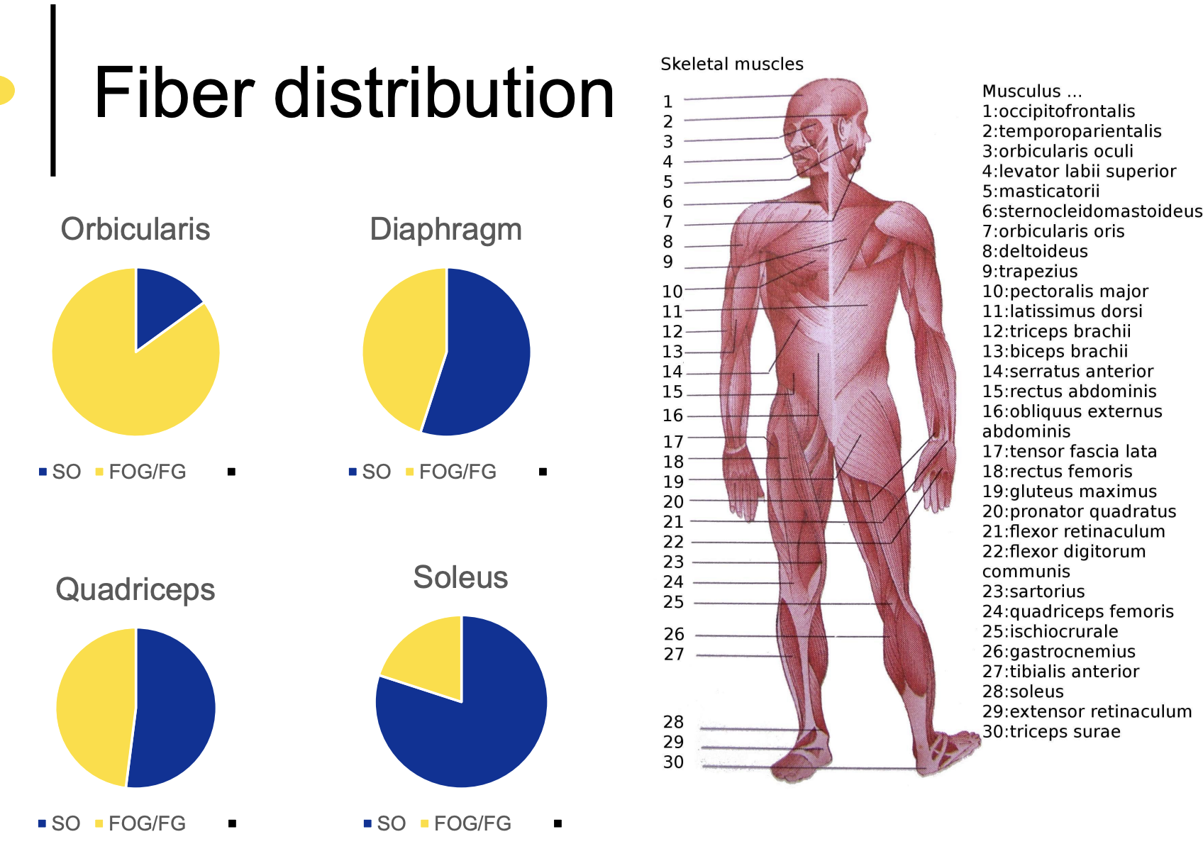
Fiber distribution
Orbicularis - Eyes
Majority FOG/FG, less SG
Fast movements
Diaphragm
~ 1:1 SO to FOG/FG, though more SO
Quadriceps
~1:1 SO to FOG/FG
Soleus - Calves, walking, all-day muscle
All-day walking muscle, mostly SO
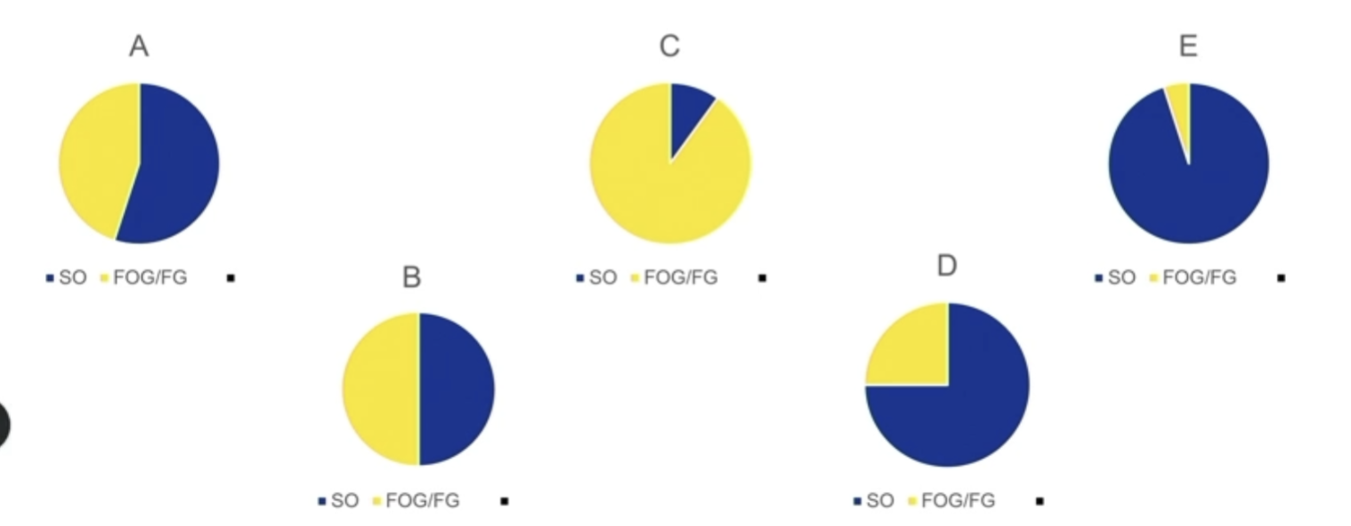
iClicker
Cardiac - E
Heart is always pumping, can speed up, but mostly same rhythm
High frequency stimulation in Cardiac muscles
High frequency stimulation causes arrhythmic cardiac muscle contractions
Heart muscles CANNOT stay in tetanus
If heart cannot relax, it cannot fill with blood and cause a contraction to send blood out
Thus : RyR channel is not connected to DHPR channel / L-Type channel
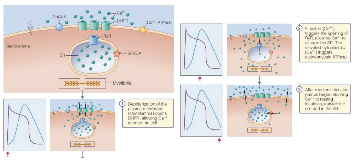
Cardiac muscle: Ca2+ Pathway
RyR channel is separated from DHPR channels
In skeletal, they are physically attached
1) AP spreads along sarcolemma and into T-tubules, reaches voltage-gated DHPR channel, opens due to DP
2) Ca2+ enters cells through the DHPR channel
3) Ca2+ level gets high enough, THEN RyR channels open → DEPOLARIZATION OPENS SARCOPLASMIC RETICULUM (RyR)
Ca2+ levels in sarcoplasm sharply rise
Increases latency period between AP and contraction, which is important for the heart to relax and refill with the next beat
Cardiac RyR is similar to a voltage-gated channel in Cardiac muscle, but not in skeletal muscle, where it is more like a mechanically gated channel
iClicker
E
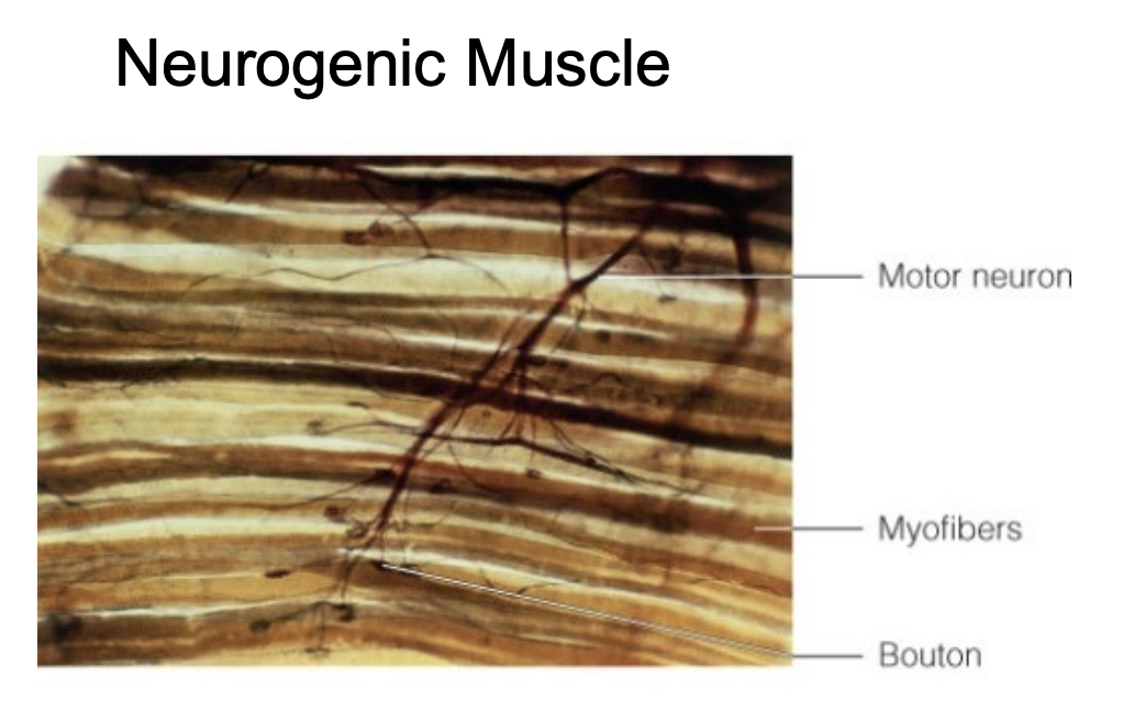
Neurogenic muscle
Muscle is triggered by nerve
Nerve sends message to NMJ → depolarization → contraction
Skeletal
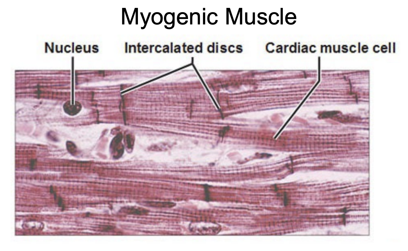
Myogenic myscle
Triggered by muscle next to it
Sometimes neurogenic muscle
GAP JUNCTIONS between myogenic muscles
Like cardiac muscles
Cardiac muscles cause adjacent cardiac muscles to contract, allowing heart to contract as one
Pacemaker cells cyclically release AP to cause heart cells to contractions
Neurogenic / Myogenic for Smooth muscles
Smooth muscle can be myogenic or neurogenic
Cardiomyocyes have unique structures that distinguish them
Nucleus is inside cardiac cells
Bi/single nucleated
Branched; not long and skinny - allows them to contract in multiple directions at the same time
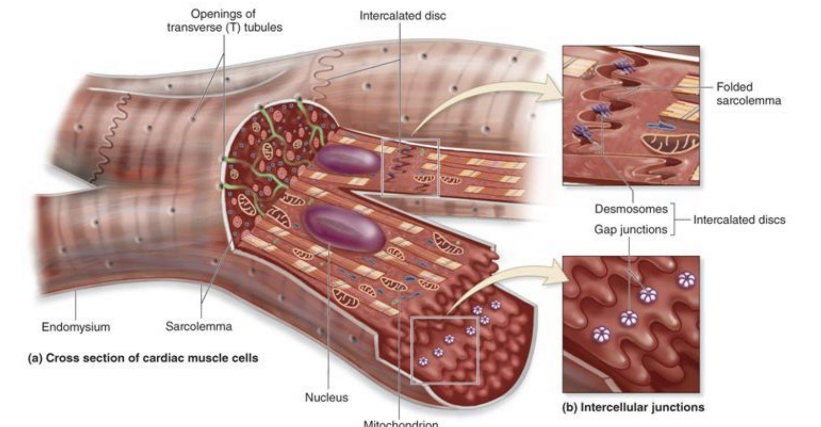
Intercalated disc - where 2 cardiomyocytes are attached and where gap junctions are
Connected via gap junctions (AP spread cell to cell) and desmosomes (velcro)
Skeletal muscles are NOT connected by gap junctions
Each muscle fiber must be stimulated separately via a motor neuron
Smooth muscles
Present in cardiovascular system, but not herat - arteries, veins; gut
Adhesion plates allow them to connect together
Myosin/Actin pull on the Adhesion plates on all directions = no pattern
Means that cell can contract in multiple directions at same time
Ex: Allows stomach to get smaller to contract, get smaller, and mechanically grind up food
Ex: Small intestine can move digested food throughout itself
Mono-nucleated
Sometimes have gap junctions
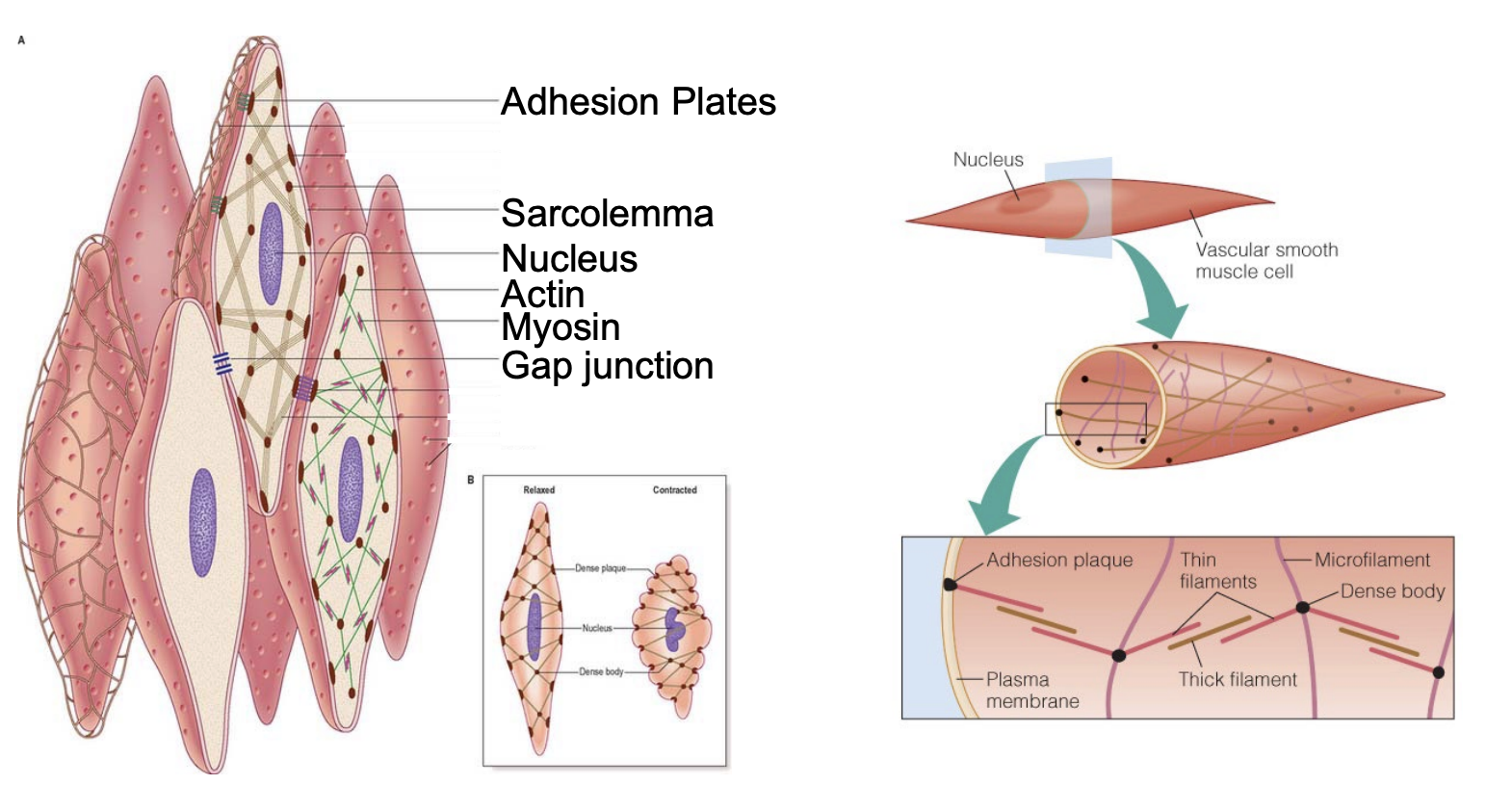
Ca2+ binding to move Actin/Myosin in SM: Calmodulin
Smooth muscles, instead of using Troponin C, use Calmodulin
Ca2+ binds to Calmodulin instead of TnC, causes movement of tropomyosin → contraction
Smooth muscle is in…
Digestive tract
Large blood vessels
Smooth muscles in organized in 2 layers:
Circular muscles, in circles
Longitudinal muscles, linearly - peristaltic contraction
Shortens and lengthens