complete Male pelvis guide for ardms
1/100
There's no tags or description
Looks like no tags are added yet.
Name | Mastery | Learn | Test | Matching | Spaced | Call with Kai |
|---|
No analytics yet
Send a link to your students to track their progress
101 Terms
Which of the following structures are components of the male pelvis? (Select all that apply)
a. Lower ureters
b. Urinary bladder
c. Urethra
d. Prostate
e. Scrotum
f. Testicles (testes)
g. Penis
h. Gallbladder
i. Spleen
a. Lower ureters
b. Urinary bladder
c. Urethra
d. Prostate
e. Scrotum
f. Testicles (testes)
g. Penis
The paired testicles begin to develop in the upper abdomen in the fetus and descend into the pelvis during which week of gestation?
a. 4th week
b. 12th week
c. 20th week
d. 28th week
a. 4th week
The fetal testicles normally descend into the scrotum during which week of gestation?
a. 4th week
b. 12th week
c. 20th week
d. 28th week
d. 28th week
Failure of the testicles to fully descend into the scrotum is known as:
a. Hydrocele
b. Varicocele
c. Cryptorchidism
d. Epididymitis
c. Cryptorchidism
The testes function as which of the following types of glands?
a. Endocrine glands only
b. Exocrine glands only
c. Both endocrine and exocrine glands
d. Neither endocrine nor exocrine glands
c. Both endocrine and exocrine glands
Each testis is surrounded by a double layer of tissue consisting of a parietal and visceral covering known as the:
a. Tunica albuginea
b. Tunica vaginalis
c. Dartos fascia
d. Cremaster muscle
b. Tunica vaginalis
Beneath the layers of the tunica vaginalis, the testis is surrounded by a dense fibrous layer of tissue called the:
a. Tunica dartos
b. Tunica vaginalis
c. Tunica albuginea
d. Cremaster muscle
c. Tunica albuginea
Hydroceles are most commonly located between which of the following structures?
a. Between the testis and epididymis
b. Between the two layers of the tunica vaginalis
c. Within the tunica albuginea
d. Within the seminiferous tubules
b. Between the two layers of the tunica vaginalis
Which of the following correctly describes the parts of the epididymis?
a. Neck, fundus, and body
b. Apex, cortex, and medulla
C. Head, body, and tail
d. Proximal, fundus, and neck
C. Head, body, and tail
The normal thickness of the scrotal wall ranges between which of the following measurements?
a. 0-2 mm
b. 2-8 mm
c. 8-12 mm
d. 10-15 mm
b. 2-8 mm
The normal mediastinum testis appears how on sonography?
a. Anechoic cyst in both planes
b. Echogenic linear structure in the sagittal plane and triangular in the transverse plane
c. Hypoechoic mass in the sagittal plane and circular in the transverse plane
d. Calcified structure with shadowing in both planes
b. Echogenic linear structure in the sagittal plane and triangular in the transverse plane
Although rare, a patient may have three or more testicles. This condition is called:
a. Cryptorchidism
b. Varicocele
c. Polyorchidism
d. Hydrocele
c. Polyorchidism
Polyorchidism is most often located on which side of the scrotum?
a. Right side
b. Midline
c. Left side
d. Equally on both sides
c. Left side
Polyorchidism is most commonly discovered how clinically?
a. Severe scrotal pain and fever
b. Asymptomatic and found incidentally
c. Acute urinary retention
d. Elevated PSA levels
b. Asymptomatic and found incidentally
A physician palpates a scrotal mass during a physical examination in a patient who has no symptoms. Sonography later reveals an additional normal-appearing testicle that was found incidentally. Which of the following conditions does this describe?
a. Cryptorchidism
b. Varicocele
c. Hydrocele
d. Polyorchidism (supernumerary testicle)
d. Polyorchidism (supernumerary testicle)
Which of the following conditions carries an increased risk for developing testicular cancer and may be discovered incidentally during an examination?
a. Hydrocele
b. Varicocele
c. Polyorchidism (supernumerary testicle)
d. Epididymal cyst
c. Polyorchidism (supernumerary testicle)
Patients with a supernumerary testicle (polyorchidism) may be at increased risk for which of the following complications?
a. Hydrocele
b. Varicocele
c. Epididymitis
d. Testicular torsion
d. Testicular torsion
Which of the following conditions is associated with infertility and an increased risk for malignancy in the testis?
a. Varicocele
b. Hydrocele
c. Cryptorchidism
d. Epididymal cyst
c. Cryptorchidism
Which of the following is the most common cancer found in an undescended testis located above the scrotum or within the inguinal canal?
a. Embryonal carcinoma
b. Teratoma
c. Seminoma
d. Choriocarcinoma
c. Seminoma
Which of the following surgical procedures is used to correct cryptorchidism (undescended testis)?
a. Orchiopexy
b. Orchiectomy
c. Vasectomy
d. Prostatectomy
a. Orchiopexy
Which of the following conditions, also referred to as spermatic cord torsion, occurs when the arterial blood supply to the testicle is cut off due to twisting of the testicular axis?
a. Epididymitis
b. Varicocele
c. Testicular torsion
d. Hydrocele
c. Testicular torsion
Testicular torsion most commonly occurs in which age group?
a. Infants under 1 year
b. between 12 and 18 years
c. Children ages 3-6 years
d. Men over 60 years
e. pediatrics
b. between 12 and 18 years
For optimal testicular salvage, testicular torsion should ideally be treated within which time frame after symptom onset?
a. 7 hours
b. 6 hours
c. 12 hours
d. 24 hours
b. 6 hours
After approximately how long are testicles usually no longer salvageable in cases of untreated testicular torsion?
a. 6 hours
b. 12 hours
c. 24 hours
d. 7 hours
c. 24 hours
Patients with which of the following conditions are more prone to developing testicular torsion?
a. Marfan syndrome
b. Klinefelter syndrome
c. Bell-clapper deformity
d. Down syndrome
c. Bell-clapper deformity
Which of the following statements correctly describes the most common form of testicular torsion?
a. Extravaginal torsion occurring only in newborns
b. Intravaginal torsion associated with a bell-clapper deformity that is often bilateral
c. Torsion caused by varicocele within the pampiniform plexus
d. Torsion caused by hydrocele within the tunica vaginalis
b. Intravaginal torsion associated with a bell-clapper deformity that is often bilateral
Which type of testicular torsion occurs during the neonatal period or in utero and is related to twisting of the spermatic cord within the inguinal canal?
a. Intravaginal torsion
b. Intermittent torsion
c. Extravaginal torsion
d. Appendix testis torsion
c. Extravaginal torsion
During which stage of testicular torsion will the testis most commonly appear enlarged and hypoechoic or heterogeneous on ultrasound?
a. Chronic stage
b. Subacute stage
c. Acute stage
d. Extravaginal torsion
e. Intravaginal torsion
c. Acute stage
Which of the following describes torsion that has lasted more than 10 days and demonstrates enlargement of the epididymis, testis, and spermatic cord?
a. Acute testicular torsion
b. Intravaginal torsion
c. Extravaginal torsion
d. Chronic testicular torsion
d. Chronic testicular torsion
Which of the following stages of testicular torsion may demonstrate areas of necrosis within the testis along with hyperemic flow around the testis?
a. Acute testicular torsion
b. Intermittent torsion
c.Chronic testicular torsion
d. Extravaginal torsion
c.Chronic testicular torsion
Which of the following stages of testicular torsion will most commonly demonstrate an enlarged testicle with absent intratesticular blood flow on Doppler?
a. Chronic testicular torsion
b. Intermittent torsion
c. Acute testicular torsion
d. Torsion of the appendix testis
c. Acute testicular torsion
Which of the following is the most common cause of acute scrotal pain in prepubertal boys?
a. Testicular torsion
b. Epididymitis
c. Torsion of the appendix testis
d. Varicocele
c. Torsion of the appendix testis
Which of the following structures are embryologic remnants of the Müllerian duct, Wolffian duct, and mesonephric duct?
a. Tunica vaginalis, tunica albuginea, mediastinum testis
b. Epididymal head, body, and tail
c. Appendix testis, appendix epididymis, and appendix vas
d. Pampiniform plexus, testicular artery, ductus deferens
c. Appendix testis, appendix epididymis, and appendix vas
The appendix testis, appendix epididymis, and appendix vas are embryologic remnants of which of the following ducts?
a. Tunica albuginea, tunica vaginalis, and dartos fascia
b. Müllerian duct, rete testis, mesonephric duct
c. Seminiferous tubules, rete testis, and mediastinum testis
d. Müllerian duct, Wolffian duct, and mesonephric duct
e. Pampiniform plexus, testicular artery, and ductus deferens
d. Müllerian duct, Wolffian duct, and mesonephric duct
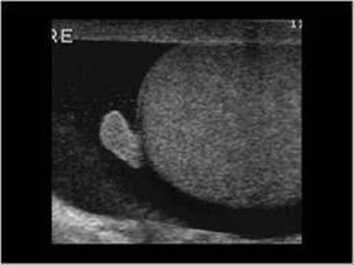
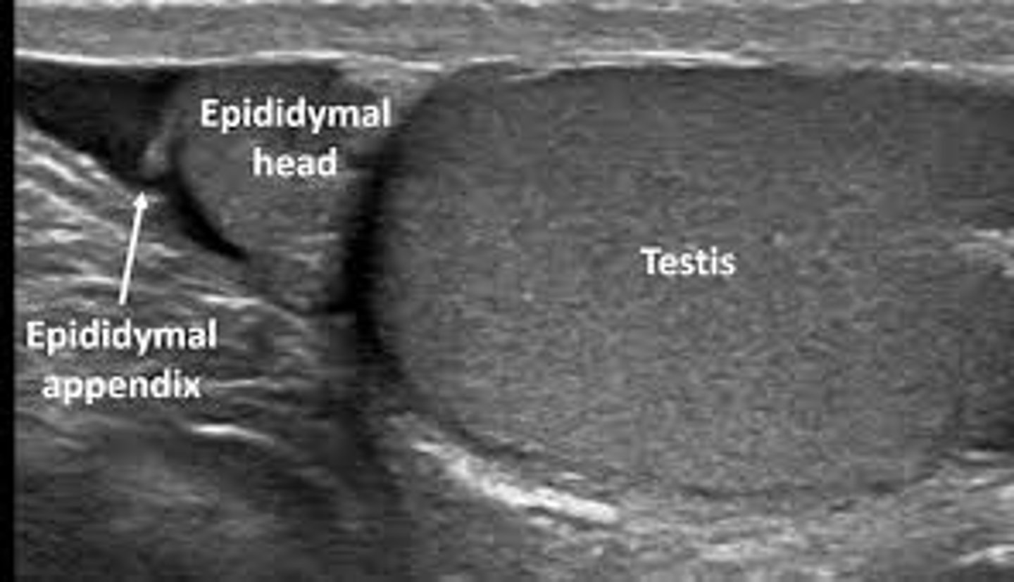
Which of the following structures is located between the head of the epididymis and the superior pole of the testis?
a. Appendix epididymis
b. Appendix vas
c. Appendix testis
d. Tunica albuginea
c. Appendix testis
Which of the following structures is located between the body and tail of the epididymis?
a. Appendix testis
b. Appendix epididymis
c. Appendix vas
d. Tunica vaginalis
c. Appendix vas
Which of the following structures is located at the head of the epididymis?
a. Appendix testis
b. Appendix epididymis
c. Appendix vas
d. Mediastinum testis
b. Appendix epididymis
epidydimal appendixWhich of the following structures is located between the body and tail of the epididymis?
a. Appendix testis b. Appendix epididymis c. Appendix vas ✅ d. Tunica vaginalis
Which of the following may result from a torsed and displaced testicular appendage that becomes free-floating within the scrotum?
a. Hydrocele
b. Varicocele
c. Scrotal pearl (scrotolith)
d. Hematocele
c. Scrotal pearl (scrotolith)
Which of the following clinical signs may be observed during a physical examination in a patient with torsion of the appendix testis?
a. Bell-clapper sign
b. Blue-dot sign
c. Cullen sign
d. Murphy sign
b. Blue-dot sign
Patient presents to his primary care doctor for focal pain within the superior pole of the testicle. The physician notices a presence of the "blue-dot sign" during a physical examination. which of the following is most indicative of the condition?
a. Testicular torsion
b. Epididymitis
c. Varicocele
d. Torsion of the appendix testis
d. Torsion of the appendix testis
Torsion of the appendix testis is typically treated with which of the following?
a. Emergency surgery
b. Antibiotics and drainage
c. Pain medication and bed rest
d. Chemotherapy
c. Pain medication and bed rest
Which of the following is the most common scrotal mass and is typically a cyst with debris found in the head of the epididymis?
a. Hydrocele
b. Varicocele
c. Spermatocele
d. Hematocele
c. Spermatocele
Which of the following findings most strongly suggests a spermatocele rather than an epididymal cyst?
a. Anechoic cyst with posterior enhancement
b. Cystic mass in the epididymal head with internal echoes or debris
c. Solid hypoechoic mass
d. Dilated tubular veins
b. Cystic mass in the epididymal head with internal echoes or debris
An epididymal cyst appears similar to a spermatocele but can be found in which of the following locations?
a. Only within the testicle
b. Only in the tail of the epididymis
c. Only in the head of the epididymis
d. Anywhere along the length of the epididymis
d. Anywhere along the length of the epididymis
Which of the following sonographic findings is most consistent with an epididymal cyst rather than a spermatocele?
a. Cystic mass in the epididymal head containing internal echoes or debris
b. Dilated tubular structures that increase with Valsalva
c. Simple anechoic cyst that may occur anywhere along the length of the epididymis
d. Solid hypoechoic intratesticular mass
c. Simple anechoic cyst that may occur anywhere along the length of the epididymis
A dilated group of veins found within the scrotum is termed which of the following?
a. Hydrocele
b. Spermatocele
c. Varicocele
d. Hematocele
c. Varicocele
Which of the following is the most common cause of correctable male infertility?
a. Hydrocele
b. Epididymitis
c. Varicocele
d. Spermatocele
c. Varicocele
Primary varicoceles are most commonly found on which side and are often palpable during a physical examination?
a. Right side
b. Bilateral sides equally
c. Midline
d. Left side
d. Left side
Secondary varicoceles are most commonly found on which side and are often palpable during a physical examination?
a. Right side
b. Bilateral sides equally
c. Midline
d. Left side
a. Right side
A right-sided (secondary) varicocele may be associated with which of the following underlying conditions?
a. Splenomegaly
b. Hepatic mass, marked hydronephrosis, hepatomegaly, or retroperitoneal neoplasm
c. Pancreatic cyst
d. Gallstones
b. Hepatic mass, marked hydronephrosis, hepatomegaly, or retroperitoneal neoplasm
Which type of varicocele warrants prompt investigation of the right upper quadrant and retroperitoneum?
a. Left-sided primary varicocele
b. Bilateral varicocele
c. Right-sided secondary varicocele
d. Intratesticular varicocele
c. Right-sided secondary varicocele
Which of the following vascular anomalies may predispose a patient to developing a varicocele?
a. May-Thurner syndrome
b. Nutcracker syndrome
c. Turner syndrome
d. Marfan syndrome
b. Nutcracker syndrome
Compression of the left renal vein between the superior mesenteric artery and the aorta (Nutcracker syndrome) may lead to a varicocele on which side?
a. Right side - secondary varicocele
b. Bilateral sides
c. Midline
d. Left side - primary varicocele
d. Left side - primary varicocele
Which of the following maneuvers is commonly performed during ultrasound to help diagnose a varicocele?
a. Trendelenburg maneuver
b. Murphy maneuver
c. Valsalva maneuver
d. Heimlich maneuver
e. McBurney point
c. Valsalva maneuver
note:
* Heimlich maneuver: Choking
* Murphy sign: Gallbladder
*McBurney point: Appendicitis
* Trendelenburg: Venous insufficiency
Which of the following venous measurements is most consistent with a varicocele on ultrasound?
a. Dilated veins measuring less than 1 mm
b. Dilated veins measuring greater than 1.5 mm but less than 2mm
c. Dilated veins measuring greater than 2 mm
d. veins should not be dilated
c. Dilated veins measuring greater than 2 mm
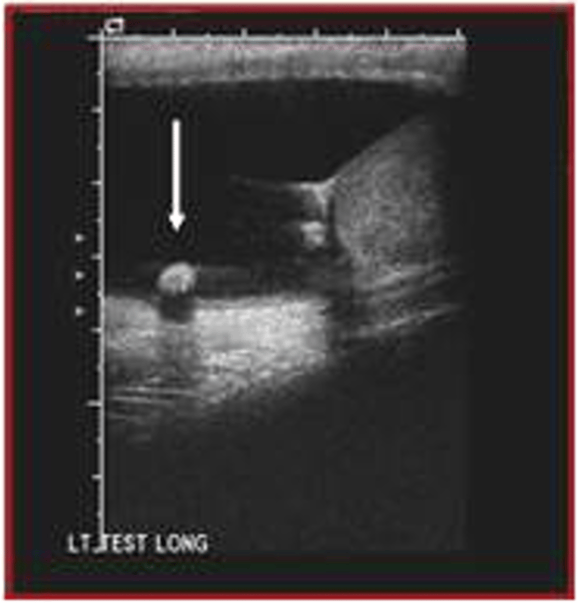
Which of the following is also known as a scrotal pearl and represents an extratesticular calculus?
a. Varicocele
b. Hydrocele
c. Scrotolith
d. Hematocele
e. Epididymitis
f. Orchitis
c. Scrotolith
Which of the following findings appears as an extremely echogenic, mobile extratesticular structure that produces posterior acoustic shadowing on ultrasound?
a. Hydrocele
b. Varicocele
c. Scrotal pearl (scrotolith)
d. Epididymal cyst
c. Scrotal pearl (scrotolith)
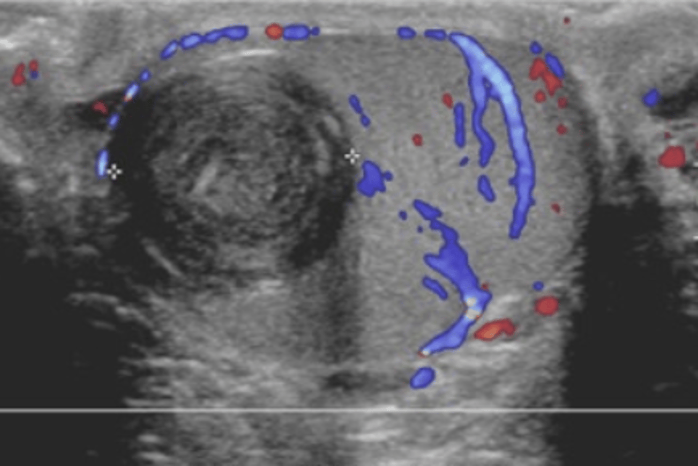
scrotal pearl / scrotolith image
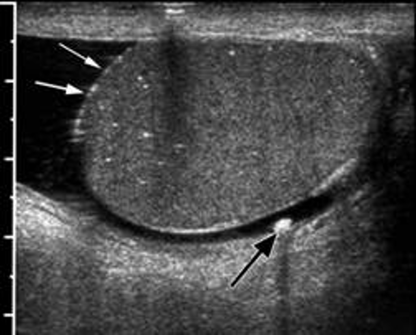
scrotolith / scrotal pearl image
Inflammation of the epididymis is called:
a. Orchitis
b. Epididymitis
c. Prostatitis
d. Varicocele
b. Epididymitis
which of the following is the most common cause of acute testicular pain in adults and present with leukocytosis, fever, dysuria, ureteral discharge and scrotal wall edema?
a. Orchitis
b. Epididymitis
c. Prostatitis
d. Varicocele
b. Epididymitis
Inflammation of the testicle is called:
a. Epididymitis
b. Orchitis
c. Hydrocele
d. Hematocele
b. Orchitis
Inflammation of both the epididymis and testicle is called:
a. Orchitis
b. Varicocele
c. Epididymo-orchitis
d. Prostatitis
c. Epididymo-orchitis
Scrotal infections typically spread in which direction?
a. From the testis outward
b. Ascending from the urinary tract or prostate
c. From the liver
d. From the pancreas
b. Ascending from the urinary tract or prostate
Common causes of epididymitis in younger men include which of the following?
a. Chlamydia and gonorrhea
b. E. coli and Salmonella
c. Tuberculosis and hepatitis
d. Influenza and measles
a. Chlamydia and gonorrhea
Which of the following sonographic findings best differentiates epididymitis from orchitis?
a. Epididymitis demonstrates decreased vascularity while orchitis demonstrates absent flow
b. Epididymitis demonstrates increased vascularity in the epididymis, whereas orchitis demonstrates increased intratesticular vascularity
c. Epididymitis appears as anechoic fluid around the testis while orchitis appears as calcifications
d. Epididymitis appears as a solid intratesticular mass while orchitis appears as dilated veins
b. Epididymitis demonstrates increased vascularity in the epididymis, whereas orchitis demonstrates increased intratesticular vascularity
Which of the following sonographic findings is most consistent with chronic epididymitis?
a. Enlarged hypoechoic epididymis with hyperemia
b. Anechoic cyst in the epididymal head
c. Enlarged hyperechoic epididymis with calcifications
d. Dilated tubular veins greater than 2 mm
c. Enlarged hyperechoic epididymis with calcifications
An intratesticular abscess most commonly results from untreated epididymo-orchitis. Which of the following sonographic findings is most consistent with a testicular abscess?
a. Simple anechoic cyst with posterior enhancement
b. Dilated tubular veins greater than 2 mm with Valsalva
c. Complex intratesticular mass with no internal flow but hyperemic flow surrounding the mass
d. Solid hypoechoic intratesticular mass with uniform vascularity
c. Complex intratesticular mass with no internal flow but hyperemic flow surrounding the mass
A complex intratesticular mass with no internal flow but hyperemic flow around its margins most likely represents:
a. Seminoma
b. Varicocele
c. Testicular abscess
d. Epididymal cyst
c. Testicular abscess
A complex hydrocele that contains pus which may have septations or loculation is termed:
a. Hydrocele
b. Hematocele
c. Pyocele
d. Spermatocele
c. Pyocele
Blood within the scrotum is termed:
a. Hydrocele
b. Varicocele
c. Hematocele
d. Pyocele
c. Hematocele
Which of the following sonographic findings is most consistent with a hematocele?
a. Complex fluid collection within the scrotum, often following trauma
b. Dilated tubular veins greater than 2 mm with Valsalva
c. Simple anechoic fluid surrounding the testicle
d. Solid hypoechoic intratesticular mass
a. Complex fluid collection within the scrotum, often following trauma
Multiple small intratesticular cysts located along the mediastinum testis that may result from dilation of the rete testis are referred to as:
a. Testicular microlithiasis
b. Tubular ectasia of the rete testis
c. Epididymal cysts
d. Scrotoliths
b. Tubular ectasia of the rete testis
Multiple echogenic foci within the testis that produce no acoustic shadowing are termed:
a. Scrotoliths
b. Testicular microlithiasis
c. Teratoma
d. Epididymal cyst
b. Testicular microlithiasis
Which of the following conditions has been associated with malignancy, infertility, Klinefelter syndrome, and cryptorchidism?
a. Hydrocele
b. Varicocele
c. Testicular microlithiasis
d. Epididymal cyst
c. Testicular microlithiasis
Which of the following sonographic findings is most consistent with an epidermoid cyst of the testicle?
a. Multiple echogenic foci with no acoustic shadowing
b. Simple anechoic cyst with posterior enhancement
c. Whorled or "onion-skin" appearance within the testicle
d. Dilated tubular veins greater than 2 mm
c. Whorled or “onion-skin” appearance within the testicle
note:
to me it looks more like a "cd"
epidermoid cyst
Which of the following conditions has been associated with malignancy, infertility, Klinefelter syndrome, and cryptorchidism?
a. Hydrocele
b. Varicocele
c. Testicular microlithiasis
d. Epididymal cyst
c. Testicular microlithiasis
Which of the following conditions resembles a mass within the testicle and is associated with congenital adrenal hyperplasia or Cushing syndrome?
a. Seminoma
b. Epidermoid cyst
c. Testicular microlithiasis
d. Adrenal rests
d. Adrenal rests
Which of the following conditions appears as bilateral, round, hypoechoic intratesticular masses most commonly located near the mediastinum testis?
a. Adrenal rests
b. Epidermoid cyst
c. Testicular abscess
d. Seminoma
a. Adrenal rests
Which of the following laboratory values are most helpful in differentiating between benign and malignant intratesticular tumors?
a. PSA and Lactate dehydrogenase (LDH)
b. Alpha-fetoprotein (AFP) and human chorionic gonadotropin (hCG)
c. TSH and T4
d. Calcium and PTH
b. Alpha-fetoprotein (AFP) and human chorionic gonadotropin (hCG)
Which laboratory value is most commonly elevated in association with embryonal cell carcinoma, teratomas, and yolk sac tumors?
a. Human chorionic gonadotropin (hCG) b. PSA
c. Alpha-fetoprotein (AFP)
d. Lactate dehydrogenase (LDH)
c. Alpha-fetoprotein (AFP)
Seminoma, a germ cell tumor of the testicle, is most commonly found in which age group?
a. Children under 10 year
b. Adolescents 12-18 years
c. Men between 30 and 50 years of age
d. Men older than 70 years
c. Men between 30 and 50 years of age
Which of the following is the most common malignant neoplasm of the testicles?
a. Embryonal carcinoma
b. Teratoma
c. Seminoma
d. Choriocarcinoma
c. Seminoma
Patients with which of the following conditions have an increased risk of developing seminoma germ cell tumor?
a. Varicocele
b. Hydrocele
c. Cryptorchidism
d. Epididymitis
c. Cryptorchidism
Which of the following tumors is typically unilateral and hypoechoic. It may replace the entire testicle, presents as a painless mass with testicular hardening, and may be associated with elevated hCG levels?
a. Teratoma
b. Embryonal carcinoma
c. Seminoma
d. Epididymal cyst
c. Seminoma
Which of the following germ cell tumors is most commonly associated with only having an elevated alpha-fetoprotein (AFP) levels?
a. Seminoma
a. Seminoma
b. Teratoma
c. Choriocarcinoma
d. Embryonal cell carcinoma
e. Yolk sac tumor
e. Yolk sac tumor
Which of the following germ cell tumors most commonly demonstrates areas of hemorrhage and necrosis on ultrasound?
a. Seminoma
b. Teratoma
c. Choriocarcinoma
d. Embryonal cell carcinoma
e. Yolk sac tumor
c. Choriocarcinoma
Which of the following germ cell tumors most commonly demonstrates cystic components on ultrasound?
a. Seminoma
b. Teratoma
c. Choriocarcinoma
d. Embryonal cell carcinoma
e. Yolk sac tumor
d. Embryonal cell carcinoma
whiWhich of the following germ cell tumors may contain hair and teeth due to the presence of multiple tissue types and be benign with malignang potential?
a. Seminoma
b. Teratoma
c. Choriocarcinoma
d. Embryonal cell carcinoma
e. Yolk sac tumor
b. Teratoma
Which of the following germ cell tumors are considered malignant? (Select all that apply)
a. Seminoma
b. Teratoma
c. Choriocarcinoma
d. Embryonal cell carcinoma
e. Yolk sac tumor
a. Seminoma
c. Choriocarcinoma
d. Embryonal cell carcinoma
e. Yolk sac tumor
Which of the following germ cell tumors may be benign but has malignant potential or malignant components?
a. Seminoma
b. Teratoma
c. Choriocarcinoma
d. Embryonal cell carcinoma
e. Yolk sac tumor
b. Teratoma
Which of the following sonographic findings is most consistent with an inguinal hernia extending into the scrotum?
a. Simple anechoic fluid surrounding the testicle
b. Dilated tubular veins greater than 2 mm with Valsalva
c. Heterogeneous mass within the scrotum demonstrating peristalsis and possibly containing air or fluid
d. Solid hypoechoic intratesticular mass with increased vascularity
c. Heterogeneous mass within the scrotum demonstrating peristalsis and possibly containing air or fluid
A patient presents with intermittent scrotal swelling, abdominal pain, and blood in the stool. Which of the following conditions is most consistent with these findings?
a. Hydrocele
b. Varicocele
c. Epididymitis
d. Inguinal hernia
d. Inguinal hernia
The penis is covered with skin and subsequently a dense fibrous tissue known as which of the following?
a. Tunica albuginea
b. Dartos fascia
c. Buck fascia
d. Tunica vaginalis
c. Buck fascia
The inner penis is composed of how many cylindrical tissue components?
a. Two
b. Three
c. Four
d. Five
b. Three
Which of the following are the three cylindrical tissue components of the inner penis?
a. Two corpora cavernosa and one corpus spongiosum
b. Two corpus spongiosum and one corpus cavernosum
c. Two seminiferous tubules and one corpus spongiosum
d. Two epididymal ducts and one corpus cavernosum
a. Two corpora cavernosa and one corpus spongiosum
The vascular supply to the penis begins from which of the following arteries?
a. Internal iliac artery
b. External iliac artery
c. Internal pudendal artery
d. Femoral artery
c. Internal pudendal artery
Which of the following arteries is a tributary of the internal iliac artery and supplies blood to the penis?
a. Testicular artery
b. Internal pudendal artery
c. Renal artery
d. Inferior epigastric artery
b. Internal pudendal artery