fetal circulation
1/32
There's no tags or description
Looks like no tags are added yet.
Name | Mastery | Learn | Test | Matching | Spaced | Call with Kai |
|---|
No analytics yet
Send a link to your students to track their progress
33 Terms
…fill most of the thoracic cavity
lungs
…serves as the lateral borders of the heart
lungs
…forms the floor of the chest
diaphragm
other structures in the thoracic cavity
heart
thymus
trachea
vessels
ribs/spine
normal thoracic cavity is what shape?
bell shaped
describe the bell shape of the thoracic cavity
ribs form lateral margins
clavicles form upper margins
diaphragm forms lower margin
…forms the lateral margins of the heart and are superior to the diaphragm
lungs
how does fetus breath in utero
fetus does not breath air in utero. amniotic fluid enters lungs
what is the most important marker for fetal viability?
lung development
describe the size of thorax
typically smaller than the abdominal cavity
large variations in size could be indicative of?
pathology
presence of oligohydramnios; resultant pulmonary hypoplasiacna lead to
a reduction in thoracic size
chest circumference measurements made in…
TRANS, level of the 4 CH view
the apex of the heart is directed towards
left (spleen)
abnormal positioning of the heart may indicate the presence of?
chest mass
pleural effusion
cardiac malformation
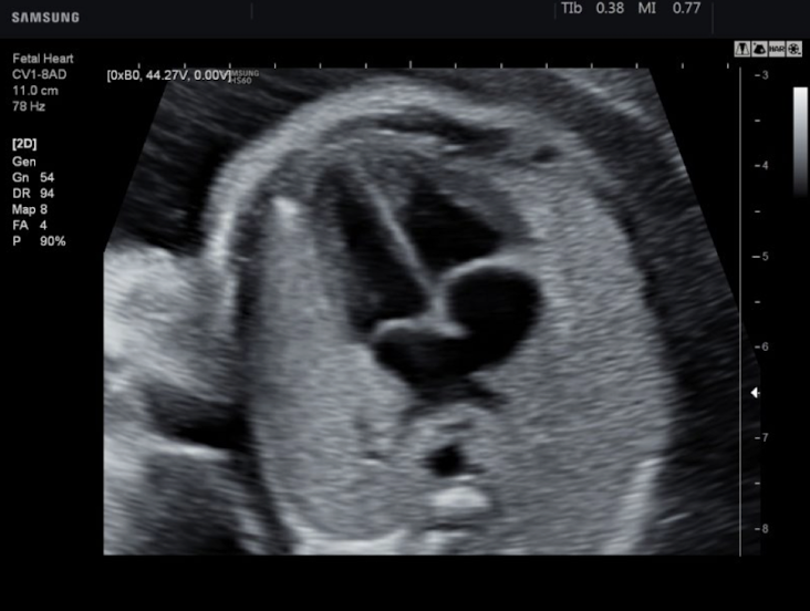
normal or abnormal
normal
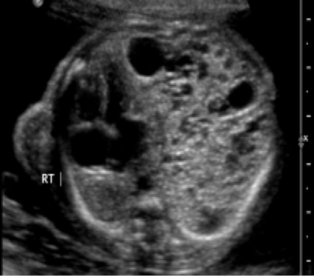
normal or abnormal
abnormal: CCAM/CPAM
fetal lungs appear?
homogenous and slightly echogenic
how does pregnancy progression affect lung echogenicity?
increases
describe the diaphragm
muscle that separates the thorax and the abdomen
hypoechoic curvilinear structure between the liver and lung
coronal abdominal view best for visualizing
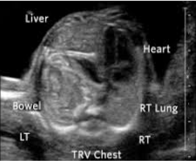
what is this?
diaphragmatic hernia
describe FB on US
seesaw movement of fetal chest or abdomen sustained for at least 20 seconds
absent if no activity during a 20 minute observation period
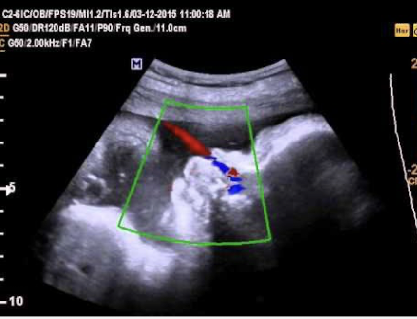
how can FB be documented?
color doppler and obtain a profile view
fetal circulation…the fetal lungs
bypasses
blood flow in the fetus differs in three ways…
ductus venosus
foramen ovale
ductus arteriosus
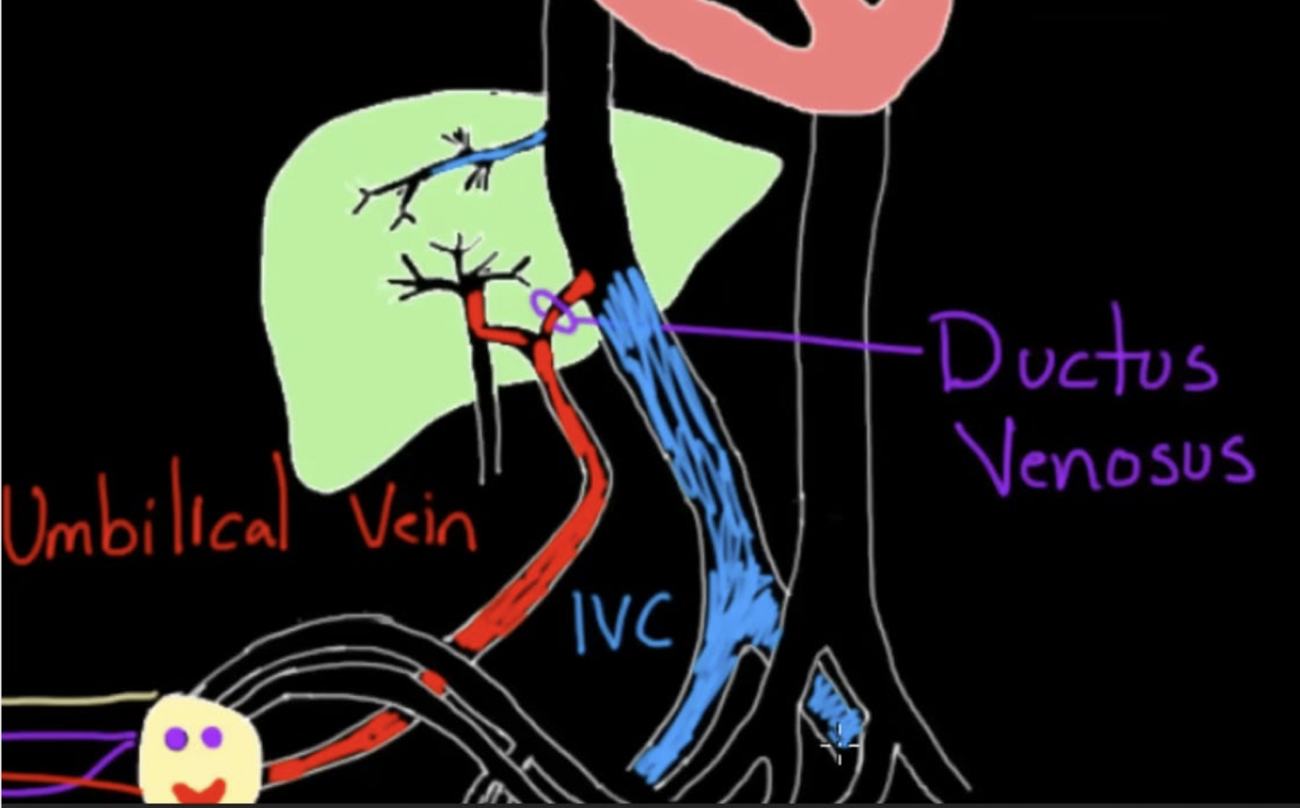
ductus venosus
connects the umbilical vein to the IVC
blood bypasses the liver
after birth forms the ligamentum venosum
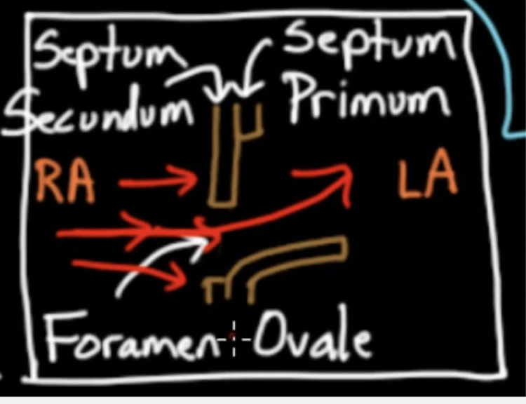
connection between the RA and LA
forament ovale
describe foramen ovale
connection between the RA and LA
allows for oxygenated blood to pass from RA supplied by the IVC to the LA
blood bypasses the lungs
what happens to the foramen ovale after birth?
usually closes after birth
describe the walls in the foramen ovale
flap in the 2 walls separating the RA and the LA
wall 1: septum primum
wall 2: septum scundum
connection between the pulmonary artery and the aorta
ductus arteriosus
ductus arteriosus allows for…
blood to be directed out of the RV and PA and into the aorta, blood bypasses the lungs
what happens to the ductus arteriosus after birth?
forms the ligamentum arteriosus