ANAT EXAM 4
1/133
There's no tags or description
Looks like no tags are added yet.
Name | Mastery | Learn | Test | Matching | Spaced | Call with Kai |
|---|
No analytics yet
Send a link to your students to track their progress
134 Terms
Muscular system
F: movement, posture, joint stability, heat (waster product during contraction)
Connective tissues
comprised of collagen and elastic fibers
F: protection, hold together, sites for blood vessels and nerves, attachment of muscle to skeleton
fascia
connective tissue that connects muscles to skin or other muscles
Fascicle
group of structurally and functionally related cells and their ECM
excitability
responds to stimulus, change in membrane potential
contractability
proteins in muscle cells draw together (sarcomere)
conductivity
electrical changes across membrane conduct membrane
action
body movement because of a muscle contraction
innervate
to supply a muscle with nerves
insertion
muscle attachment point that will move the structure to which it is attached when the muscle contracts
origin
relatively stationary attachment points of a muscle
agonist
muscle that contracts to produce a particular movement
antagonist
muscle that opposes the action of its agonist partner
synergist
muscles that aid the action of the agonist by guiding the movement to ensure a smooth action
fixators
muscles that stabilize the bone during an action
Sternocleidomastoid
Manubrium of sternum, medial portion of clavicle
Mastoid process of temporal bone
Together: flex head; individual: flex and rotate head toward the opposite side. Accessory muscles of inspiration
Trapezius
External occipital protuberance, cervical vertebra, spinous processes of thoracic vertebra
Lateral clavicle & spine and acromion of scapula
elevates scapula, rotates scapula superiorly
Rectus abdominus
superior aspect of pubis bones
costal cartilages of inferior ribs
flexes the trunk, compresses abdominal cavity
Pectoralis major
Medial clavicle, sternum, costal cartilages 1-7
Greater tubercle & lateral lip of intertubercle sulcus of humerus
Flexes and adducts the arm, rotates the arm medially
deltoid
Acromion & spine of scapula; lateral clavicle
Deltoid tuberosity of humerus
Abducts the arm; secondarily flexes and extends arm
triceps brachii
olecranon process of ulna
extends the forearm
biceps brachii
Supraglenoid tubercle & coracoid process of scapula
radial tuberosity of radius
flexes the forearm, supinates the forearm
gluteus maximus
Posterior & lateral portions of ilium, sacrum & coccyx
Gluteal tuberosity of femur
Rotates the thigh laterally, abducts the thigh, extends the thigh
Adductor longus
Body of pubis
Linea aspera of femur
adducts the thigh, flexes the thigh, rotates the thigh medially
Gastrocnemius
Medial and lateral condyles of femur
Posterior calcaneus
Plantarflexes the food, flexes the leg
Tibialis anterior
Lateral condyle of tibia primarily
Tarsal and 1st metatarsal
dorsiflex the food, invert food
Rectus femoris
Anterior inferior iliac spine, superior margin of acetabulum
Patella and tibial tuberosity
Extends the leg, flexes the thigh at hip
Biceps femoris
Ischial tuberosity & distal half of posterior femur
Head of fibula; lateral condyle of tibia
Extend the thigh, flexes the leg
skeletal muscle
sarcomere
stimulated by nervous system for contraction
cardiac muscle
gap junctions in intercalated discs - coordinate contraction
autorhythmic - generate muscle impulse without nervous stimulation
sarcomere
smooth muscle
thin filaments attached to dense bodies
cells physically coupled at dense bodies
contraction: myosin heads opposite facing, long and thin cells to fat globs
Gap junctions
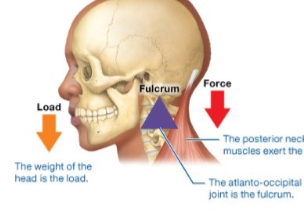
First class lever
Fulcrum in middle
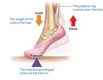
Second class lever
Load in middle
Fulcrum close to load
Mechanical advantage because large load is moved with small force
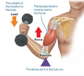
Third class load
Force in middle
Most motion
Sensory receptors
perceive stimuli
effectors
carry out effects of nervous system
Sensory (afferent)
carry signals from sensory receptors to CNS
Motor (efferent)
carry signals from CNS to muscles or glands
Interneuron
Between sensory and motor neurons
Neuroglia
Nerve glue
specialized support cells for neurons
Astrocytes
anchors neurons and blood vessels, clean up excess chemicals, facilitate formation of blood brain barrier CNS
oligodendrocytes
form myelin sheath CNS
microglia
phagocytes, eat stuff CNS
ependymal cells
produce and circulate cerebrospinal fluid CNS
Schwann cells
form myelin sheath PNS
Satellite cells
surround and support cell body PNS
Nerves
Bundle of neuron axons packed with blood vessels and myelin sheath
F: carry signals to and from CNS
sensory nerves
contain sensory neurons
F: carries signal from sensory receptor to CNS
Motor nerves
Contain mostly motor neurons
F: carry signal from CNS to muscles or glands (effectors)
PNS Somatic Sensory division (voluntary afferent)
sensory neurons carry general sensory signals from muscles, bones, joints, and the skin as well as special sensory signals to CNA
PNS Visceral Sensory division (involuntary afferent)
Sensory neurons carry signals from organs (viscera) to CNS
PNS Somatic Motor division (voluntary efferent)
Motor neurons carry signals from CNS to skeletal muscles
PNS Visceral Motor Division / Autonomic Nervous System (involuntary efferent)
parasympathetic and sympathetic motor neurons carry signals from CNS to smooth muscle, cardiac muscle, and glands (viscera)
Sympathetic
Fight or flight, maintains homeostasis during exercise, emotions, emergency
Parasympathetic
digest and rest, homeostasis of maintenance functions such as digestion and formation of urine during rest
Acetylcholine
regulates muscle contraction, parasympathetic NS, some brain functions
norepinephrine / epinephrine (adrenaline)
influences heart rate, blood pressure, digestion, regulates sleep / wake cycle, attention, feeding
dopamine
coordinate movement, involved in emotions and motivation
serotonin
mood regulation, affects emotion, attention, cognition, motor behavior feeding behavior, daily rhythms
histamine
regulation of arousal and attention, mediates allergic responses
neuromodulation
alter synaptic transmission (strengthen or weaken effect of neurotransmitter)
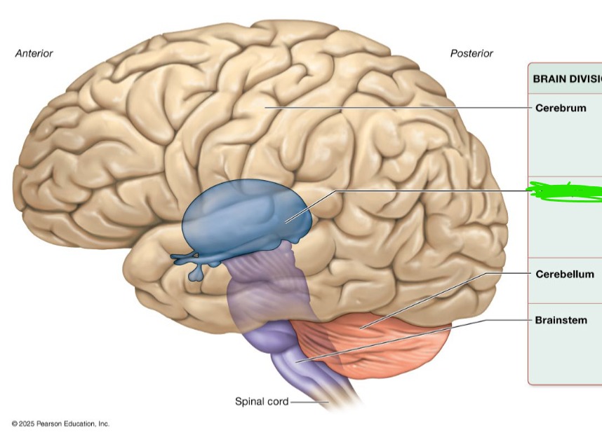
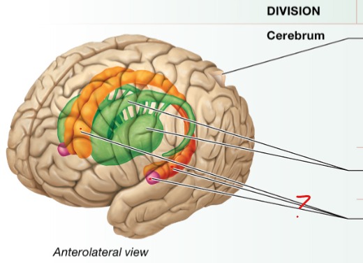
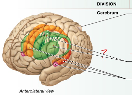
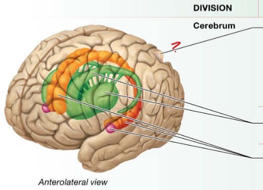
Cerebrum
Performs higher mental functions
Interprets sensory stimuli
Plans and initiates movement
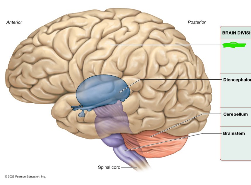
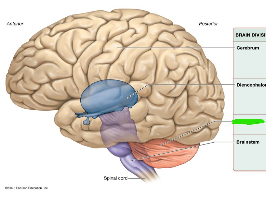
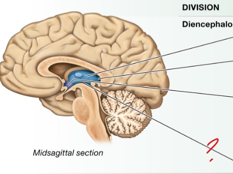
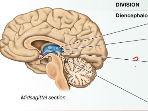
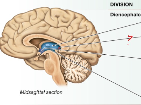
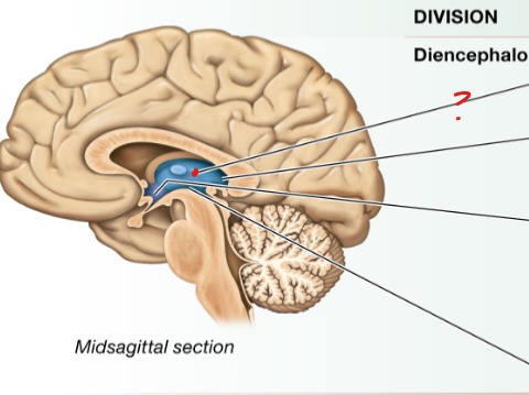
Diencephalon
Processes, integrates, and relays info
Maintains homeostasis
Regulates biological rhythms
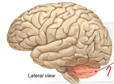
Cerebellum
Monitors and coordinates movement
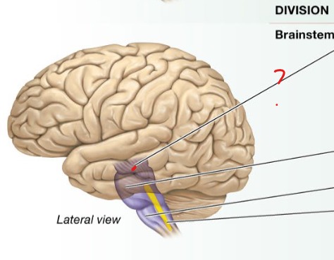
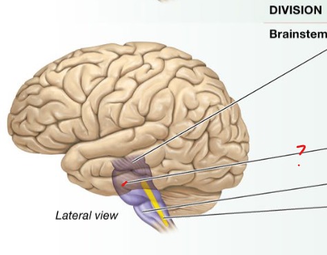
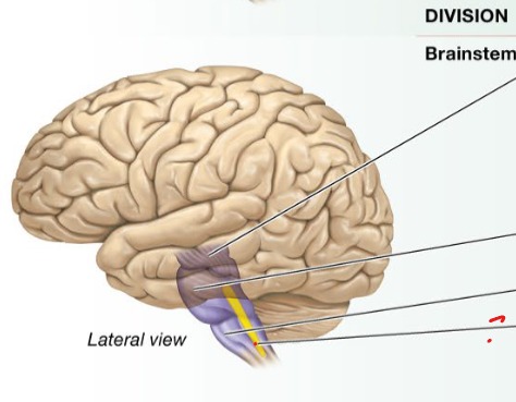
Brainstem
Maintains homeostasis
Controls certain reflexes
Monitors movement
Integrates and relays information

Dura Mater
Superficial (outer) meningeal layer
Connects to bone
Meninges
connective tissue surrounding brains and spinal cord
F: protection
Arachnoid Mater
Middle loose meningral layer
Pia Mater
Deepest thin meningeal layer vascular, clings to grooves of cortex
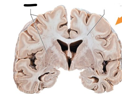
Grey Matter
Cell bodies, dendrites, interneurons
Generally forms the cortex
F: process information
Develops by 20s
No myelin
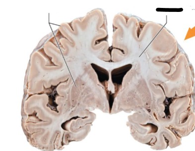
White matter
Myelinated axons
deep to brain structures
F: communication to and from grey areas and between gray areas and rest of body
developments peaks middle age
Ventricles
F: hollow spaces in brains filled with cerebrospinal fluid
Lateral Ventricle (first and second)
horns

Third ventricle
head
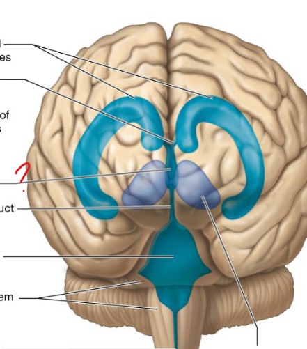
founrth ventricle
neck
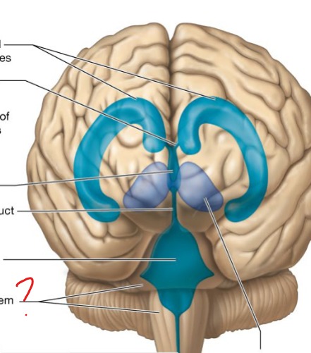
Cerebrospinal Fluid
F: Provides buoyancy, protects brain, regulates environment of CNS (nutrient and waste transport)
Blood brain barrier
barrier between blood and neurons in brain
property of capillaries in brain
only lets certain things in
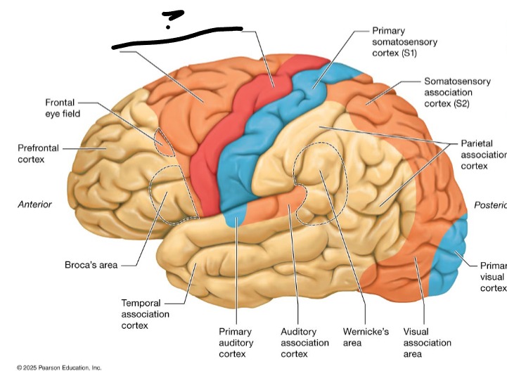
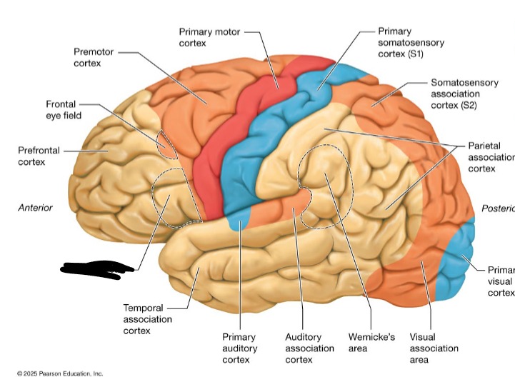
Frontal Lobe
voluntary motor function
Parietal Lobe
General sensory function
Temporal Lobe
Hearing and smell
Occipital lobe
process incoming visual info and stores visual memories
Insula Lobe
Memory and interpret taste (inside of frontal and temporal)
Somatosensory cortex
interprets incoming somatic sensory stimuli (temperature and touch)
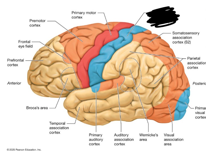
Primary motor cortex / premotor cortex
plans and executes movement (complex = premotor)
Upper motor neurons
plan movement
Wernicke’s area
language comprehension
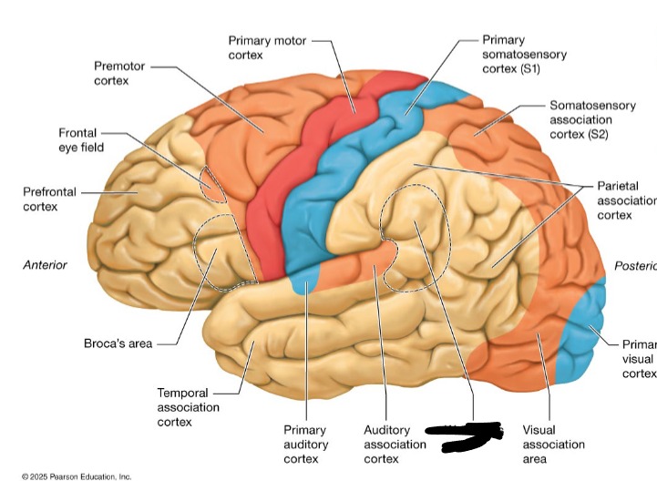
Broca’s Area
Language production
Limbic System
Hippocampus: Memory and learning
Amygdala: Behavioral expression and emotion
Basal nuclei
regulates voluntary movement
Cerebral cortex
Primary motor cortex: plans and executes voluntary moments \
Primary sensory cortices: receive and process different types of sensory input
Multiple task association areas: integrate sensory and motor info from a variety of different primary cortex
Subthalamus
Regulates voluntary movement
Hypothalamus
Regulates autonomic NS
Regulates sleep/wake cycle
Regulates thirst and hunger
Regulates body temp
Controls secretion from pituitary gland
Epithalamus
Produces melatonin
Thalamus
Controls sensory information entry into the cerebral cortex
Edits, sorts and routs stimuli
Cerebellum
Coordinates ongoing voluntary movement to reduce motor error
Midbrain
Processes and routs visual and auditory stimuli to the thalamus
Carries motor fibers from the cerebral cortex
Monitors movement with the basal nuclei
Mediates reflexes
Pons
Regulates breathing and sleep/wake cycle
Medulla oblongata
regulates autonomic functions (HR, breathing BP)
Nuclei of cranial nerves

Reticular formation
Inside of medulla oblongata, involved in sleep and arousal, pain transmission
Plays a role in mood regulation
Maintains homeostasis of many variables
All regions of Brainstem
Contain tracts of white matter involved in movement and sensation, route for ascending and descending tracts between cerebellum and spinal cord