head and neck anatomy ch 7-9 exam
 Call Kai
 Learn
Learn Practice Test
 Spaced Repetition
 Match
Match Flashcards
 Knowt Play
1/86

There's no tags or description
Looks like no tags are added yet.
Name | Mastery | Learn | Test | Matching | Spaced | Call with Kai |
|---|
No analytics yet
Send a link to your students to track their progress
87 Terms
duct
passageway to carry secretion from the exocrine gland to set location
exocrine
gland with associated duct serving as passageway
all major and minor salivary glands or sweat glands
only glands that have ducts
endocrine
ductless gland with secretions being poured directly into vascular system
what they make is secreted directly into bloodstream
adrenal glands
primary lymph nodes
organs include the bone marrow and thymus
create special immune system cells called lymphocytes
secondary lymph nodes
organs include the lymph nodes, the spleen, the tonsils and certain tissue in carious mucous membrane layers in the body
lymph only comes from a primary node
do not
unlike blood vessels, lymph vessels __ __ have valves to prevent backflow from occurring
parotid gland
the largest salivary gland
is encapsulated
innervated by the 9th cranial nerve
empties around the 2nd maxillary molar which is a site of calculus build-up
another name for parotid duct
stenson duct
percentage of saliva excreted from parotid duct
25%
serous secretion
mumps is a result of this enlarged saliva gland
parotid
submandibular gland
is encapsulated
innervated by the 7th cranial nerve
gland drains into the submandibular lymph nodes
most common site for salivary stones
seen externally as the sublingual caruncle
another name for submandibular gland
Wharton’s duct
“men go to war”
percentage of saliva secreted from submandibular gland
65%
mixed secretions, mostly serous
sublingual gland
located below the tongue
is the only gland not encapsulated
innervated by the 7th and 9th cranial nerve
seen externally as sublingual caruncle
percentage of saliva excreted by sublingual gland
10%
mixed secretions but mostly mucous
minor salivary glands
they are most numerous but generally smaller
located on soft palate, hard palate, and every type of mucosa
all secrete mucous saliva EXCEPT Von Ebner
thyroid gland
2 lobes
is the largest endocrine gland in the body
located below the thyroid cartilage where the larynx meets the trachea
not visible in healthy patients
deals with metabolism and hormones
thyroxine (T4)!!
hormone secreted by thyroid
thyroxine
what regulated the thyroid
pituitary gland
where does the thyroid gland drain
into superior deep cervical lymph node
arteries that supplies the thyroid
inferior and superior thyroid arteries
parathyroid
4 small bean shaped glands on the back of the thyroid gland
cannot be palpated
produces and secretes the parathyroid hormone which regulates calcium and phosphorus levels
where do parathyroid veins drain
into the thyroid vein plexus
where do parathyroid lymphatic vessels drain
into the deep cervical lymph nodes
lacrimal glands
located within the orbit above the lateral end of the eye
sits in lacrimal fossa
tears flow down to the medial canthus and drain into the inferior nasal meatus
(runny nose when crying)
innervated by the 7th cranial nerve
composed of epithelial and lymphatic tissue
cervical lymph nodes
receive all the lymph from the head and neck
either directly or indirectly via the superficial lymph nodes
these lie along the internal jugular vein
thymus
primarily to produce and mature T-cells
superficial and lateral to the trachea and deep to sternum (cannot palpate)
attached to lobes of thyroid gland
innervated by the 10th cranial nerve
supplied by inferior thyroid and internal thoracic arteries
shrinks after puperty
T-cells
a type of white blood cell that helps the body fight infection and disease
nerve
bundle of neural processes outside the CNS
parts of peripheral nervous system
nervous system
extensive, intricate network of structures that activates, coordinates, and controls all functions of body
neuron
cellular component of nervous system
composed of a cell body and neural processes
afferent nerve
SENSORY nerve carry info from body periphery TO brain or spinal cord
periphery → brain/spinal cord
efferent nerve
MOTOR nerve carrying info AWAY from brain or spinal cord to periphery
brain/spinal cord→ periphery
dental plexus
network of nerves within both maxillary and mandibular arches
synapse
junction between 2 neurons or between neuron and effector organ that transmits through electrical or chemical means
the two components of nervous system
central nervous system
peripheral nervous system
central nervous system (CNS)
brain and spinal cord
peripheral nervous system (PNS)
composed of afferent and efferent nerves
automatic nervous system
somatic nervous system
automatic nervous system
involuntary
sympathetic nervous system
parasympathetic nervous system
somatic nervous system
voluntary actions and movements
muscle innervation
sympathetic nervous system
fight or flight
parasympathetic nervous system
rest and digest
CN I- Olfactory
afferent only
responsible for sense of smell
passes through cribriform plate of ethmoid bone
( cribs smell)
CN II - Optic
afferent only
responsible for vision
enters optic canal of sphenoid bone
both R and L nerves join at the optic chiasma where the fibers cross to contralateral sides before entering the brain
CN III - oculomotor
efferent only
controls most eye muscles and pupil constriction
passes through superior orbital fissure of sphenoid bone
CN IV - Trochlear
efferent only
controls superior oblique muscles of the eye
exits skull through superior orbital fissure of sphenoid bone
CN V - Trigeminal
both sensory and motor roots
afferent- teeth, tongue, oral cavity, lacrimal gland, nasal cavities, and skin of face
efferent - muscle of mastication
three divisions
opthalmic (V1)
maxillary (V2)
mandibular (V3)
opthalmic division of trigeminal nerve
sensory
enters at superior orbital fissure
maxillary branch of trigeminal nerve
sensory
enters at foramen rotundum
mandibular branch of trigeminal nerve
motor AND sensory
enters at foramen ovale
CN VI - Abducens
efferent only
supplies muscles for eye movement and innervates lateral rectus muscle of eye
exits through superior orbital fissure of sphenoid bone
1st nerve affected by serious sinus infections
CN VII - Facial
both afferent and efferent
efferent - facial expression muscles
afferent - taste anterior 1/3 of the tongue
passes through stylomastoid foramen
gives motor innervation to stylomastoid muscles
bells palsy
paralysis of facial nerve
chorda tympani
branch of facial nerve
CN VIII -Vestibulocochlear
afferent only
helps with hearing and balance
passes through internal acoustic meatus
damage causes vertigo, hearing loss, tinnitus
CN IX- Glossopharyngeal
both motor and sensory
efferent - stylopharyngeus muscle, parotid gland
afferent - taste and sensation to posterior 1/3 of tongue
includes gag reflex
passes through jugular foramen
CN X- Vagus
both motor and sensory
efferent - soft palate, pharynx, larynx, thoracic organs
afferent - taste sensation for epiglottis
passes through jugular foramen
helps with speech and swallowing
CN XI - Accessory
motor only
provides innervation for trapezius and sternocleidomastoid muscles, muscle of soft palate, and muscles of pharynx
passes through jugular foramen
damage may cause shoulder droop and difficulty turning head
CN XII- Hypoglossal
motor only
controls tongue movements
aids in speech
passes through hypoglossal canal of occipital bone
branches we block for trigeminal nerve
V2 Maxillary
V3 Mandibular
maxillary arch local anethetic blocks
PSA
MSA
ASA
NP
GP
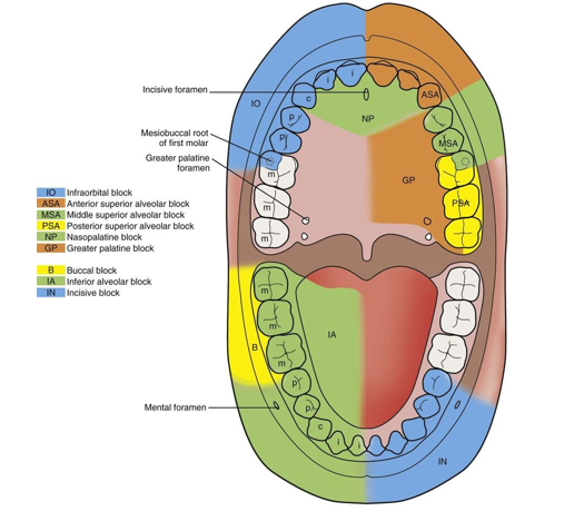
Mandibular arch local anethetic blocks
IA
Long buccal nerve block
mental/ incisive nerve block
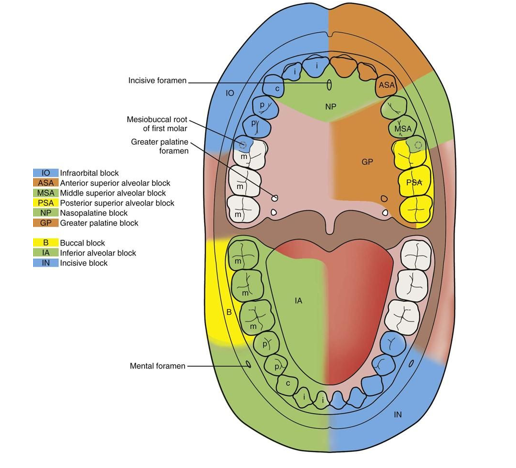
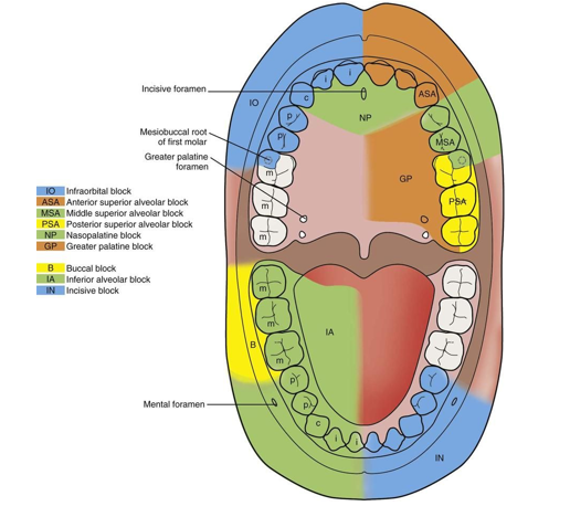
posterior superior alveolar nerve block (PSA)
insertion: mucobuccal fold above the DB root of the 2nd molar
target is maxillary tuberosity
can cause hematoma if injected into pterygoid plexus
teeth anesthetized: maxillary molars except the MB root of the 1st molar
soft tissue anethetized: buccal gingiva of the maxillary molars
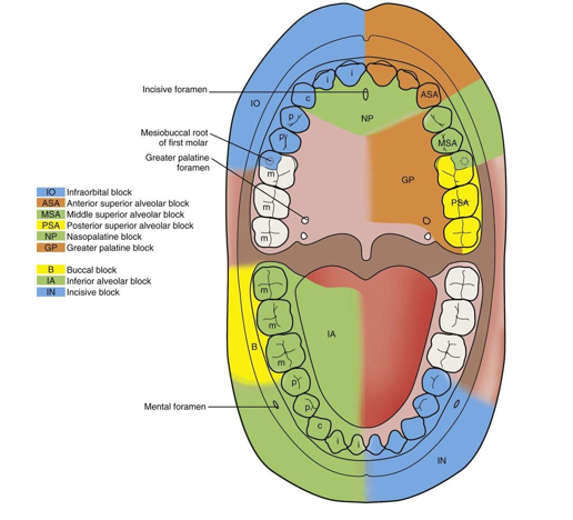
middle superior alveolar nerve block (MSA)
insertion: mucobuccal fold above 2nd premolar
teeth anesthetized: maxillary premolars and MB root of the 1sst molar
soft tissue anethetized: buccal gingiva of maxillary premolars
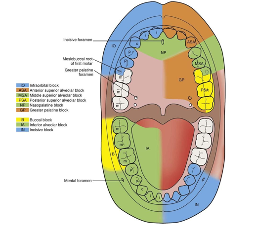
anterior superior alveolar nerve block (ASA)
insertion: height of the mucobuccal fold near mesial aspect of maxillary canine
teeth anesthetized: maxillary incisors/cuspids and MSA teeth
soft tissue anesthetized: maxillary incisors and cuspid gingiva
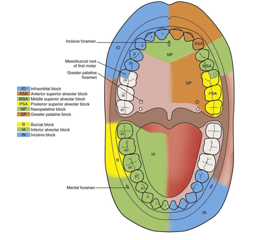
Nasopalatine nerve block (NP)
insertion: palatal mucosa just lateral to the incisive papilla
soft tissue anethetized: lingual gingiva of the maxillary incisor/cuspids
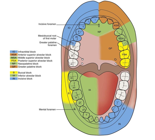
greater palatine nerve block (GP)
Insertion: apply pressure to slightly bow needle at junction of alveolus and hard palate of the greater palatine foramen and inject anterior to the cotton tip applicator
locate the foramen before injecting an utilize pressure anesthesia!
soft tissue anesthetized: lingual gingiva of the maxillary molars/ premolars
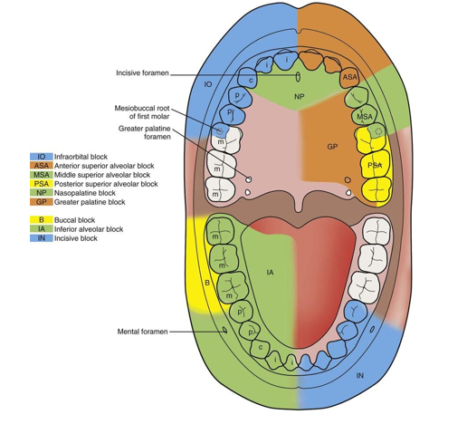
Inferior alveolar nerve block (IA)
mandibular block
most commonly used block in dentistry
anesthetizes: mandibular teeth to midline, associated lingual periodontium, gingiva, and facial periodontium to the midline
target: IA nerve exiting the mandibular foramen on medial surface of the mandibular ramus
insertion: 2/4 distance from coronoid notch to deepest part of the pterygomandibular raphe, 6-10 mm above mandibular occlusal plane
can cause hematoma if injected into pterygoid plexus
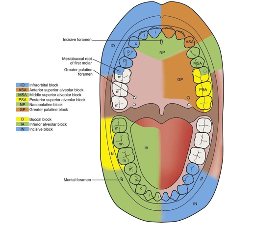
long buccal nerve block
anesthetizes: long buccal nerve, associated buccal periodontium and gingiva of mandibular molars within
insertion: mucous membrane distal and buccal to last molar and parallel to occlusal plane
long buccal block + IA = complete mandibular quadrant anesthesia
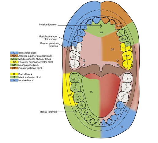
mental / incisive nerve block
anesthetizes: mental nerve, associated periodontium, and gingiva of mandibular anterior teeth and premolars to midline
includes lower lip and chin
insertion: mucobuccal fold, just anterior to mental foramen, parallel to long axis of tooth
right between premolars
Vazirani-Akinosi nerve block
anesthetizes: IA, lingual, mental, incisive, and mylohyoid nerves (within pterygomandibular space), and long buccal nerve 75% of the time
mandibular teeth to midline and associate periodontium, and gingiva
more similar to IA block in coverage
Gow Gates
anesthetizes: IA nerve, lingual nerve, mental, incisive, mylohyoid, auriculotemporal, and long buccal nerve
considered the true mandibular block because it anesthetizes entire mandibular nerve and its branches
mneumontic for cranial nerve names
Oh
Once
One
Takes
The
Anatomy
Final
Very
Good
Vacations
Are
Heavenly
mneumonic for sensory motor or both
some
say
marry
money
but
my
brother
says
big
boobs
matter
more
SAME mneumonic
Same/Afferent
Motor/ Efferent
foramen location of the olfactory nerve
cribriform plate of the ethmoid bone
foramen location of the optic nerve
optic canal of sphenoid bone
eventually crosses over in the optic chiasm
foramen location of the oculomotor nerve
superior orbital fissure of sphenoid bone
foramen location of the trochlear nerve
superior orbital fissure of the sphenoid bone
foramen location of the trigeminal nerve
different for each branch
opthalmic = superior orbital fissure
maxillary = rotundum
mandibular = ovale
foramen location of the abducens nerve
superior orbital fissure of the sphenoid bone
foramen location of the facial nerve
exits skull via stylomastoid foramen
“need style”
foramen location of the vestibulocochlear nerve
internal acoustic meatus
foramen location of the vagus nerve
jugular foramen
foramen location of the glossopharyngeal nerve
jugular foramen
foramen location of the accessory nerve
jugular foramen
foramen location of the hypoglossal * nerve
hypoglossal canal