ATAR Psychology Year 11
1/247
Earn XP
Description and Tags
Bio-Psy and Research Methods so far
Name | Mastery | Learn | Test | Matching | Spaced | Call with Kai | Chat |
|---|
No analytics yet
Send a link to your students to track their progress
248 Terms
Peripheral Vs Central Nervous System (how is it divided?)
The nervous system is divided into;
•Central Nervous System (CNS): consisting of the brain and spinal cord
•Peripheral Nervous System (PNS): made up of the nerve tissue outside of the CNS that connects the CNS with receptors, muscles and glands
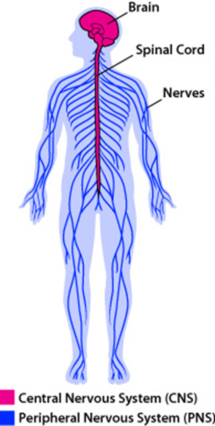
Brain (definition)
organ made up of nerve tissue that controls body functioning
Spinal cord (definition)
a cylindrical structure that runs down the bony vertebral column.
The spinal cord connects the brain to the peripheral nervous system.
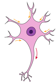
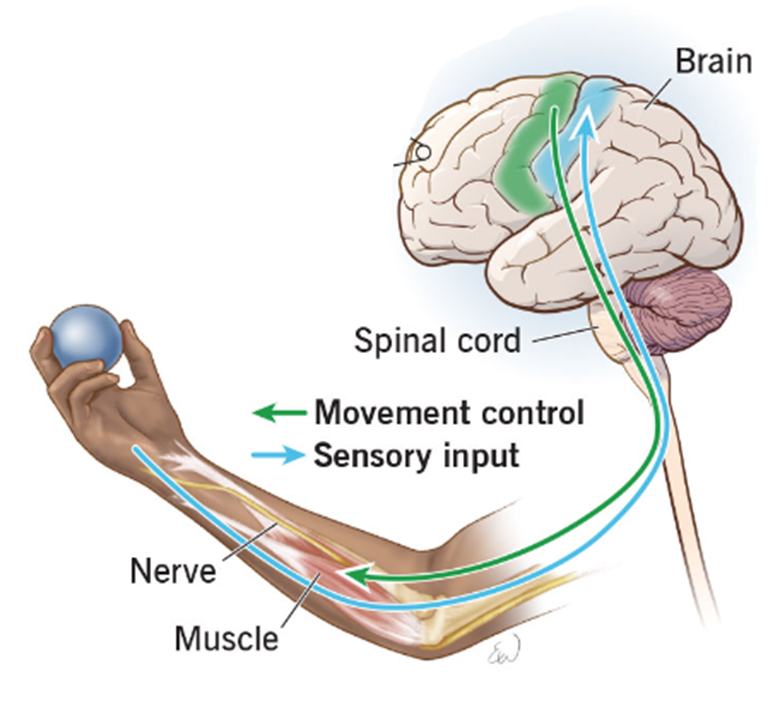
Sensory neurons (function)
transmit nerve impulses from receptors
to the CNS
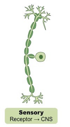
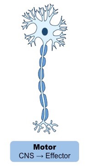
Neurons (definition)
Microscopic nerve cells - highly specialised cells that transmit nerve impulses (action potentials) from one neuron to another.
Motor neurons (function)
transmit nerve impulses from CNS to effectors (muscles or glands.
Interneurons (function, where are they found?)
transmit nerve impulses from sensory neurons to motor neurons. These are found within the spinal cord and brain.
The PNS (makeup/composition)
•Neurons are nerve cells
•Neurons are arranged into nerve fibres
•Nerve fibres are bundled into nerves
•Nerves of the PNS can contain
oJust sensory neurons
oJust motor neurons
oA mix of sensory and motor neurons
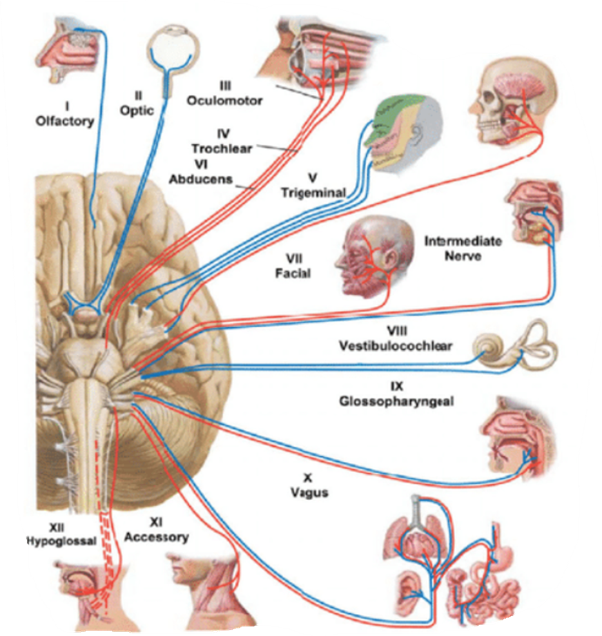
Cranial nerves of the PNS (types)
Motor nerve containing motor neurons sending information from the CNS to effectors
Sensory nerve containing sensory neurons transmitting information from receptors to the CNS
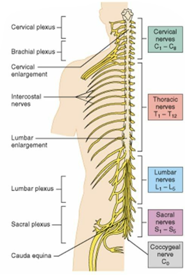
Spinal nerves of the PNS (diagram)
Each spinal nerve consists of both motor and sensory fibres
Somatic NS vs Autonomic NS (diagram)
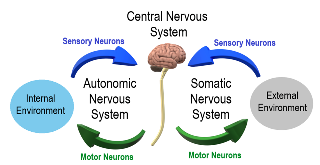
Somatic Nervous System (definition, explanation)
Every deliberate, conscious action a person makes involves neurons in the somatic nervous system.
Effectors of the somatic nervous system = voluntary skeletal muscles.
Nerves of the somatic nervous system contains sensory neurons which relay messages from receptors in ligaments, tendons and muscles to the central nervous system (CNS)
Nerves in the somatic nervous system contains motor neurons which relay nerve impulses from the CNS to stimulate voluntary muscle contraction.
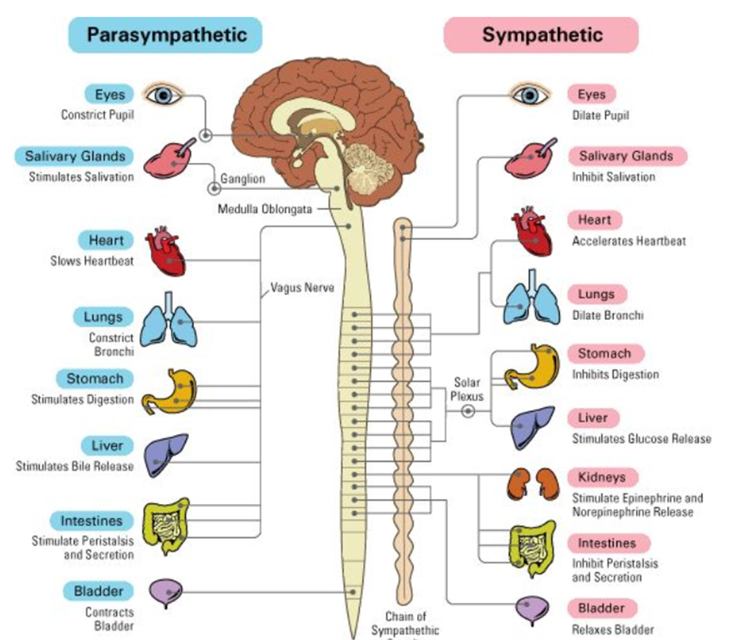
Autonomic Nervous System (purpose, composition)
Regulates the function of internal involuntary organs such as
•Heart
•Digestive tract
•Lungs
•Liver
•Bladder
•Pupils
•Salivary glands
Sweat glands
Contains sensory neurons which relay messages from receptors about the internal environment of the body to the central nervous system (CNS)
Contains motor neurons which relay messages from the CNS to stimulate involuntary muscle contraction.
Somatic NS vs Autonomic NS (table)
Comparison | Autonomic Nervous System | Somatic Nervous System |
Effectors it Activates | Involuntary muscles and glands | Voluntary skeletal muscles |
Primarily concerned with | Regulation of Internal environment for optimal body function | Responding to external environment |
Actions are | Unconscious / involuntary | Conscious / voluntary |
Part of the | Peripheral nervous system | Peripheral nervous system |
Sympathetic and Parasympathetic NS (function)
The Autonomic NS divides into the sympathetic NS and the parasympathetic NS.
Sympathetic = Excitation - stress response, “fight or flight”
Parasympathetic = Inhibition - normal functioning or a relaxed state, “rest and digest”
Fight or Flight response
An automatic physiological reaction, activated by the sympathetic nervous system, to an event that is perceived as stressful or frightening, that prepares the body to fight or flee.
Fight or Flight responses are evolutionary adaptations to increase chances of survival in threatening situations.
Effects of the Sympathetic NS on the body (table)
Body System | Physiological effect | Consequence |
Heart | Increased heart rate | Increase in blood flow to cells |
Circulation | Dilation of blood vessels serving skeletal muscles | Increased availability of oxygen and glucose to skeletal muscles |
Lungs | Dilation of bronchi | Increased availability of oxygen in blood |
Liver | Increased conversion of stored energy to increase blood glucose | Increased availability of glucose in skeletal muscle and brain cells |
Skin | Skin becomes pale as blood flow to skin is reduced, sweat glands increase sweat production | Increased blood flow to skeletal muscles and away from non-essential parts of the body. Sweat cools the body. |
Eyes | Dilation of the pupils | Allows in more light so that visual acuity is improved to scan nearby surroundings |
Sympathetic vs Parasympathetic NS effects on the body (image)
Nerves (definition)
Bundles of neurons (nerve cells) are called nerves.
Parts of a neuron (with functions, diagram)
dendrites often have many branches. they receive messages from other neurons and carry them towards the cell body.
the cell body contains the nucleus and many other cell organelles.
there is only one axon. It is often long and is unbranched for most of its length and end in an axon terminal. Axons carry nerve impulses away from the cell body.
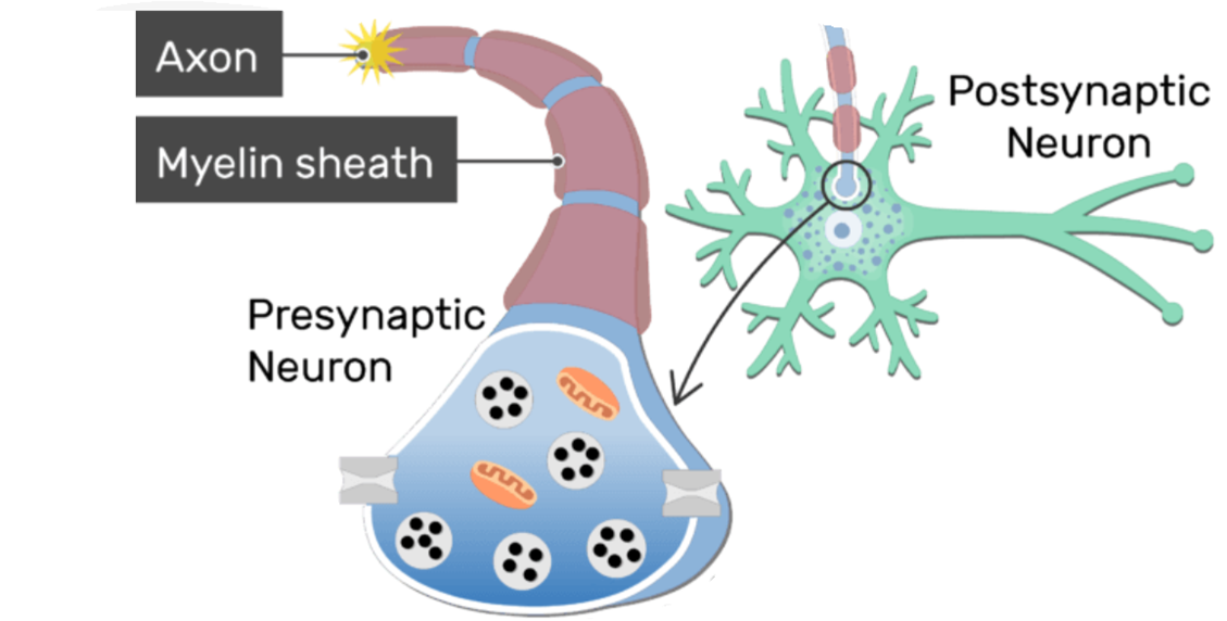
myelin sheath: fatty material that wraps around the axon of sensory and motor neurons.
the axon terminal is the end of the axon that contacts the dendrites of the next neuron, or the effector that the neuron activates
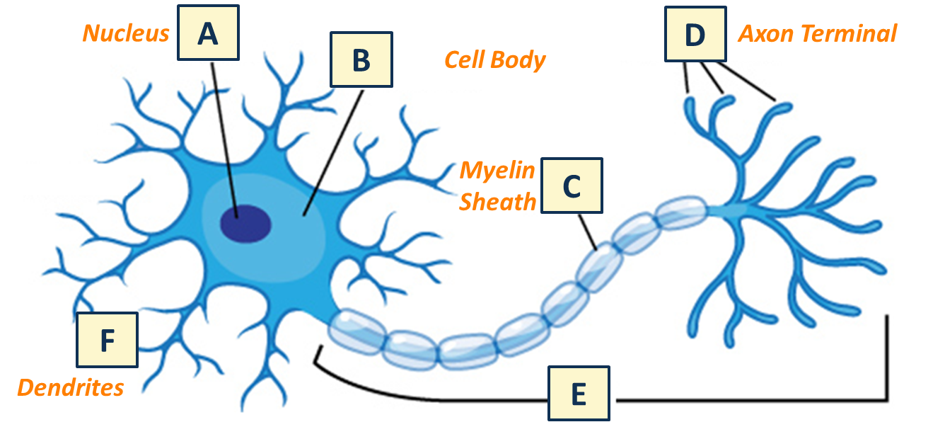
Myelin Sheath (definition, function)
Fatty material that wraps around the axon of sensory and motor neurons.
Function:
protects neuron
insulates the neuron
speeds up transmission of the nerve impulse
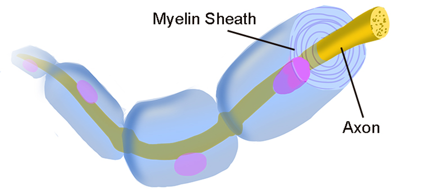
Axon terminal (purpose, composition)
The end of the axon that contacts the
•dendrites of the next neuron
•or the effector that the neuron activates
Axon terminal contains vesicles which house chemicals called neurotransmitters
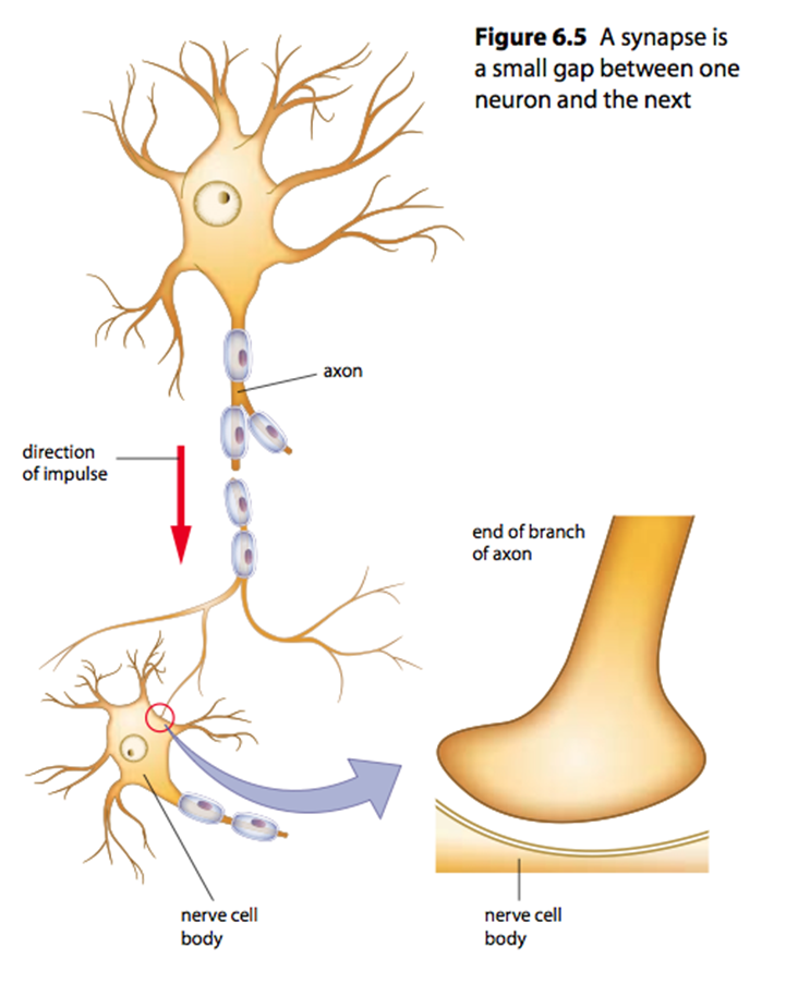
Nerve impulse direction of transmission
Messages, also called nerve impulses, run down neurons like electricity at very high speeds. Dendrite → cell body → axon.
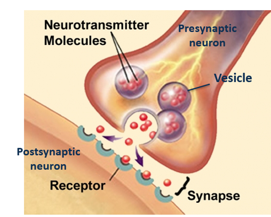
The synapse (definition, where it is, makeup)
A junction between two neurons.
The neurons do not touch, there is a very small gap between them.
Synapses occur between the axon terminal of the presynaptic neuron (neuron before the synapse) and dendrites of the post synaptic neuron (neuron after the synapse).
On the presynaptic neuron, there are vesicles containing neurotransmitters. On the post-synaptic neuron, there are receptors on the dendrite.
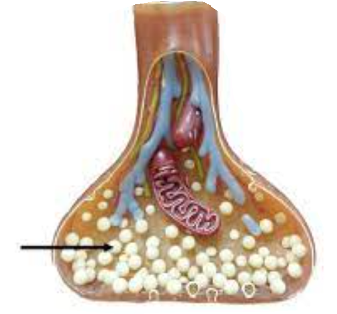
Neurotransmitters (location, description)
The axon terminal of the presynaptic neuron contains vesicles.
Vesicles contain chemicals called neurotransmitters.
Neurotransmitters are chemicals that transmit the electrical action potential across the synapse = electrochemical transmission.
Transmission across the Synapse (step-by-step)
1. The action potential (nerve impulse) travels down the axon of the pre-synaptic neuron and reaches the axon terminal.
2. The vesicles in the axon terminal release their neurotransmitter into the synapse.
3. Neurotransmitters diffuse across the synapse and bind to receptors on the dendrites of the post synaptic neuron.
4. An action potential is generated in the post synaptic neuron.
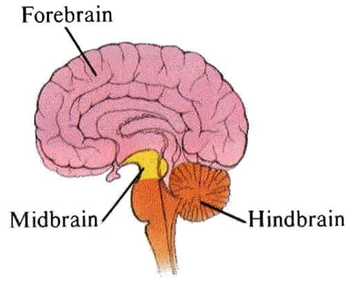
3 major divisions of the brain (and what they contain)
Forebrain
hypothalamus
thalamus
cerebrum
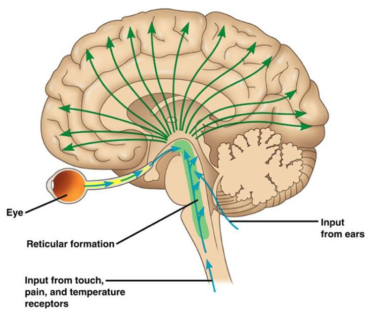
Midbrain
reticular formation
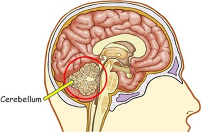
Hindbrain
cerebellum
medulla oblongata
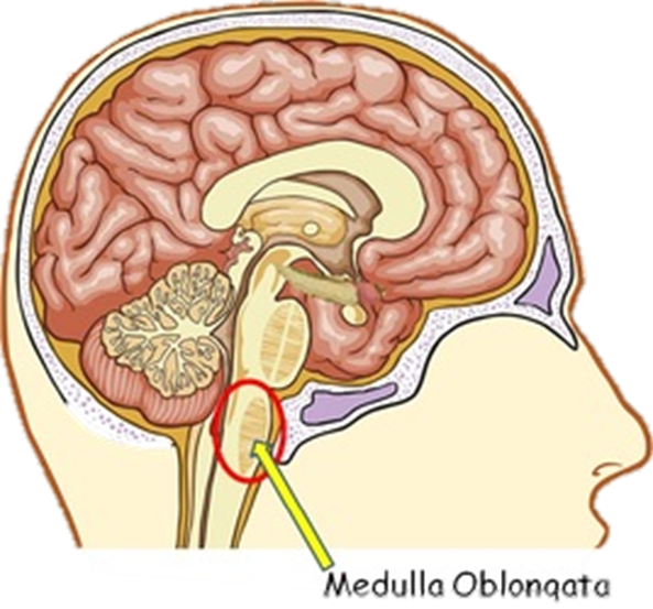
Hindbrain (location, what it contains)
·Located at the back of the head at the base of the brain
·looks like an extension of the spinal cord
·Contains the medulla oblongata and cerebellum
·Also known as the brainstem
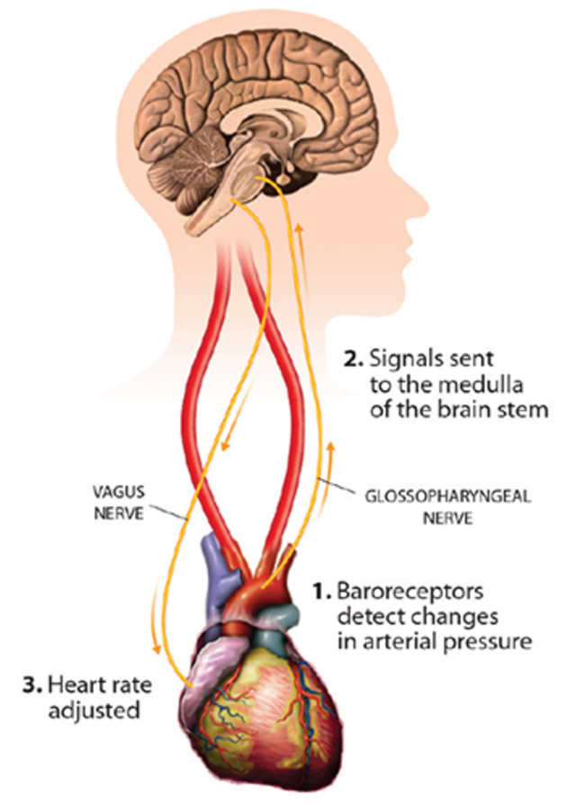
Hindbrain: Medulla oblongata (function, if damaged?)
Controls the involuntary processes of the autonomic nervous system, involved in:
respiratory centre (breathing regulation)
cardiac centre (heart rate, blood pressure)
digestion and reflexes like swallowing, choking and sneezing
If damaged, a person will require life support machines to regulate breathing and heart function → “brain dead”
Hindbrain: Cerebellum (input, functions, if damaged?)
Input: receives messages from muscles, tendons, joints, and structures in our ear
Functions:
balance, posture and coordination of muscles
muscle memory and motor learning
everyday voluntary but automatic tasks, i.e. walking and writing
If damaged, a person may move in uncoordinated ways
affected by alcohol
Midbrain: Reticular formation (location, function)
Located in the centre of the brain, in between the forebrain and hindbrain.
It is a complex network of brainstem nuclei and neurons that serve as a major integration and relay center for many vital brain systems to coordinate functions necessary for survival.
a net-like, diffuse network of nuclei located throughout the brainstem that regulates arousal, consciousness, and sleep-wake cycles
Forebrain (definition, structures)
The largest and most highly developed part of the brain - the top “brain-looking” part
Structures:
thalamus
hypothalamus
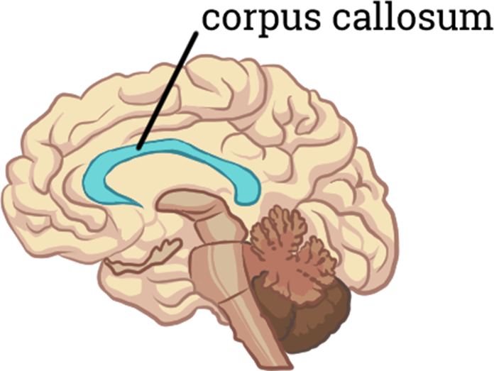
cerebrum (which includes the cerebral cortex and carpus callosum)
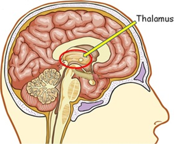
Forebrain: Thalamus (function)
Function: relay sensory and motor signals to and from the cerebral cortex
a link between sensory & motor signals and the cortex
(contains the grey matter of the cerebrum)
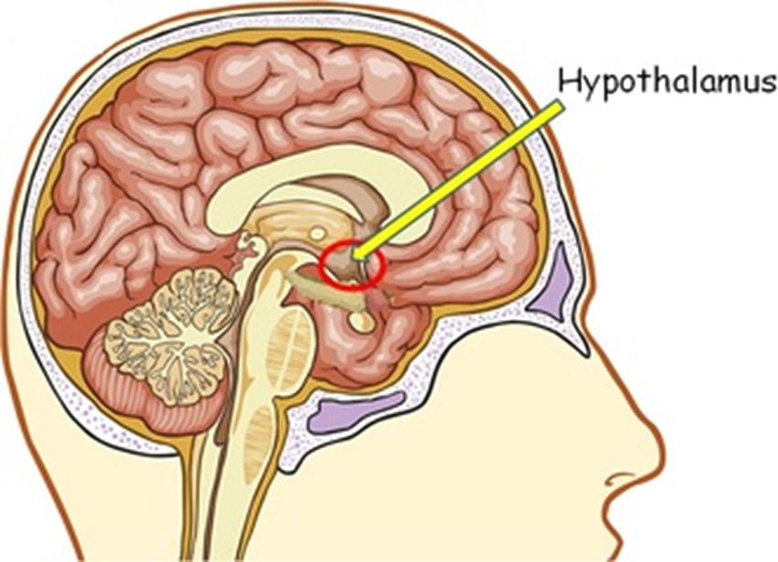
Forebrain: Hypothalamus (function)
Functions: regulates
body temperature
water levels
circadian rhythm
libido
food intake
the activity of the pituitary gland
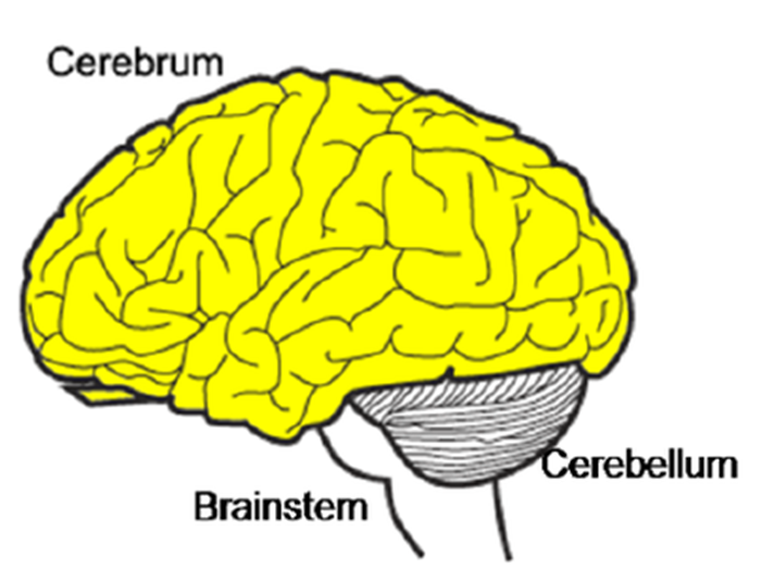
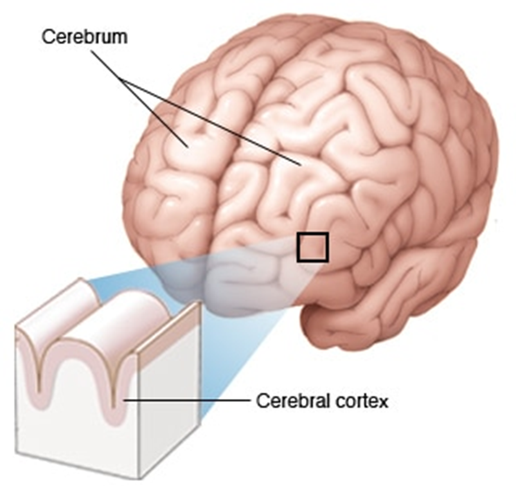
Forebrain: Cerebrum (subparts, function)
Subparts:
cerebral cortex
corpus callosum
Main function:
primary control centre for voluntary muscle movements
primary processing centre of sensory information
higher functioning - memory, logic, personality
Reticular formations vs thalamus vs hypothalamus
Reticular formation - the space - canal that allows fluids that allow the neurons and diff parts to connect to part of the brain
FLUIDS
Corpus callosum - two hemisphere
Reticular formation - all different parts of the brain
Thalamus and hypothalamus - specific autonomic functions
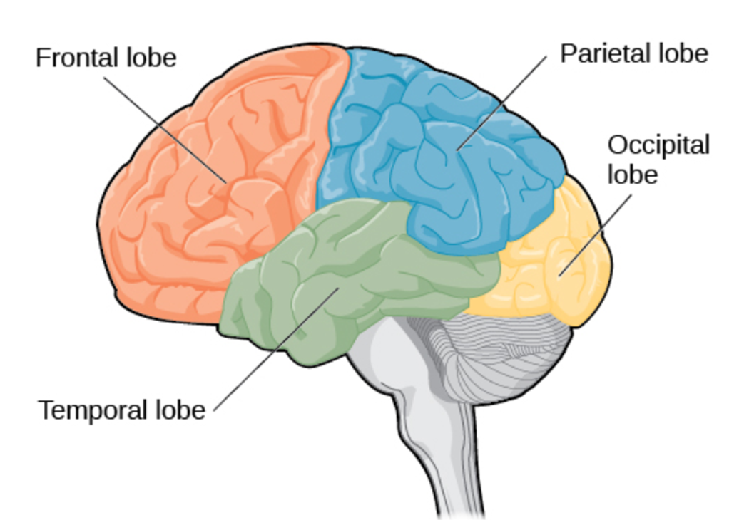
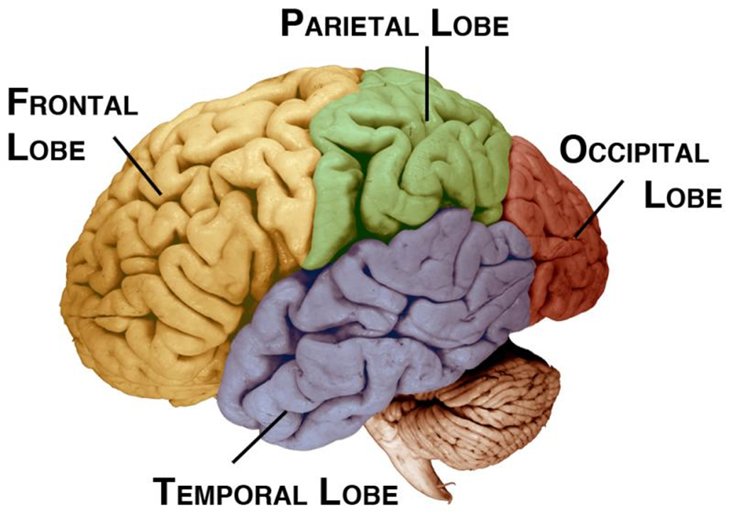
Lobes of the Cerebrum (diagram)
Grey and White Matter (definitions)
Grey Matter: Contains the cell bodies, dendrites and axon terminals of neurons, so it is where all synapses are.
White Matter: Made of axons (including fatty myelin sheath) that connect different parts of grey matter to each other.
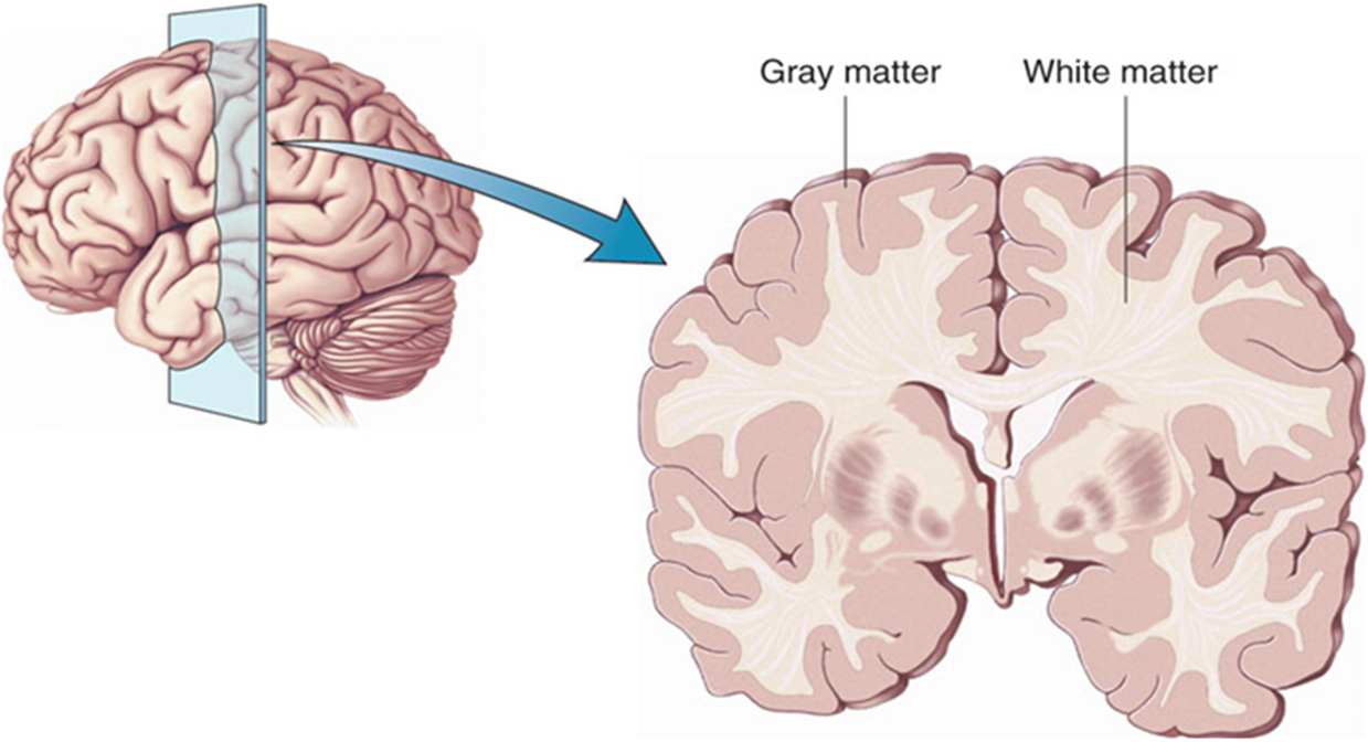
Cerebrum: cerebral cortex (definition, function)
Outermost layer of the cerebrum. Comprises of folded grey matter, where all the thinking happens.
Forebrain: Corpus callosum (function)
Function: Connects the right and left hemispheres of the brain and allows them to communicate.
The lobes of the Cerebral Cortex (names)
The cerebral cortex is subdivided into lobes. The lobes are defined by deep grooves (fissures) in the cerebral cortex. They are:
frontal lobe
temporal lobe
occipital lobe
parietal lobe
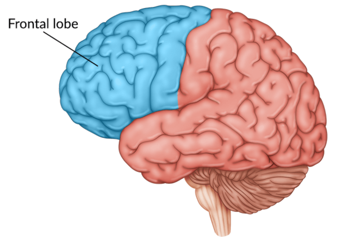
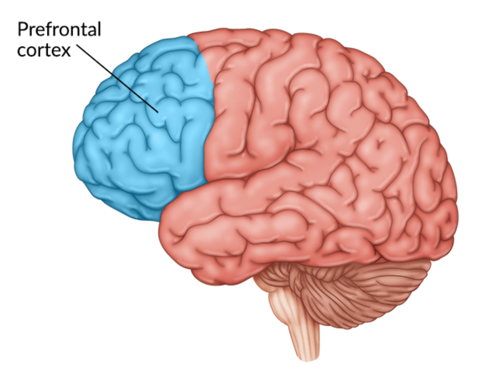
Frontal lobe (subparts)
It is made up of:
prefrontal cortex
primary motor area
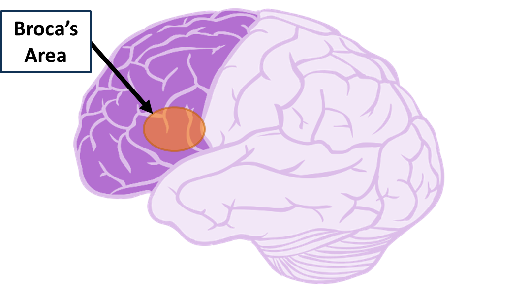
Broca’s area (LEFT HEMISPHERE ONLY)
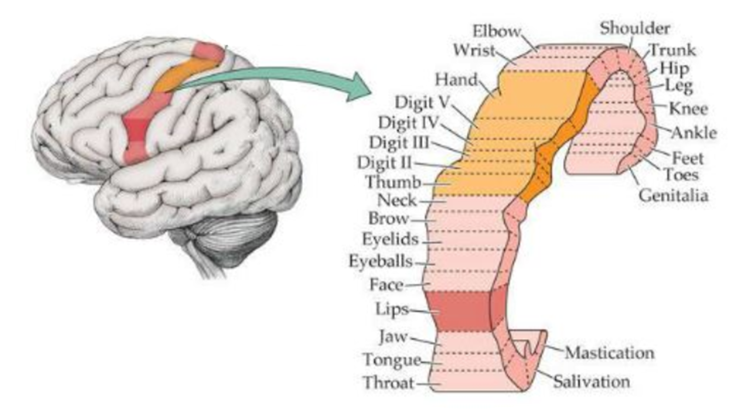
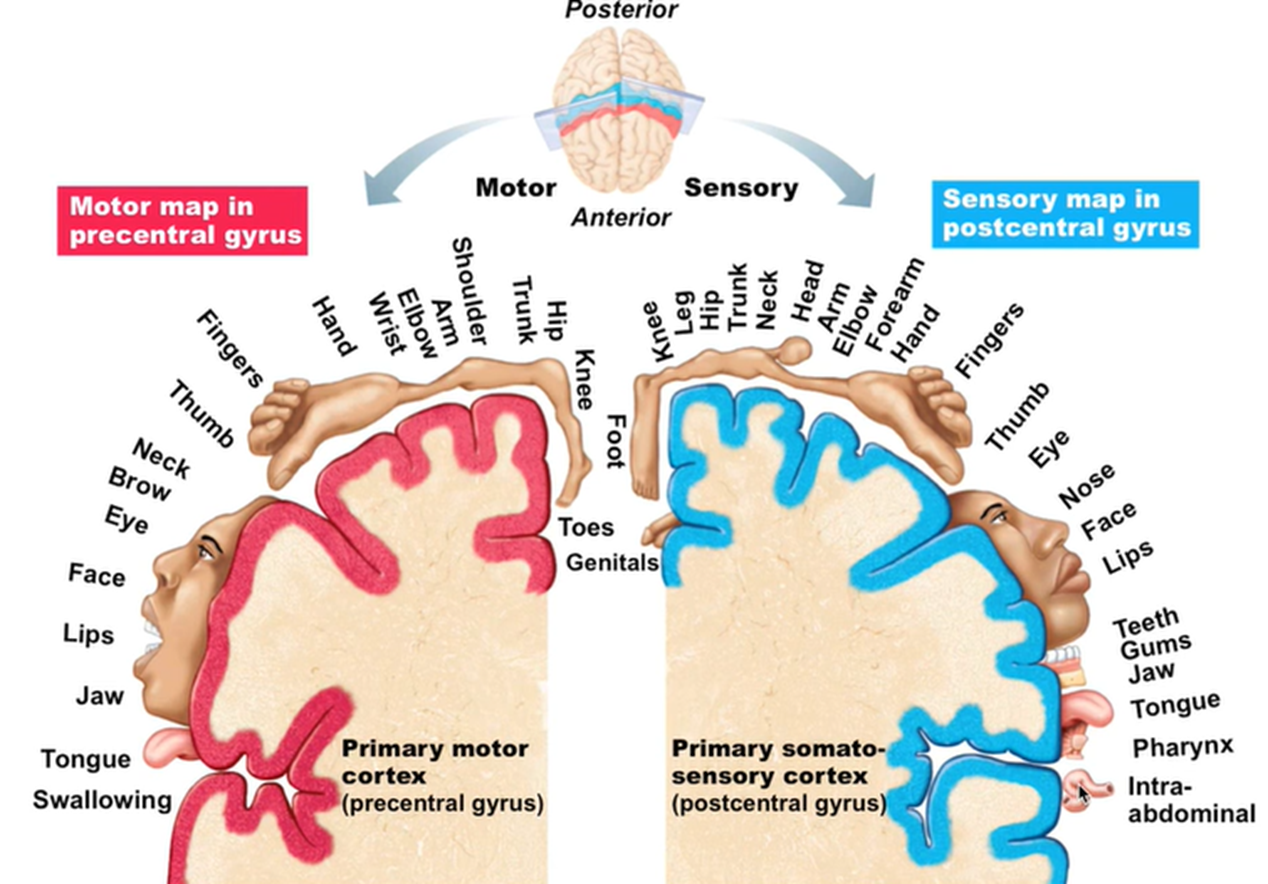
Frontal lobe: Primary motor cortex (function, how it works and looks)
It generates neural impulses that control the execution of voluntary movement.
When performing a voluntary movement, action potentials are sent from the primary motor cortex via fast myelinated motor neurons which activate groups of skeletal muscles to achieve movement.
The amount of brain matter devoted to any body part represents the amount of control that the primary motor cortex has over that body part.
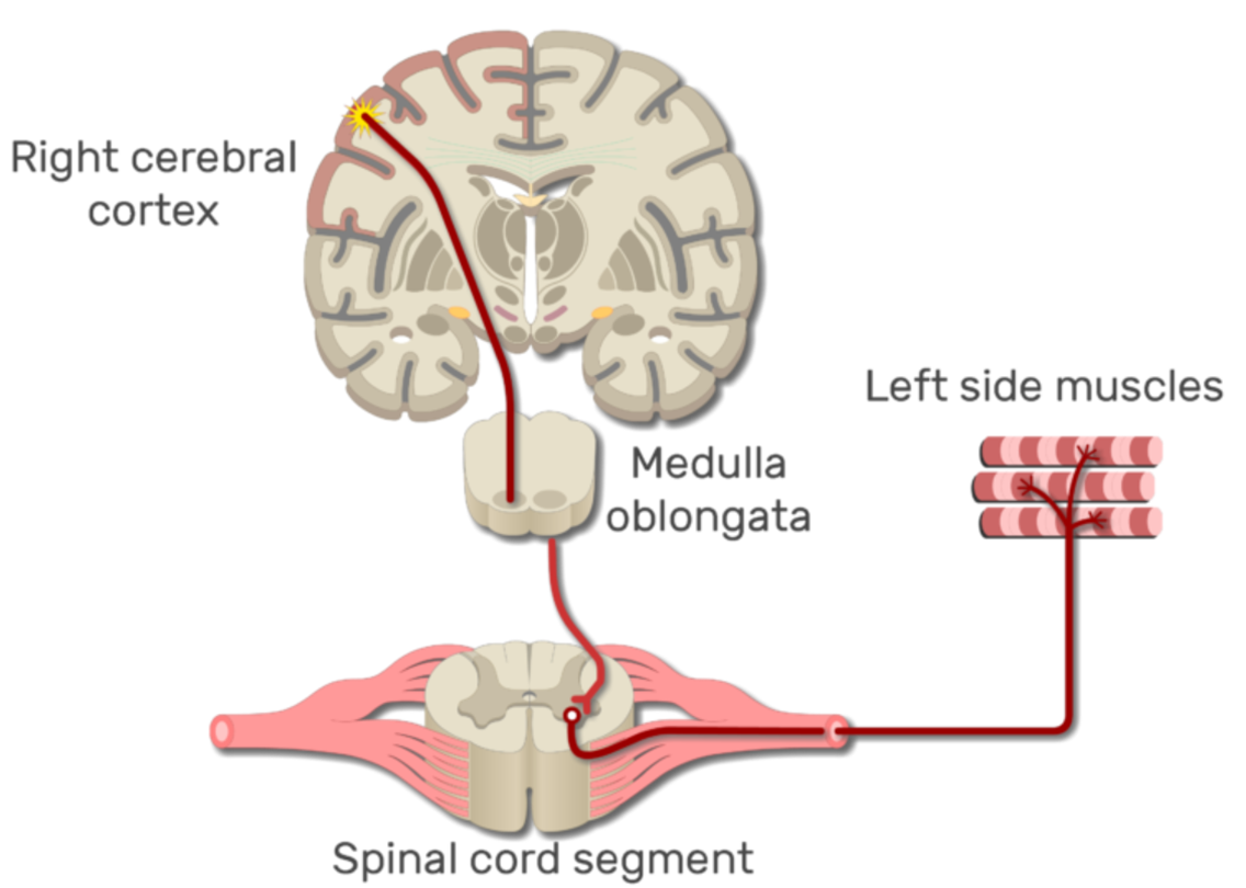
Contralateralisation (contralateral control) (how it works)
Nerve impulses cross the body’s midline to activate skeletal muscles on the opposite side of the body. The left hemisphere of the cerebrum controls voluntary movement on the right side of the body and vice versa.
Frontal lobe: Prefrontal Cortex (function)
Responsible for executive functioning:
Abstract thinking
Decision making/problem solving
Goal-oriented behaviour/planning/motivation
Logic
Judgement and reasoning
Emotional regulation
Personality
Frontal lobe: Broca’s Area (left hemisphere) (definition, function)
Motor speech area - adjacent to the primary motor cortex in the frontal lobe only
Function: controls the fine muscles responsible for clear (articulate) speech production e.g. tongue, cheek, lips and jaw and larynx muscles
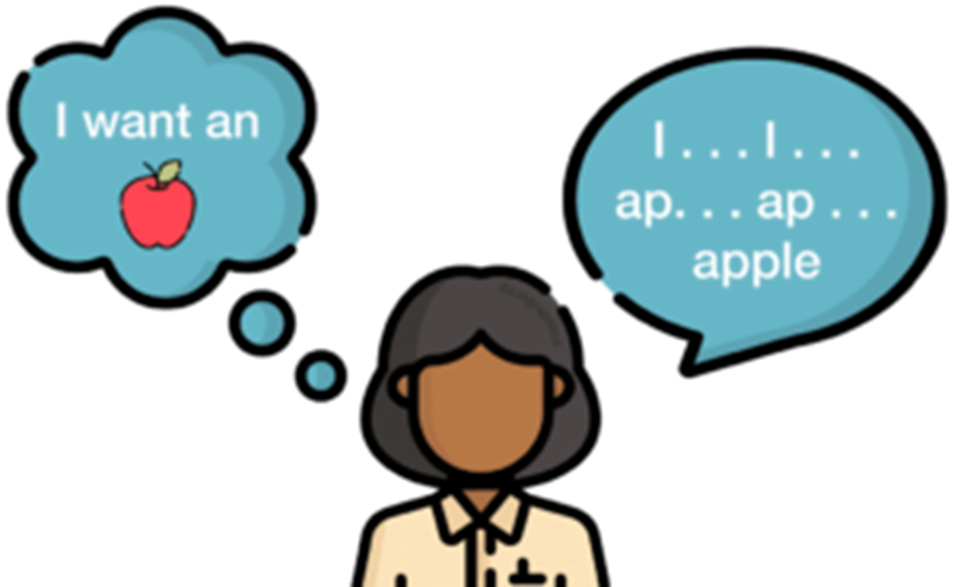
Broca’s aphasia (definition)
When there is an issue in the Broca’s area, a patient can sequence words to make meaningful speech but has difficulty producing the words.
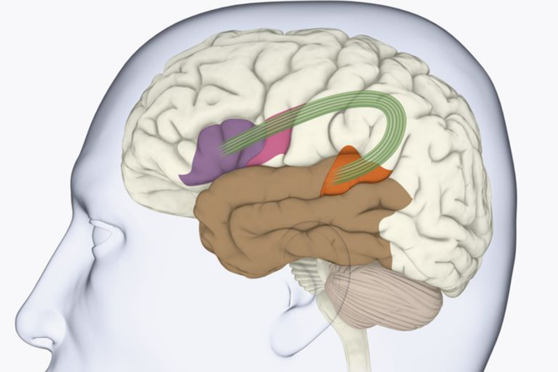
Broca’s and Wernicke’s Areas (and nerve fibres)
Nerve fibers (green) connect:
The Broca's area in the frontal lobe (purple) required for the motor movements for word production
To the Wernicke's area in the temporal lobe (orange) for language comprehension and word sequencing.
Occipital lobe (diagram, what it contains)
Has the primary visual cortex
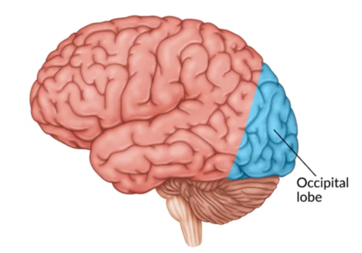
Occipital lobe: Primary Visual Cortex (location, function, how it works)
Location: The primary visual cortex is found in the occipital lobe in both cerebral hemispheres.
Function: Receives and processes visual information e.g. colour, shape, motion and depth
Both hemispheres of the cerebrum have a visual cortex that gets data from the opposing eye.
The right cortical areas interpret messages received from the left eye, whereas the left cortical areas interpret data from the right eye.
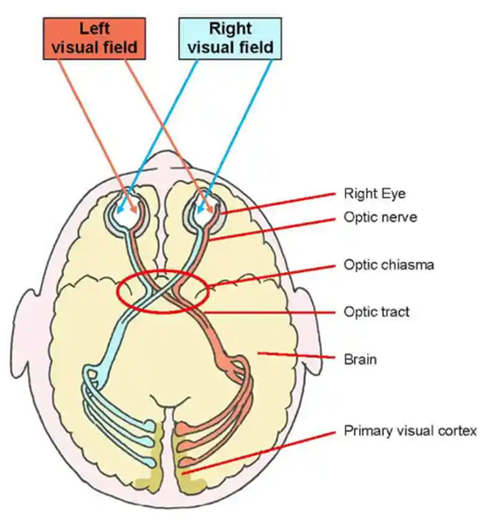
The Organ of Vision: The Eye (how it processes information)
1. Light enters the eye
2. Light is detected by the photoreceptors of the sensory neurons located in the retina (rods for black and white vision, cones for colour vision).
3. The axons of the sensory neurons make up the optic nerve.
4. The optic nerve carries visual information to the Primary Visual Cortex of the Occipital lobe
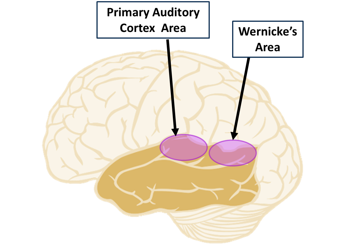
Temporal lobe (diagram, what it contains)
Contains the primary auditory cortex and the Wernicke’s area
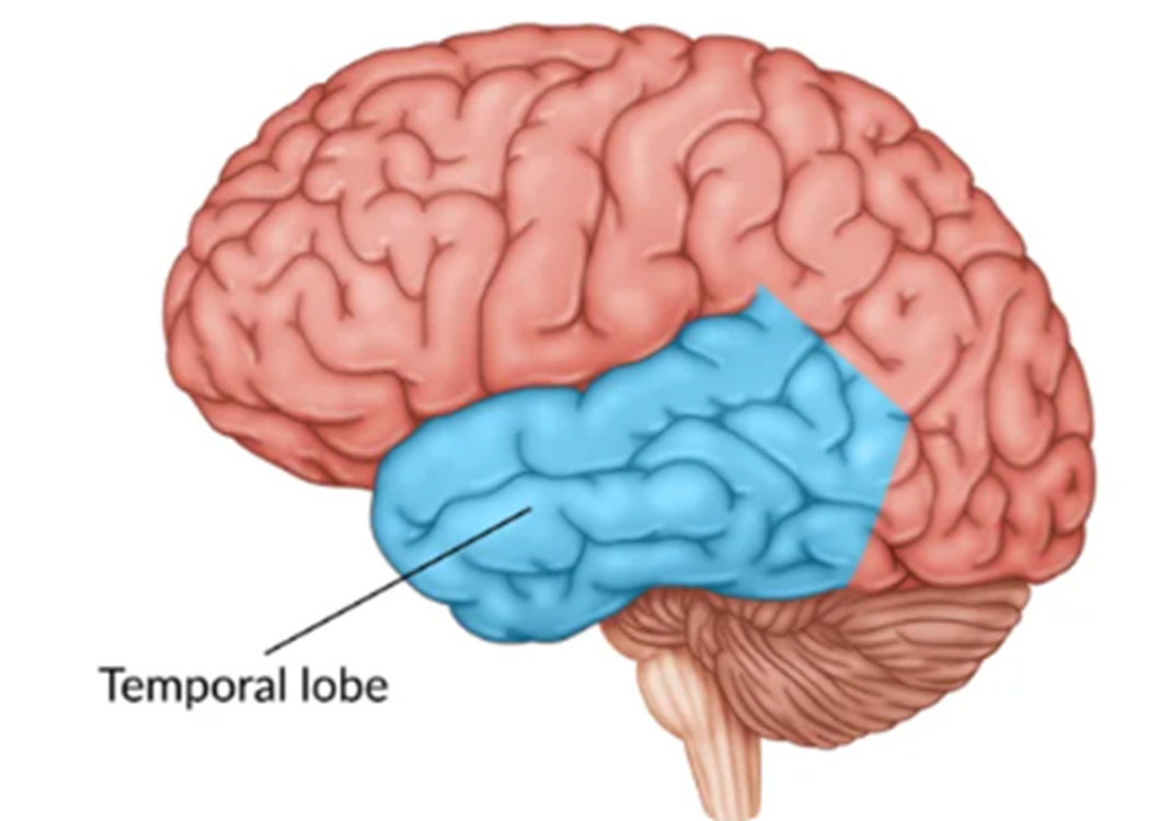
Cerebrum: Temporal Lobe (areas)
Two areas of the Temporal Lobe are:
primary auditory cortex: processing of auditory (sound) stimuli
Wernicke’s Area - allows for the sequencing of words so that sentences make sense
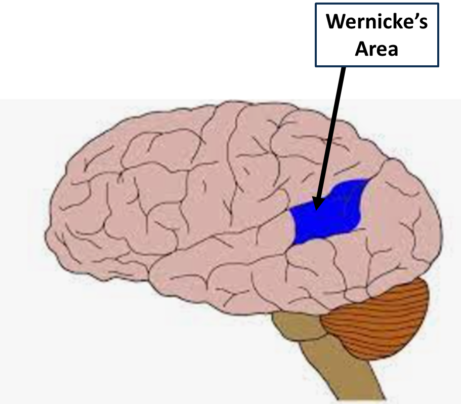
The Temporal Lobe - Wernicke’s Area (LEFT cerebral hemisphere) (function, diagram)
Enables the sequencing of words so that sentences make sense. Allows for the selection of the correct words to express meaning.
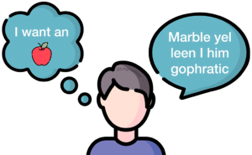
Wernicke’s aphasia (definition)
Damage to Wernicke’s area results in a patient who can produce fluent speech but cannot form sequences of words that make sense to others.
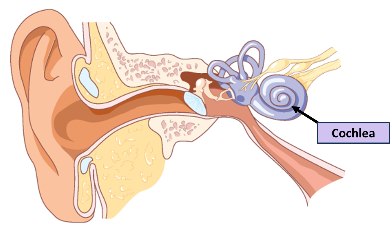
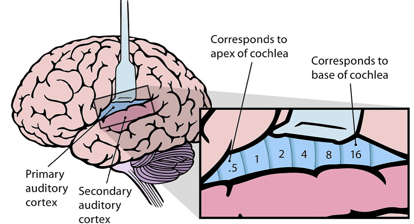
Organ of HEARING - Cochlea of the Ear (how it works)
Auditory nerve contains sensory neurons that convert sound waves into nerve impulses that are transmitted to the primary auditory cortex of the temporal lobe for processing.
Temporal Lobe: Primary Auditory Cortex (how it works)
Neurons in the primary auditory cortex are organised according to the frequency of sound to which they respond best. Neurons at one end of the auditory cortex respond best to low frequencies (low pitch); neurons at the other respond best to high frequencies (high pitch).
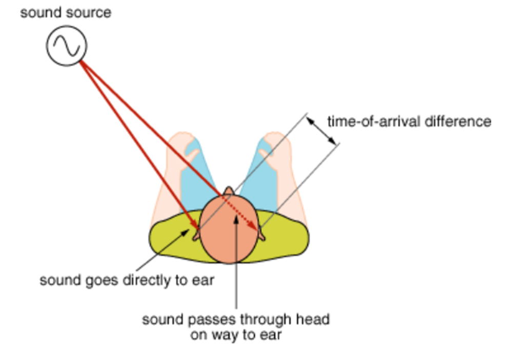
Damage to the Primary Auditory Cortex (how it works)
Destruction of one side of the primary auditory cortex affect one’s ability to localise the source of a sound, because comparative signals in both cortices are required to detect the direction a sound is coming from.
Destruction of both primary auditory cortices greatly reduces hearing sensitivity.
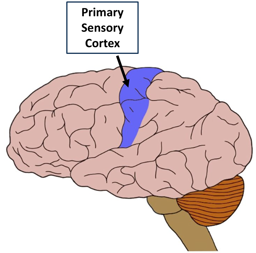
Parietal lobe (diagram, what it contains)
Contains the primary sensory cortex
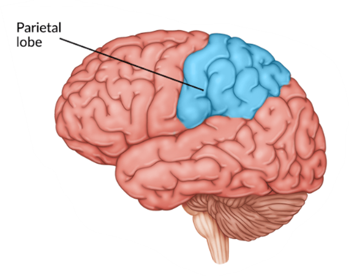
Parietal lobe: Primary sensory cortex (function)
It:
receives general sensory information from skin and proprioceptors of skeletal muscle, joints and tendons
is capable of spatial discrimination - the identification of body region being stimulated
When such receptors detect one of these sensations, the information is sent via the thalamus to the primary sensory cortex of the Parietal lobe.
Homunculus: Primary sensory cortex (blue) and primary motor cortex (red)
Case studies as a research design (what it involves)
Involves the collection of detailed data based on observation and study of an individual, group of individuals or an event.
Case studies: strengths
provide detailed (rich qualitative/descriptive) information
provides insight for further research
allows for investigation of otherwise impractical (or unethical) situations
Case studies: limitations
findings cannot be generalised to the wider population
researchers’ own subjective feeling may influence the case study
difficult to replicate
time-consuming and expensive to conduct
Phineas Gage case study (overview)
was a brain injury survivor - 25yo foreman on the railroad (was capable, active, well organised, reasonable and calm)
an accident forced an iron rod into the left side of Gage’s face under his cheek bone, through his frontal lobe and out the top of his skull
changed his behaviour
and personality, become argumentative and angry, knew no social inhibitions
provided the first evidence that the prefrontal cortex was involved in personality and impulse control.
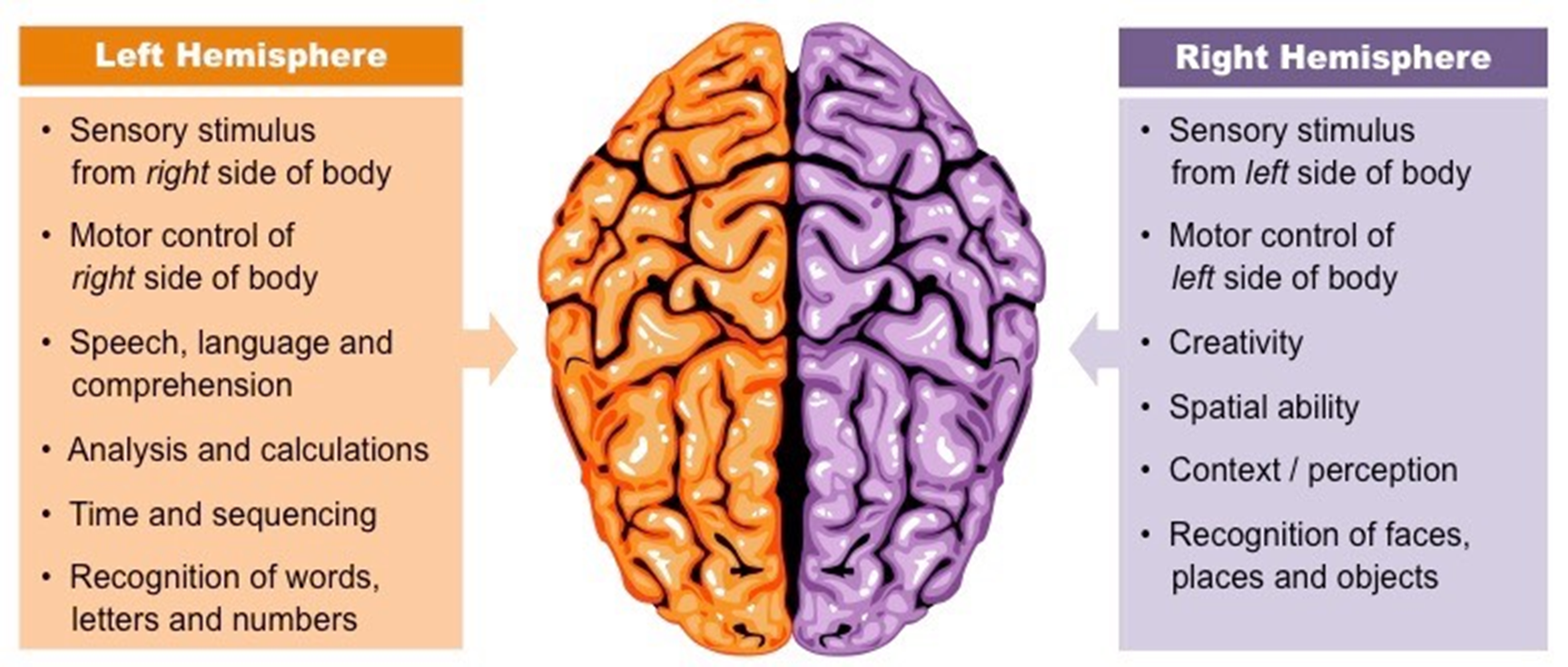
Cerebrum: Hemispheric Lateralisation (definition, what it controls)
Hemispheric laterialisation is the idea that each hemisphere is responsible for different functions
Cerebrum: Corpus callosotomy on Epileptics (overview)
Split Brain surgery disconnects the two hemispheres of the brain by cutting the corpus callosum - historically performed on patients with uncontrollable epileptic seizures, limiting the spread of epileptic activity (communicated of misfired neurons) between the two hemispheres of the brain.
Roger Sperry: Split Brain Experiments (overview)
studied patients that had split brain surgery
presented visual information to one hemisphere or the other in isolation
research showed the two hemispheres of the cerebrum are responsible for processing different information (called hemispheric laterialisation)
Sperry discovered that the left hemisphere of the brain was responsible for language understanding and articulation, while the right hemisphere could recognize a word, but could not articulate it.
Sperry’s split brain experiments (method)
Sperry’s sample used people who had previously had their corpus callosum cut, so the two hemispheres of the cerebrum could not communicate with each other.
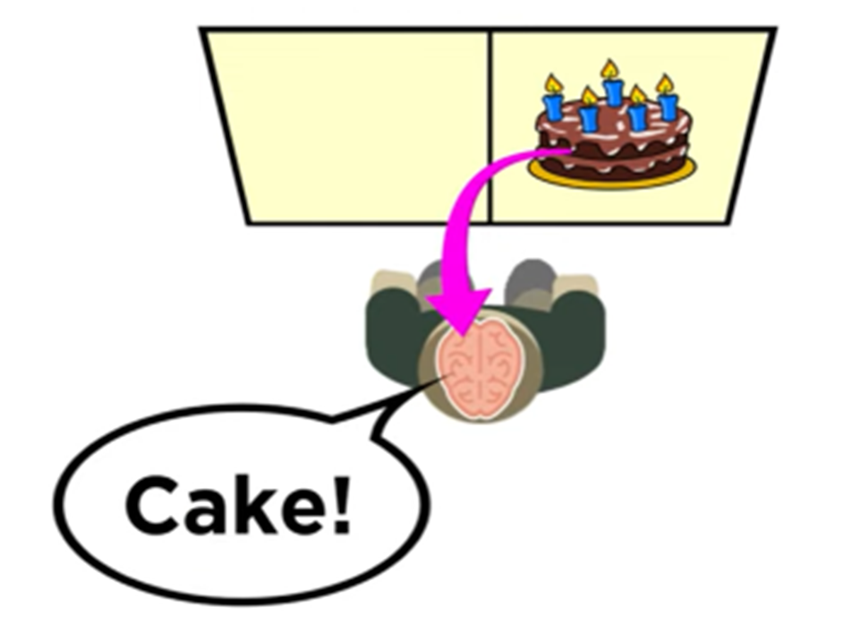
Cake image presented to the left hemisphere in isolation and the patient is asked what they saw:
Cake image from the right visual field of view goes to the left cerebral hemisphere.
Language is localized in the left hemisphere.
Participant says cake.
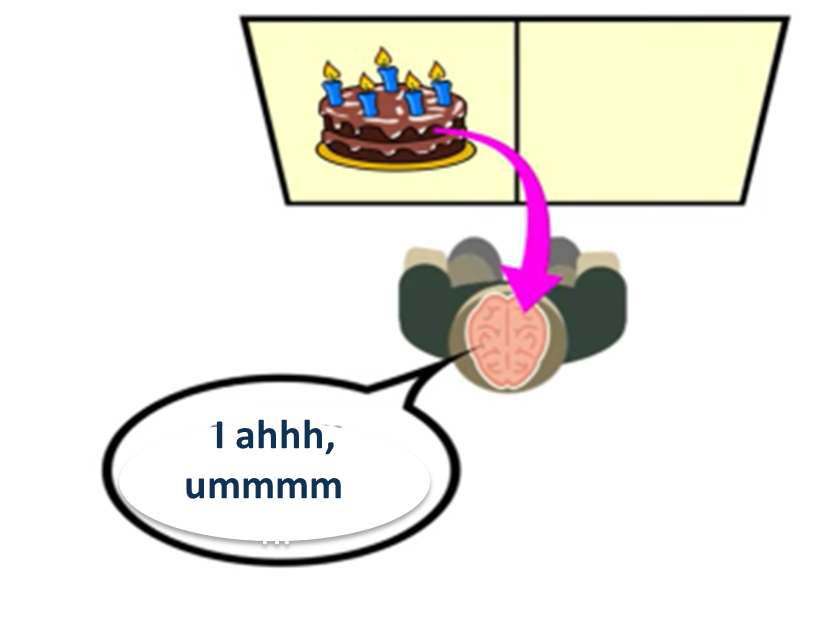
Cake image presented to the right hemisphere in isolation and the patient is asked what they saw:
Cake image from the left visual field goes to the right cerebral hemisphere.
Language is localized in the left hemisphere.
Because of the severed corpus callosum the hemispheres are unable to share information.
Participant can’t say what they see.
Participant is unable to say what they saw but they could draw it - Right hemisphere is for spatial processing and visual motor tasks.
Ethical guidelines (definition, explanation)
Codes of practice to be followed by people involved in psychological research. They allow people to deem what actions are morally right and wrong.
Ethical guidelines are used by organisations such as the Australian Psychological Society, universities and research hospitals to define what actions are morally right and wrong.
Guidelines are used by members as a code with which them to perform their duties.
Ethics Committee (definition, explanation)
A professional body that reviews research proposals planning to use human participants to see if they meet Australian guidelines in the Australian Code of Ethics.
·There are more than 200 human research ethics committees in Australia, most are attached to research organisations e.g. universities, hospitals
·Prior to the commencement of research, the researcher submits a research plan to an ethics committee for approval
Role of ethics committees (purpose, explanation)
Ethics committees assess if the potential benefits of the research to society are justifiable in the light of the possible risk of physical or psychological harm to the participants.
They decide whether there has already been similar completed research the nullifies the current proposal.
Then deny or grant approval for the study to be conducted.
Monitor the ethical conduct throughout the study to ensure ethical guidelines are being followed.
Ethical Guidelines: Protection from Harm (definition and meaning)
Researchers must protect the physical and psychological wellbeing of participants, following the ethical guidelines to achieve this.
Researcher must ensure if vulnerable groups are used, they receive suitable care e.g., children, people with disability.
Debriefing participants once the research has been completed may reduce any psychological harm done.
Ethical Guidelines: Informed Consent (definition)
Written, informed agreeance to participate in the research must be obtained from each participant before commencement of the research.
A guardian must sign if participant is under 18 years.
What must participants be informed about in a consent form? (ethical guidelines: informed consent)
·Purpose of the study
·What the participant will be required to do and the required time investment of the participant
·Any potential risks of participating in the study
·What information will be collected and recorded: privacy
·How confidentiality of the participants personal information will be maintained
·The participant’s withdrawal rights, and that participation is voluntary
Ethical guidelines: Withdrawal Rights (definition)
The participant can end their participation in the study at any time; or have their results removed during or at completion of the study, for any reason incurring no penalty or pressure to continue.
Withdrawal rights should be explained in the consent form.
Ethical Guidelines: Deception (definition and description)
Deception is used when the participants knowing the true purpose of the study would act as a demand characteristic - clues in an experiment which lead participants to change their behaviour because think they know what the researcher is looking for, thus affecting the results.
Deception violates the principle of informed consent, but ethical standards recognize that deception is sometimes required to achieve a research purpose.
At the end of the study, that uses deception, the participants must be fully debriefed about the “what and why” of the deception. As the participant agreed take part without actual knowledge of what they were consenting to they can request their results be removed from the study.
Ethical Guidelines: Privacy (definition and description)
The right of protection from unwanted intrusion by the Government or other into one’s affairs - protected by privacy Commonwealth laws.
Privacy is concerned with what participant information is collected; only information relevant to the study should be collected from participants.
Ethical Guidelines: Confidentiality (definition, description, how to achieve it)
Adopting of procedures to ensure that personal information pertaining to the participant is not revealed to those outside of the study.
Confidentiality is concerned with how participants’ information is collected and safeguarded.
How to achieve confidentiality:
store participants’ information securely
ensure only researchers have access to participant information
dispose of participant information when no longer required
do not include names or identifying information of participant in published reports; written consent from the participant or guardian must be obtained before publishing.
Ethical Guidelines: Voluntary Participation (definition)
People are willing participants in the research, meaning coercion or pressure cannot be applied to the individual to achieve participation, and negative consequences cannot be applied if they chose not to participate.
Ethical Guidelines: Debriefing (definition and when it is needed)
An explanation of the research given to the participants at the conclusion of the study.
It should include the following where applicable:
When deception is used: explanation about the true purpose of the experiment, what and why of the deception employed, along with an offer for the participant to remove their data from the study as it was gathered under false pretenses.
Use of a control group: What group the participant was in - control (placebo) or experimental should be divulged to the participant
Psychological harm done: offer of counselling to the participant
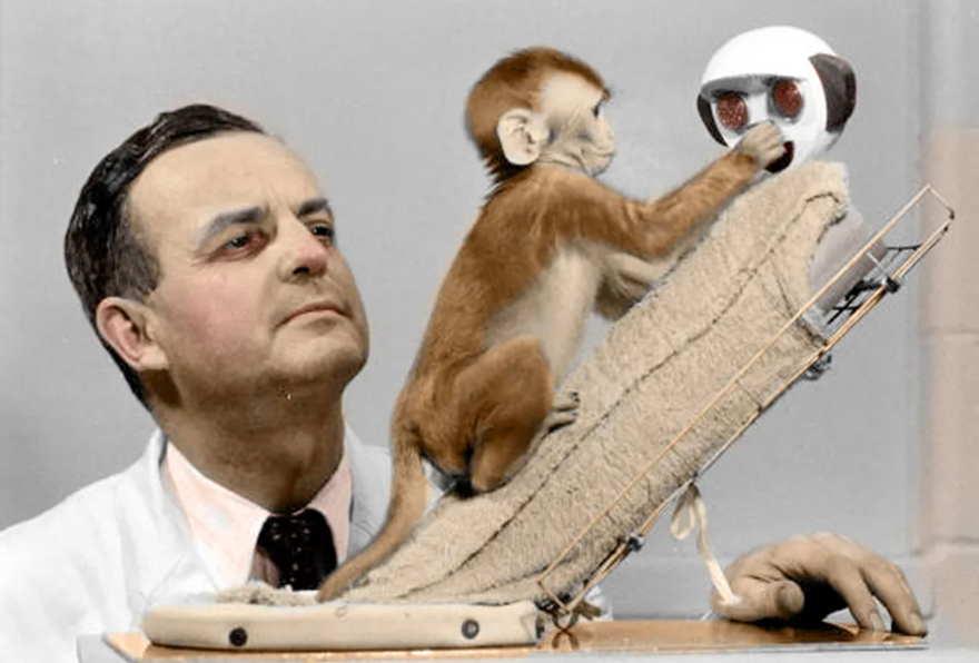
Why are animals used in research? (and how much?)
Psychology is a study that is designed to understand human behavior and how a person’s mind works
The underlying assumption for the use of animals in research is that (to some degree) laws of behavior are the same for all species and knowledge gained by studying rats, dogs, cats and other animals can provide insight into humans
7-8% of psychological research involves the use of animals
90% of animals used in research are rodents and birds
Only about 5% animals used are monkeys and primates
Using Animals in Research: Strengths
Studying other species attempts to avoid some of the complex ethical problems involved in studying humans. E.g.,
looking at the effects of maternal deprivation by removing infants from their mothers
isolation experiment on humans would not be possible
Some human behaviours are like other species. E.g., territoriality, courtship rituals, defending our young, aggressive when threatened, engage in play. Parallels can be inferred between ourselves and other mammals with complex forms of social organisation.
Using Animals in Research: Limitations
Animals are not humans. Humans have a more sophisticated intelligence than other species and comparatively more of our behaviour is due to conscious decision than instinct or drive.
Conclusions based on animal research cannot be generalised to humans.
Humans are the only animal to develop language. Other animals communicate using signs, we use symbols; our language enables us to communicate about past and future events as well as about abstract ideas.
Ethical breaches - human subjects can give or withhold their consent, while animals used in problematic experiments didn’t have that choice. They cannot provide informed consent and cannot withdraw.
APA Ethics code to minimise physiological and psychological harm to animals (outline)
•Receive instruction in the care, maintenance and handling of the species being used.
•Make reasonable efforts to minimise the discomfort, illness and pain of animal subjects.
•Use procedures subjecting animals to pain, stress or privation only when an alternative procedure is unavailable, and the goal is justified by its prospective scientific, educational or applied value.
•Perform surgical procedures under appropriate anaesthesia and follow techniques to avoid infection and minimize pain during and after surgery.
•Proceed rapidly when it is appropriate that an animal's life be terminated, with an effort to minimise pain.
Australian code for the care and use of animals (identification)
Three Rs
Replace (animal studies with non-animal methods)
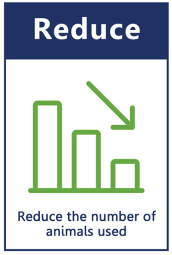
Reduce (as few animal studies as required and necessary)
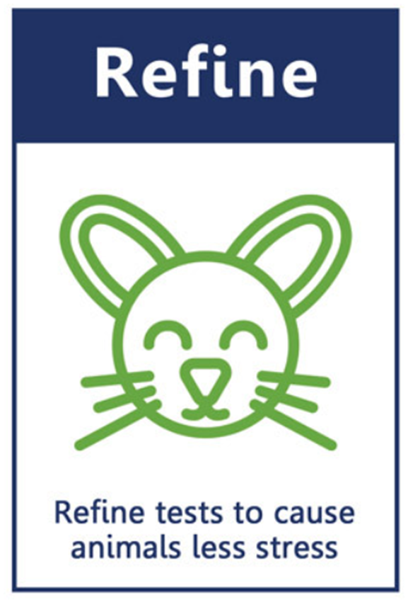
Refine (minimise stress of study animals)
Three Rs: Replacement (definition)
The use of alternative methods that eliminate the need for animals in the study.
Three Rs: Reduction (definition)
The use of methods that result in obtaining comparable levels of information from the use of fewer animals in scientific procedures or for obtaining more information from the same number of animals.
Three Rs: Refinement (definition)
Use methods that alleviate or minimise potential pain and distress and enhance animal wellbeing e.g. using anaesthetics to minimise pain, and house animals in enriched environments.
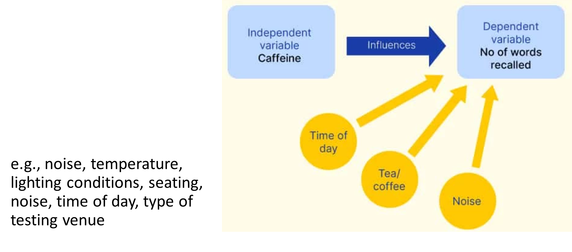
Independent Variable (definition and explanation)
The variable manipulated by the researcher to observe its effects on the dependent variable.
the variable being changed
Some variables cannot be manipulated by a researcher, these variables cannot be studied experimentally. Some examples-
-Premature birth – the researcher cannot induce mothers to give birth early
-Addiction- the researcher cannot addict people to cocaine to research the drug’s effects on the body
Dependent Variable (definition, explanation)
The variable that responds to the change made by the experimenter.
the variable being tested.
The experimenter develops a measurement tool to measure the dependent variable.
Dependent variable vs Measurement tool:
Dependent Variable | Measurement Tool |
Goal accuracy | Number of goals in 2 minutes |
Level of depression | Becks depression inventory |
Memory | Recall of words from a list of 20 |
Child’s exploration of environment | Number of toys interacted with |
Obedience to an authority figure | Number of volts perceived to be delivered to a learner to teach them |
Hunger levels | Number of Kilojoules consumed daily |
Controlled variables (definition, types, often made mistake)
Variables, other than the IV, that may influence the DV and therefore are kept constant across the whole experiment.
The types are:
Controlled Participant Variables
•Participants must not have preexisting medical conditions other than allergies
•Participants must have an average 8-hour sleep pattern
Controlled Environmental variables
•The antihistamine is to be taken before food on waking in the morning
•Method of drug delivery – tablet form
Often made mistake: students claim that the amount of drug must be controlled – implying all the participants in the 5 mg group need to get 5mg of antihistamine, and so on. This is correct, but dosage is the IV and is manipulated across the experiment into four conditions- 0mg, 5mg, 10mg and 15mg. Dosage therefore is not kept constant across the whole experiment and is not a controlled variable.
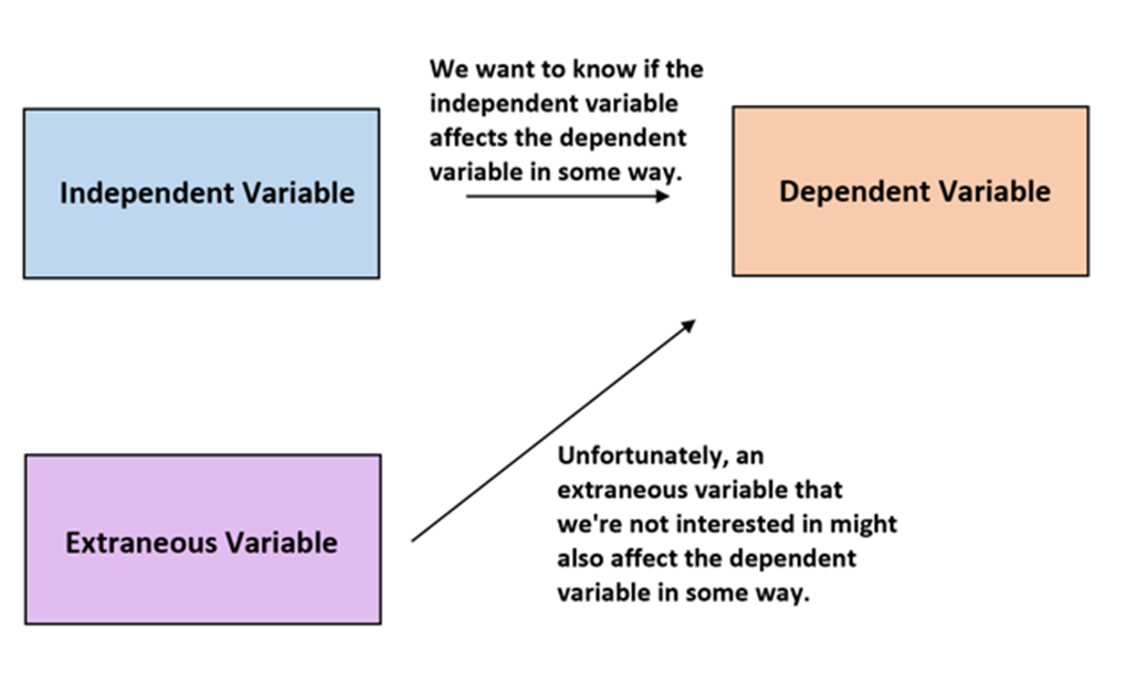
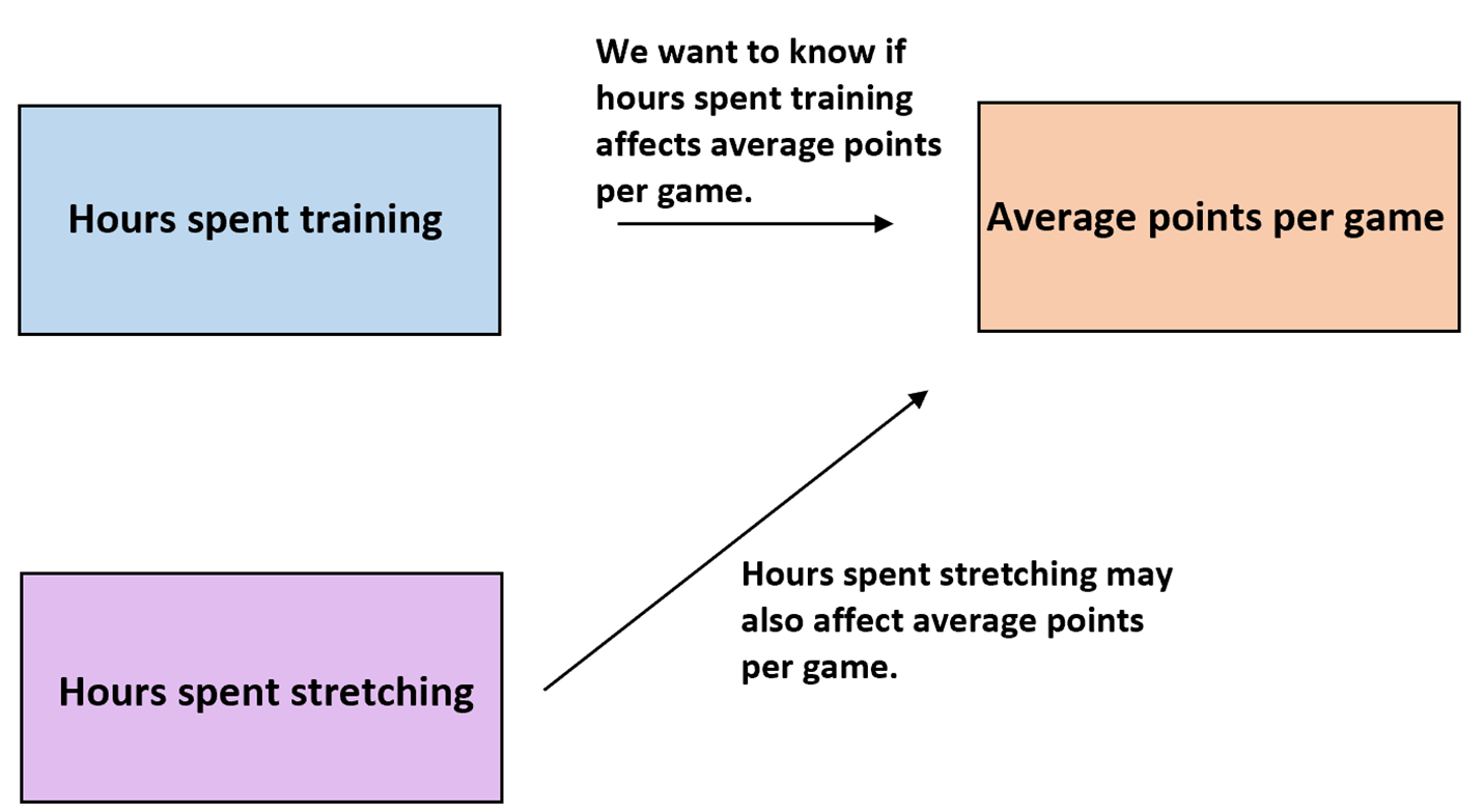
Extraneous variables (definition, explanation, example)
An unwanted variable that may impact the dependent variable.
The potential influence of an extraneous variable on the dependent variable requires that the variable be controlled during experiments so that it does not confound the results.
Extraneous variables that are identified and controlled in an experiment become controlled variables.
Extraneous variables that are not identified and controlled in an experiment may become confounding variables.
Extraneous Participants Variables (definition, explanation, examples)
An extraneous variable related to the characteristics of the individual.
Characteristic or aspect of a participant’s background that could affect study results, even though it’s not the focus of an experiment.
e.g. memory, culture, mood, anxiety, intelligence, physical skills, gender, motivation, education, diet, mood, anxiety, intelligence, self-esteem, awareness, confidence, abnormal sleep patterns, fed state, pre-existing medical conditions
Extraneous Environmental Variables (definition, examples)
An extraneous variable related to the environment that the research takes place in and how it affects the participant responses.
e.g., noise, temperature, lighting conditions, seating, noise, time of day, type of testing venue
Extraneous Researcher Variables (definition, examples and explanation)
An extraneous variable relating to the personality, characteristics, appearance and conduct of the researcher that unintentionally impact how the participant responds.
Unintentional clues: A researcher might unintentionally suggest subtle clues to the participant about what the experiment is about of how a participant should behave.
Personal Attributes of the Researcher: Age, gender, accent manner politeness, personality, opinions of the researcher
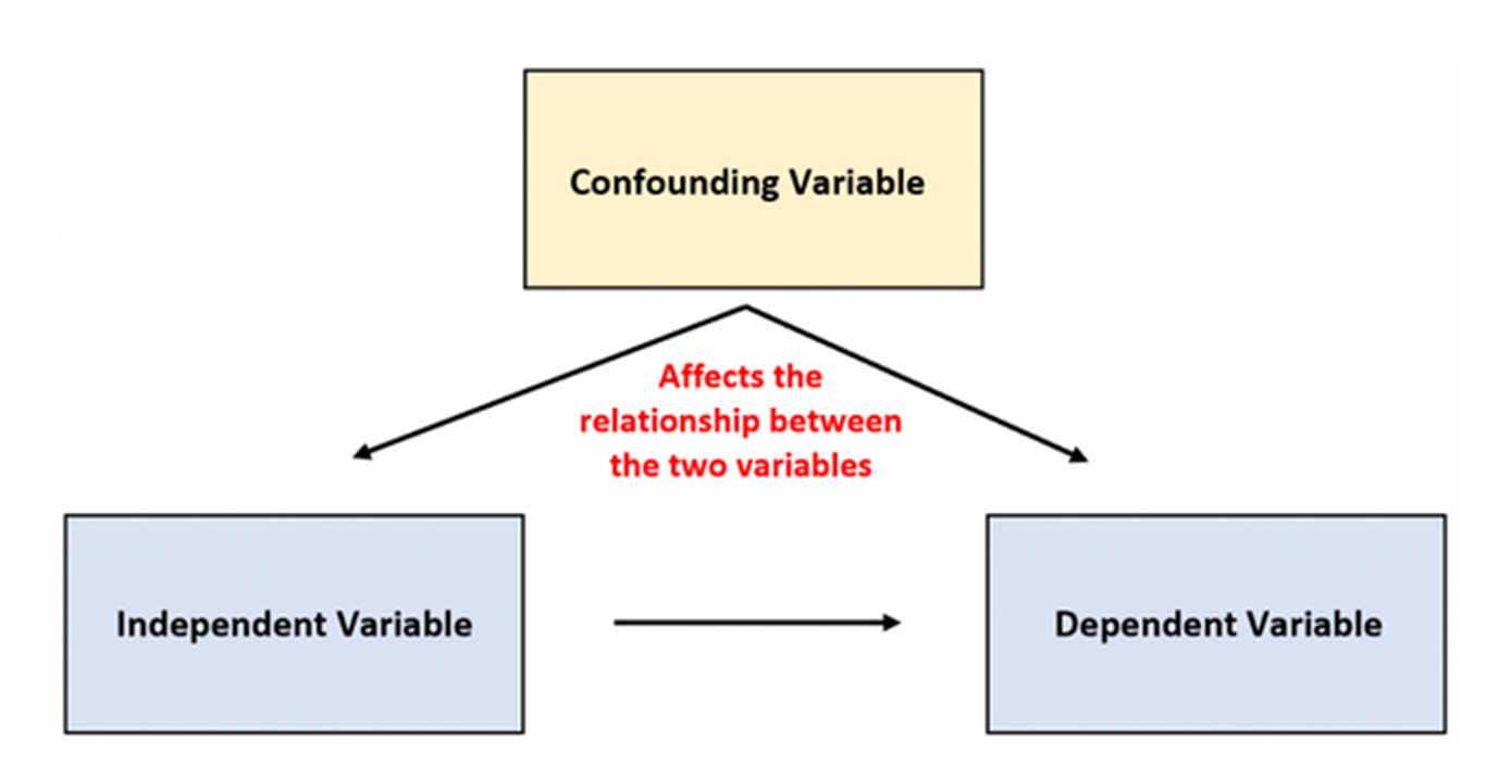
Confounding variables (definition, explanation, example)
Variables, other than the independent variable, that impact the dependent variable, as they have not been controlled by the researcher.
Confounding variables can make it difficult to determine if the results are due to the influence of the independent variable, the confounding variable, or an interaction of the two.
Extraneous variables that are not controlled may become confounding variables.
Example: Annually murder rates and ice cream sales rise together.
•Possibility #1: Murders cause people to purchase ice cream.
•Possibility #2: Purchasing ice cream causes people to murder or get murdered.
•Possibility #3: A confounding variable—which causes the increase in BOTH ice cream sales AND murder rates
Wintery weather: people stay at home rather than go outside and murder people. They probably don’t eat a lot of ice cream.
Summery weather: people spend more time outside interacting with each other, are more likely to get into situations that lead to murder. They probably buy ice cream as a cooling treat.
Sources of Extraneous and Confounding Variables (identify)
Experimenter Effect
Demand Characteristics
Placebo Effect
Sources of Extraneous/Confounding Variables: Experimenter Effect (definition, explanation, examples)
The expectations and behaviours of the researcher that may bias the results. This can include:
Unintentional errors in observations- E.g., only seeing what the experimenter expects to see.
Making inaccurate measurements, recordings analysis or interpretations of the data. E.g., failure to record data the experimenter doesn’t want to include.
Presenting the experiment to the participants in a biased way. E.g., wording, question order, tone, expression, emphasis, elaboration of a certain point.
Treating participants in the different trial groups differently. E.g., giving verbal and non-verbal feedback, as well as biased presentation.
EXAMPLE: Study Aim: To check how well participants can recall a text read out loud to them as an audio book opposed to text that they read silently to themselves.
The experimenter, while giving instructions, dismisses the part where participants must read the text carefully and let's slip that they should pay close attention to what is being said by the narrator in the audio book.
Based on this one clue, the subjects automatically focus more on the audio book test and score a better recall result than on the text that they read silently to themselves―thus negating the validity of the study.
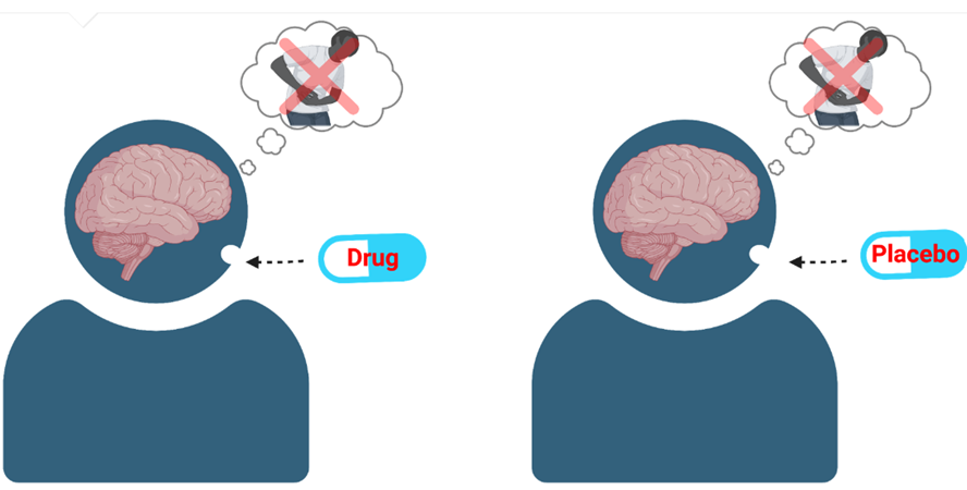
Sources of Extraneous/Confounding Variables: Placebo Effect (definition, explanation)
A phenomenon where people report real or perceived improvement after taking a substance or treatment with no therapeutic value.
The belief or expectation that a treatment will work is enough to relieve symptoms along a variety of clinical conditions like depression, sleep disorders, menopause and allergies. One of the strongest placebo effects occurs during the treatment of pain.